SURGICAL TECHNIQUES FOR THORACIC, ABDOMINAL, PELVIC, AND EXTREMITY DAMAGE CONTROL

Published on 10/03/2015 by admin

Filed under Critical Care Medicine

Last modified 22/04/2025

Print this page

rate 1 star rate 2 star rate 3 star rate 4 star rate 5 star
Your rating: none, Average: 0 (0 votes)

This article have been viewed 1356 times

CHAPTER 60 SURGICAL TECHNIQUES FOR THORACIC, ABDOMINAL, PELVIC, AND EXTREMITY DAMAGE CONTROL

Injury severity and spectrums of injury have continually evolved, resulting in greater and different challenges for the modern trauma surgeon. High energy blunt trauma, with resultant multisystem organ injury, as well as increasingly sophisticated firearms with greater wounding capacity, has resulted in greater severity of injury. Despite the fact that these injury patterns are more likely to result in the death of a patient, improvements in prehospital transport and trauma resuscitation have allowed more moribund patients to reach the hospital alive but in extremis. Damage control surgery, addressing the life-threatening injuries immediately but delaying definitive repair until the metabolic and physiologic perturbations have been corrected, has evolved to address this population of patients.

Damage control has three separate and distinct phases of management. The first phase includes aggressive volume expansion, rapid control of hemorrhage, and the minimization of contamination, often associated with placement of packing to tamponade bleeding. Next, contamination from hollow viscus injuries is quickly obtained. Temporary wound management strategies are implemented. The second phase, which occurs in the intensive care unit (ICU), involves the aggressive rewarming of the hypothermic patient, with correction of coagulopathy, and ongoing resuscitation with crystalloids and blood. Once normal physiology has been re-established, the third phase involves definitive operative management of the patient’s injuries.

Indications for damage control strategy are multiple. The goal of the damage control procedure is to preserve life in the face of devastating injuries with profound hemorrhagic shock. As described by Moore et al., they include an inability to achieve hemostasis resulting from ongoing coagulopathy, a technically difficult or inaccessible major venous injury, a time-consuming procedure in the face of under-resuscitated shock, and a need to address other life-threatening injuries. These indications have been expanded to include hemodynamically unstable patients with high-energy blunt torso trauma or multiple penetrating injuries, or any trauma patient presenting in shock with hypothermia and coagulopathy (Table 1). Most commonly applied to the abdomen, damage control approaches have now been applied successfully to both devastating thoracic and orthopedic injuries.

Table 1 Indications for Damage Control Procedures

Adapted with permission from Rotondo MF, Zonies DH: The damage control sequence and underlying logic. Surg Clin North Am 77:761–777, 1997.

PREDISPOSING FACTORS

The goal of damage control procedures is the early termination of surgical intervention before the development of irreversible physiologic derangement. Uncontrolled hemorrhage results in global ischemic injury, while resuscitation subjects the patient to further injury resulting from the period of reperfusion. Inevitably the lethal triad of hypothermia, coagulopathy, and acidosis develops. While each of these complications is potentially life threatening in and of themselves, the combination of the three together results in an exponential increase in morbidity, contributing to a downward spiral that eventually results in the patient’s demise if not corrected. Damage control surgery, by abbreviating surgical intervention and returning the patient to the ICU for correction of the lethal triad, has resulted in improved mortality over time.

Hypothermia is defined as a core temperature of less than 35° C. The reasons for hypothermia after trauma include aggressive resuscitation with unwarmed fluids, exposure at the time of the primary and secondary survey with radiant heat loss to the environment, and evaporative loss from exposed peritoneal and pleural surfaces in the operating room. The incidence of clinical hypothermia after trauma laparotomy is 57%. Hypothermia has clearly been shown to increase mortality, increasing significantly in patients with a core temperature less than 34° C, and approaching 100% in patients with a core temperature less than 32° C.

Hypothermia has systemic effects, including negative impact on hemodynamics (decreased heart rate and cardiac output, increased systemic vascular resistance), renal function (decreased glomerular filtration rates), and central nervous system depression. Hypothermia also independently affects the coagulation cascade, as clot formation relies on a series of temperaturedependent enzymatic reactions. Studies have clearly demonstrated increases in prothrombin time (PT), thrombin time, and partial thromboplastin time (PTT). Hypothermia also affects platelet function, leading to sequestration in the portal circulation and prolonged bleeding times.

Metabolic acidosis occurs after hemorrhagic shock because of the switch from aerobic to anaerobic metabolism during periods of hypoperfusion. The detrimental effects of acidosis include depressed myocardial contractility and a diminished response to inotropic medications. Acidosis predisposes the myocardium to ventricular dysrhythmias and can worsen intracranial hypertension. Finally, acidosis has been shown to worsen coagulopathy by independently prolonging PTT and decreasing Factor V activity. Acidosis has been linked to the development of disseminated intravascular coagulation (DIC) and may exacerbate a consumptive coagulopathy.

Hypothermia, acidosis, and coagulopathy develop secondary to the dilutional effects when massive resuscitation initially is comprised of crystalloid and packed red blood cells without the addition of clotting factor replacement. Exposed tissue factor secondary to tissue injury also contributes to the development of coagulopathy through the activation of the clotting cascade and resultant consumption of clotting factors.

PHASE I: DAMAGE CONTROL OPERATION

Damage Control Laparotomy

Most damage control procedures are performed in the abdomen. In fact, the earliest descriptions of what subsequently became known as damage control surgery were in patients with major hepatic injuries, in whom the placement of perihepatic packing and staged operative management resulted in decreased morbidity and mortality. The most common injuries that trigger a damage control approach are major liver injuries and major vascular injuries.

The primary method of controlling hepatic hemorrhage is packing. The technique of pack placement is of the utmost importance and depends on the anatomic nature of the liver injury. Major hepatic lacerations require complete mobilization of the liver and inflow occlusion (the Pringle maneuver) to minimize blood loss. Direct ligation of bleeding vessels in the depth of the laceration is necessary to obtain surgical hemostasis. Once major vessel bleeding has been controlled, perihepatic packs should be placed anteriorly and posteriorly, compressing the liver between the two beds of packs and providing tamponade. In penetrating injuries, balloon tamponade of the missile tract, in conjunction with perihepatic packing, can be life saving.

There are several options available for the management of major vascular injuries. Some venous injuries will respond to packing. Ongoing bleeding, however, requires direct surgical intervention. Many abdominal vascular injuries can be managed with simple ligation of the bleeding vessel (Table 2). Ligation, however, is not tolerated in aortic or proximal superior mesenteric artery injuries, and is not technically feasible in retrohepatic caval injuries. These injuries are typically initially approached by an attempt at repair or the placement of a temporary intraluminal shunt, with planned repair at a second operation (phase 3). Commonly, Argyle carotid shunts and Javid shunts have been used for this purpose. Chest tubes may be used when larger conduits are necessary. The shunts should be secured using umbilical tapes, vessel loops, or suture, and do not require anticoagulation to maintain patency. Another technique for the management of exsanguinating vascular injury is the use of endoluminal balloon catheters to obtain proximal and distal control of hemorrhage. The catheters are inserted into the vessel at the site of injury, and the balloon inflated. This technique allows repair of the injured vessel in a relative dry operative field.

Table 2 Abdominal Vessel Ligation and Expected Complications

Vessel Complication Recommendations
Celiac axis None  
Splenic artery None if the short gastric vessels are intact  
Common hepatic artery None if the portal vein is intact, possible gallbladder ischemia Cholecystectomy (may be done at second look)
Superior mesenteric artery Bowel ischemia Second-look procedure
Superior mesenteric vein Bowel ischemia Second-look procedure
Portal vein Bowel ischemia Second-look procedure
Suprarenal inferior vena cava Possible renal failure Wrap and elevate legs, assess for compartment syndrome
Infrarenal inferior vena cava Lower extremity edema Wrap and elevate legs, assess for compartment syndrome
Left renal vein (proximal) None  
Right renal vein Renal ischemia Nephrectomy
Common and external iliac artery Lower extremity ischemia Ipsilateral calf and sometimes thigh fasciotomies or extra-anatomic bypass
Common and external iliac vein Lower extremity edema Wrap and elevate legs
Internal iliac artery None  
Internal iliac vein None  

Adapted with permission from Shapiro MB, Jenkins DH, Schwab CW, Rotondo MF: Damage control: collective review. J Trauma 49:969–978, 2000.

Candidates for damage control surgery often have associated hollow viscus injury. The goal in management of these injuries is the control of contamination. Intestinal lacerations may be controlled by linear stapling or by stapled resection. After enterectomy, the gastrointestinal tract is left in discontinuity, and the decision to perform an anastomosis or stoma is postponed until the patient is stabilized and able to return to the operating room for definitive management (phase 3). Associated biliary or pancreatic injuries can often be managed with judicious placement of closed suction drains, with plans to address the injury at the second procedure (phase 3).

Hemodynamic instability caused by splenic lacerations should be managed with expeditious splenectomy. Similarly, hemorrhagic shock resulting from extensive renal lacerations or pedicle injuries should be managed by nephrectomy, especially if attempts at perinephric packing have failed. Ureteral injuries diagnosed during the damage control procedure should be ligated or drained with a percutaneous urostomy. Intraperitoneal bladder injuries should be rapidly oversewn with definitive management delayed until the second operation.

Given the volume of resuscitation after a damage control procedure, and the edema that results from reperfusion of previously ischemic tissue, formal closure of the abdomen is not an option. Forced closure of the abdomen is associated with a prohibitively high risk of subsequent abdominal compartment syndrome, adult respiratory distress syndrome (ARDS), and multisystem organ failure (MSOF). Multiple options exist for the temporary closure of the abdomen. The goals of temporary closure should be containment of the abdominal viscera, control of abdominal ascites, maintenance of tamponade on areas that have been packed, and optimization of later fascial closure.

Simple techniques involve closure of only the skin, with suture or towel clips. This technique will facilitate the tamponade of bleeding while coagulopathy, acidosis, and hypothermia are corrected. However, it does not expand abdominal volumes significantly and is often associated with development of secondary abdominal compartment syndrome. When this occurs, it can be managed in the ICU by simply reopening the skin. Techniques which expand the abdominal volume include coverage of the viscera with the Bogota bag, a 3-liter urologic bag that is stapled or sewn to the skin. The benefit of the Bogota bag over the vacuum-assisted techniques described below is that it allows visualization of the underlying viscera and assessment of viability, which may be important after the use of shunting or ligation for a major vascular injury.

Vacuum-assisted techniques for temporary abdominal closure have been well described. The vacuum-assisted wound coverage is constructed beginning with a nonadherent fenestrated drape placed over the viscera, followed by the application of a sterile surgical towel. Following placement of two closed suction drains, the wound is sealed with an adhesive plastic sheet applied to the skin. The benefits of this technique include maintenance of some tension on the abdominal wall fascia to minimize the risk of loss of domain and to facilitate subsequent delayed fascial closure at a later time. Commercial devices capable of similar functions are available. Another potential technique for temporary closure involves the use of Velcro, which is sutured to the fascia and sequentially tightened. In the acute setting, there is little role for prosthetic material for temporary closure of the abdomen. As there are no prospective trials comparing methods of temporary abdominal closure, the method used is at the discretion of the trauma surgeon and is often institution specific.

Damage Control Thoracotomy

Trauma patients with penetrating thoracic injuries who present in extremis should undergo emergency department (ED) thoracotomy. ED thoracotomy is rarely successful in patients with blunt trauma or with extrathoracic penetrating injury.

ED thoracotomy is classically performed through a left anterolateral thoracotomy at the level of the fifth intercostal space. This exposure permits rapid evacuation of the thoracic cavity as well as inspection of the pericardial sac to rule out tamponade. On entering the chest, any hemothorax is evacuated, and the pericardium inspected. If tamponade is present, a longitudinal pericardiotomy is performed from cephalad to caudad and avoiding the phrenic nerve. In the patient in extremis, the inferior pulmonary ligament is divided, allowing access to the posterior mediastinum, where aortic cross-clamping can be performed.

Cardiac lacerations may be temporarily controlled using a number of methods. Digital pressure over the hole is often quite effective, although the finger should not be inserted into the hole to avoid extending the laceration. A Foley catheter can be carefully passed through the injury into the ventricle and gentle traction applied after inflating the balloon to control hemorrhage. Finally, simple cardiac injuries can be repaired in the ED using a skin stapler to reapproximate the lacerated myocardium.

Exsanguination from pulmonary injuries may be controlled in several ways, including direct pressure, packing, hilar cross-clamping, or lung torsion at the hilar axis. Once in the operating room, penetrating pulmonary injuries are often amenable to a technique described by Asensio as stapled pulmonary tractotomy. A linear-cutting gastrointestinal stapler is placed within the injury tract and fired. With serial applications of the stapler, this will expose the base of the tract, permitting direct ligation of bleeding sites. Air leaks can be selectively oversewn. Patients undergoing tractotomy have a significantly lower mortality than patients undergoing formal resection.

Following damage control thoracotomy, it is not always possible to formally close the chest without prohibitive compromise of airway and ventilation mechanics. Temporary closure of the thoracic cavity can be accomplished should packing be necessary for control of medical bleeding. Following insertion of a chest tube, a skin-only closure may be fashioned to provide adequate tamponade. Routine closure of the incision can be accomplished in a delayed fashion at 48–72 hours (phase 3).

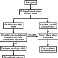
Damage Control Orthopedics

Damage control orthopedics is characterized by primary, rapid, temporary fracture stabilization. This concept applies particularly to femoral shaft fractures and pelvic fractures as these have an increased risk of adverse outcome. Both fractures are associated with significant soft tissue injury and blood loss. The impetus for the development of damage control orthopedics was the observation that patients with severe thoracic, abdominal, and head injuries had worse outcomes when subjected to extended operative procedures for fracture stabilization with ongoing evidence of hypothermia, coagulopathy, and acidosis.

The most common approach to fractures of the lower extremity is the application of an external fixator device to provide temporary fracture stabilization. External fixation is not a time-consuming procedure, and can safely be performed either in the operating room or ICU. The surgeon first obtains alignment of the extremity, and then places two pins above and two pins below the fracture site, avoiding neurovascular structures. The external fixator bars are then applied to the pins, spanning the fracture and providing stability. Although concerns have been raised over the increased risk for deep tissue infection caused by pin site sepsis, no significant increase in local infection has been reported.

Pelvic ring disruptions continue to be a significant source of both morbidity and mortality after trauma. Pelvic fractures account for 3%–8% of all skeletal fractures and most commonly result from high-energy trauma, with motor vehicle crashes being the predominant mechanism of injury. During the acute phase, the goal of treatment should be the control of hemorrhage. The retroperitoneum if intact can contain up to 4 liters of blood, and bleeding from the associated vascular injuries will continue until physiologic tamponade is obtained. However, if the retroperitoneal space is disrupted, pelvic fractures can lead to uncontrolled hemorrhage and increase the risk of exsanguination.

Decreasing the intrapelvic volume by restoring the normal pelvic anatomy remains the first step in the damage control management of the bleeding associated with pelvic fractures. This can be accomplished by multiple methods, including tying a bed sheet around the pelvis or using several commercially available pelvic slings and belts. External fixators and pelvic C clamps may also be used for this purpose and are ideal for “open-book” disruptions of the pelvic ring. The external fixator involves placement of two percutaneous pins per iliac crest. The pins are then connected with a frame that maintains reduction of the fracture and decreases pelvic volume. The C clamp consists of two pins applied on the posterior ilium in the region of the sacroiliac joints. The application of this device is relatively contraindicated in patients with fractures of the ilium and transiliac fracture dislocations.

If the patient remains hemodynamically labile after the application of an external stabilization device, ongoing bleeding from branches of the internal iliac arteries must be considered the source of hemorrhage. Arterial bleeding occurs in only 10% of patients with unstable pelvic fractures. In this patient population, pelvic arteriography with embolization of the bleeding vessel and collaterals is the optimal method of management. In situations where interventional radiology is not available, exploratory laparotomy with ligation of the ipsilateral internal iliac artery and pelvic packing is a viable alternative, although this procedure has a reported mortality rate of 25%.

PHASE 2: RESUSCITATION IN INTENSIVE CARE UNIT

After damage control procedures, patients are returned to the ICU for correction of their physiologic abnormalities, including hypothermia, acidosis, and coagulopathy. Aggressive correction of hypothermia is of paramount importance. External rewarming techniques include warming of the room and ventilator circuits, and application of a Bair Hugger. All fluids and blood products should be given through a Level I fluid rewarmer. In patients with severe hypothermia, defined as a temperature less than 32° C, consideration should be given to the use of continuous arteriovenous rewarming, as described by Gentilello et al. The technique involves the insertion of 8.5F femoral arterial and venous catheters, creating an AV fistula that then diverts a portion of the cardiac output through a heat exchanger, usually a Level 1 fluid warmer. The effectiveness of the technique is based on the patient’s cardiac output, and is capable of raising a patient’s core temperature approximately 4° C per hour. The patients who undergo continuous arteriovenous rewarming require less fluid during resuscitation and have significantly less early mortality than those warmed by conventional external methods.

Patients who are acidotic after damage control procedures are most likely under-resuscitated, and they require optimization of oxygen delivery for correction. Invasive monitoring, including central venous pressure monitoring or Swan-Ganz monitoring, may be necessary in the immediate postoperative period to guide resuscitative efforts. Persistent acidosis in a patient felt to be adequately resuscitated may be caused by a hypochloremic acidosis, which can follow aggressive resuscitation with normal saline. An anion gap can be used to differentiate lactic acidosis from hyperchloremic acidosis. Hyperchloremic acidosis results in a narrowed anion gap, while lactic acidosis results in a widened anion gap.

While correction of acidosis and hypothermia will aid in the correction of the coagulopathy seen after damage control procedures, these patients will require ongoing transfusion of fresh frozen plasma, cryoprecipitate, and platelets. All products should be delivered through a Level 1 fluid warmer to minimize the time to rewarming. Recombinant human Factor VII has also been shown to be useful in this population, as it corrects coagulopathic defects rapidly.

PHASE 3: DEFINITIVE OPERATIVE MANAGEMENT

Following normalization of physiologic parameters, with particular attention to the correction of hypothermia, acidosis and coagulopathy, the patient should be returned to the operating room for definitive management of previously temporized injuries. The timing of reoperation has not been standardized, but most trauma surgeons will return to the operating room within 48–72 hours, as re-exploration prior to 72 hours is associated with decreased morbidity and mortality.

During re-exploration, a formal exploration is performed to identify any injuries that may have been missed. Previously placed packing is removed, and bleeding sites controlled. Intestinal continuity is re-established. Small bowel injures may be safely managed with primary repair or with anastomosis, while colon injuries may be primarily repaired, anastomosed, or exteriorized, depending on the extent of the injury and the surgeon’s level of concern. Consideration should be given to delayed primary closure of the abdominal wall fascia, although this may not be feasible if significant edema persists. Most patients (80%) can have their fascia primarily closed within 5–7 days of injury. Those who cannot are candidates for splitthickness skin grafting of their open abdominal wound, with delayed closure of their planned ventral hernia at 6–12 months.

Following damage control thoracotomy, the patient may safely be returned to the operating room after correction of all physiologic derangements. Chest drainage (tube thoracostomy) and definitive thoracic closure are possible after removal of packing and control of bleeding or air leaks.

There are multiple data sets in the literature that suggest that the timing of definitive orthopedic stabilization should be delayed longer than 72 hours. It has been shown that days 2–4 after the initial damage control procedure are not optimal for definitive stabilization of orthopedic injuries, as this period is marked by ongoing systemic inflammatory response and generalized edema. A statistically significant increase in multisystem organ dysfunction has been reported in patients undergoing stabilization at 2–4 days compared with those patients whose fractures stabilized at 6–8 days.

COMPLICATIONS FOLLOWING DAMAGE CONTROL SURGERY

Immediate

The most common complication after damage control laparotomy is abdominal compartment syndrome (ACS). Despite liberal use of temporary abdominal closure methods, intra-abdominal hypertension may occur because of increased visceral swelling, ongoing bleeding, and the mechanical effects of intra-abdominal packing. Signs of ACS include a distended abdomen, increasing peak airway pressures, worsening acidosis, oliguria progressing to anuria, and decreased cardiac output with resultant systemic hypotension. The onset of ACS may be insidious or acute. Therefore, it is important to measure bladder pressures as a surrogate of intra-abdominal pressures. A pressure of greater than 25 mm Hg with evidence of physiologic compromise mandates abdominal decompression. Decompression should lead to immediate improvements in visceral perfusion, renal perfusion, cardiac function, and ventilatory mechanics.

Unplanned reoperation may be necessary in the patient with ongoing postoperative hemorrhage despite aggressive resuscitation and correction of the lethal triad. Indications for return to the operating room within the first 24 hours after a damage control procedure are listed in Table 3.

Table 3 Indications for Early Return to Operating Room

SUGGESTED READINGS

Asensio JA, Demetriades D, Berne JB, et al. Stapled pulmonary tractotomy: a rapid way to control hemorrhage in penetrating pulmonary injuries. J Am Coll Surg. 1997;185(5):486-487.

Asensio JA, McDuffie L, Petrone P, et al. Reliable variables in the exsanguinated patient which indicate damage control and predict outcome. Am J Surg. 2001;182:743-751.

Asensio JA, Petrone P, Roldan G. Has evolution in awareness of guidelines for institution of damage control improved outcome in the management of the post-traumatic open abdomen? Arch Surg. 2004;139:209-214.

Abikhaled JA, Granchi RS, Wall MJ, et al. Prolonged abdominal packing is associated with increased morbidity and mortality. Am Surg. 1997;63:1109-1113.

Barker DE, Kaufman HJ, Smith LA, et al. Vacuum pack technique of temporary abdominal closure: a 7-year experience with 112 patients. J Trauma. 2000;48:201-206.

Garner GB, Ware DN, Cocanour CS, et al. Vacuum-assisted wound closure provides early fascial reapproximation in trauma patients with open abdomens. Am J Surg. 2001;182:630-638.

Gentilello LM, Cobean RA, Offner PJ, et al. Continuous arteriovenous re-warming: rapid reversal of hypothermia in critically ill patients. J Trauma. 1992;32:316-327.

Gentilello LM, Jurkovich GJ, Stark MS, Hassantash SA, O’Keefe GE. Is hypothermia in the victim of major trauma protective or harmful? A randomized, prospective study. Ann Surg. 1997;226:439-449.

Giannoudis PV, Pape H-C. Damage control orthopaedics in unstable pelvic ring injuries. Injury. 2004;35:671-677.

Granchi T, Schmittling Z, Vasquez J, et al. Prolonged use of intraluminal arterial shunts without systemic anticoagulation. Am J Surg. 2000;180:493-497.

Gregory JS, Flancbaum L, Townsend MC, et al. Incidence and timing of hypothermia in trauma patients undergoing operations. J Trauma. 1991;31:795-800.

Jurkovich GJ, Greiser WB, Luterman A, et al. Hypothermia in trauma victims: an ominous predictor of survival. J Trauma. 1987;27:1019-1024.

Karmy-Jones R, Jurkovich GJ, Shatz DV, et al. Management of traumatic lung injury: a Western Trauma Association multi-center review. J Trauma. 2001;51:1049-1053.

Martin RR, Byrne M. Postoperative care and complications of damage control surgery. Surg Clin North Am. 1997;77:929-942.

Moore EE, Burch JM, Franciose RJ, et al. Staged physiologic restoration and damage control surgery. World J Surg. 1998;22:1184-1191.

Offner PJ, de Souza AL, Moore EE, et al. Avoidance of abdominal compartment syndrome in damage-control laparotomy after trauma. Arch Surg. 2001;136:676-681.

Pape H-C, Stalp M, Griensven M, Weiberg A, Dahlweit M, Tscherne H. Optimal timing for secondary surgery in polytrauma patients: an evaluation of 4314 serious-injury cases. Chirurg. 1999;70:1287-1293.

Raeburn CD, Moore EE, Biffl WL, et al. The abdominal compartment syndrome is a morbid complication of postinjury damage control surgery. Am J Surg. 2001;182:542-546.

Reed RLII, Bracey AW, Hudson JD, et al. Hypothermia and blood coagulation: dissociation between enzyme activity and clotting factor levels. Circ Shock. 1990;32:141-152.

Rotondo MF, Zonies DH. The damage control sequence and underlying logic. Surg Clin North Am. 1997;77:761-777.

Scalea TM, Boswell SA, Scott JD, Mitchell KA, Kramer ME, Pollak AN. External fixation as a bridge to intramedullary nailing for patients with multiple injuries and with femur fractures: damage control orthopedics. J Trauma. 2000;48:613-623.

Schreiber MA. Damage control surgery. Crit Care Clin. 2004;20:101-118.

Shapiro MB, Jenkins DH, Schwab CW, Rotondo MF. Damage control: collective review. J Trauma. 2000;49:969-978.

Wittmann DH, Aprahamian C, Gergstein JM. Etappenlavage: advanced diffuse peritonitis managed by planned multiple laparotomies using zippers, slide fastener and velcro analogue for temporary abdominal closure. World J Surg. 1990;14:218-226.