Surgical problems

Published on 03/06/2015 by admin

Filed under Neonatal - Perinatal Medicine

Last modified 22/04/2025

Print this page

rate 1 star rate 2 star rate 3 star rate 4 star rate 5 star
Your rating: none, Average: 0 (0 votes)

This article have been viewed 1648 times

CHAPTER 23 Surgical problems

Bile

Bile may appear in vomitus or in aspirates from gastric tubes. Bilious vomiting should always be considered as potentially serious, especially in a previously well baby when an acute volvulus should be suspected and ruled out.

Generally speaking, the presence of bile that has made its way into the stomach (and is seen in vomitus or gastric tube aspirates) indicates that the gut is obstructed. This obstruction may be complete or partial, and may be anatomical or functional (i.e. with a still patent lumen).

For anatomical causes of obstruction see pages 1313.

Functional gut obstruction can occur in babies in the following circumstances:

Sometimes bile is seen in aspirates when the feeding tube sits at or has passed through the pylorus — this can be confirmed on an abdominal X-ray. In an otherwise well baby, this can be fixed by pulling the tube back so that its tip lies in the body of the stomach.

Gastrointestinal perforation

Extremely preterm infants are prone to gastrointestinal perforation, which can occur in isolation or, more commonly, in association with necrotising enterocolitis (NEC; see below). If intestinal perforation is found, it does not automatically follow that NEC is present. It can be difficult to distinguish the two conditions pre-operatively, and risk factors are similar.

The management of infants with gastrointestinal perforation should include:

Necrotising enterocolitis (NEC)

This inflammatory disorder of the bowel has multifactorial causes. The extremely preterm infant is at greater risk.

There is a wide spectrum of clinical features in NEC. Signs can include feeding intolerance, vomiting, lethargy, temperature instability, abdominal distension, diarrhoea with or without frank blood, abdominal wall erythema and shock.

The Bell criteria are often used to classify the severity of the illness:

In general terms, large-bowel NEC is a much more benign illness than small-bowel NEC.

The characteristic radiographic findings are pneumatosis intestinalis and portal venous gas. Pneumatosis intestinalis represents gas in the submucous layer produced by intestinal bacteria.

Treatment of NEC consists of:

Gut obstruction — anatomical causes

Gut obstructions are usually anatomical and congenital. The presenting features of gut obstruction will depend on the anatomical level of the obstruction. In general: the higher the obstruction, the earlier is the presentation; and the lower the obstruction, the more impressive is the abdominal distension.

Some obstructions (e.g. oesophageal atresia, imperforate anus, duodenal atresia) are often associated with other congenital anomalies or may occur as part of recognised syndromes (e.g. VATER, CHARGE, Down syndrome). Many anatomical obstructions, particularly those in the mid to lower small bowel, are thought to result from in-utero vascular events. Also, the presence of one gut atresia makes a second atresia more likely in the same baby. Always examine the baby carefully for associated anomalies.

Proximal bowel obstruction

Proximal intestinal obstructions present with early onset of vomiting and distension of the upper abdomen. The vomiting may be bile-stained or non-bile-stained, depending on whether the obstruction is distal or proximal to the ampulla of Vater.

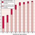
Delayed passage of meconium

The normal time to passage of meconium increases with decreasing gestational age (GA).

Common causes of delayed passage of meconium include anorectal malformations, Hirschsprung disease and meconium ileus. All can present also with vomiting and abdominal distension.

Diagnosis

Always examine the perineum and anus carefully.