FIG. 7.12 Organization of the male reproductive organs. Sagittal section of pelvis showing placement of male reproductive organs. (From Patton KT, Thibodeau GA: The human body in health & disease, ed 6, St Louis, 2014, Mosby.)
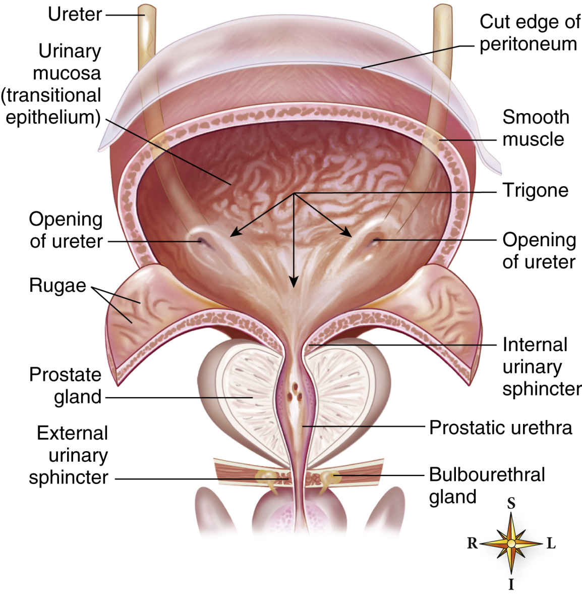
FIG. 7.13 Urinary bladder. Male bladder cut to show the interior. In the male, a large prostate gland surrounds the urethra as it exits from the bladder. (From Patton KT, Thibodeau GA: The human body in health & disease, ed 6, St Louis, 2014, Mosby.)
Go ahead and use the template available on the Evolve Resources site to try mapping the remaining procedures:
• Cystoscopy
• Cystoscopy TURBT (transurethral resection of bladder tumor)
• Urethrotomy
Penile and Testicular Procedures
Procedures
• Hydrocelectomy
• Orchiopexy
• Circumcision (Fig. 7.14)
• Orchiectomy
• Hypospadias and epispadias repair
• Penile prosthesis placement
• Vasvasotomy
• Vasectomy
• Varicocelectomy
Basic Equipment Required
• Genitourinary minor instrument set
• Electrocautery (needle tip)
• Two #15 knife blades
Additional Facts to Remember
• Repair of hypospadias, in which the urethral opening of the penis is on the underside rather than at the tip, may require multiple procedures performed in stages.
• In epispadias, the urethra ends in an opening on the upper aspect (dorsum) of the penis.
• Because of the importance of preserving skin for use as grafts in repair procedures, circumcision is not performed in infants with defects.
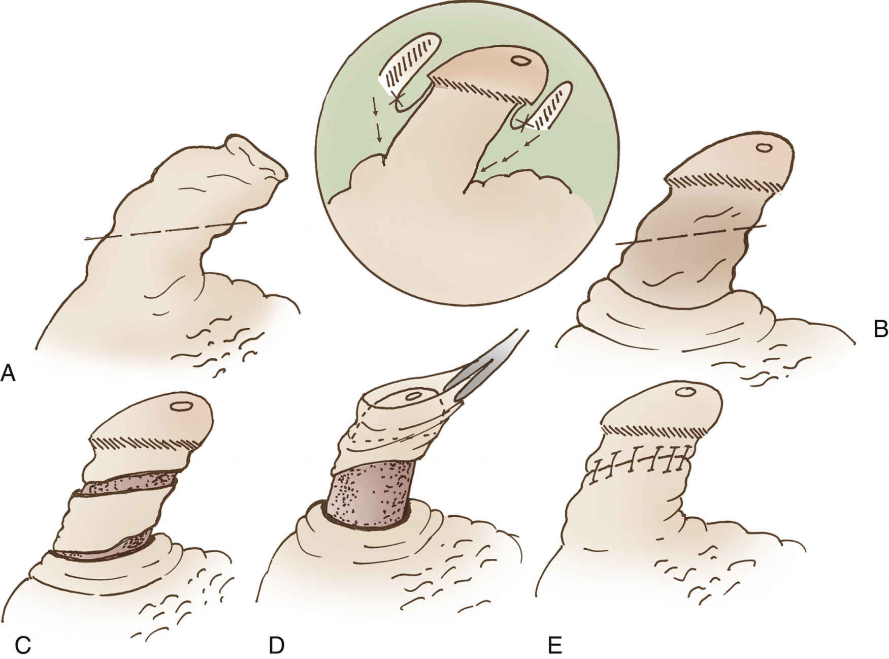
FIG. 7.14 Circumcision. A, Initial incision made in the shaft. B, Second incision made in subcoronal sulcus. C, Amount of tissue to be removed. D, Removal of tissue. E, Shaft skin sutured to subcoronal skin. (From Holcomb GW, Murphy JP: Pediatric surgery, ed 5, Philadelphia, 2010, Saunders.)
• Urethral catheters of 10 to 12 Ch/F for adult women and 10 to 16 Ch/F for adult men are generally chosen; hence, any size smaller than 8F is used for pediatric patients when avoiding damage to the urethra is of major concern.
Mapping
Here’s the mapping on two GU procedures that you’re likely to encounter in the operating room:
And now it’s your turn; try mapping the remaining procedures, using the template available on the Evolve Resources site:
• Hydrocelectomy
• Orchiopexy
• Circumcision
• Orchiectomy
• Penile prosthesis insertion
• Vasvasotomy
• Vasectomy
• Varicocelectomy
Kidney, Ureter, and Bladder Procedures
See Fig. 7.15 for more information on the urinary system.
Procedures
• Wilms tumor excision
• Nephrectomy (Fig. 7.16)
• Kidney transplant
• Pyelolithotomy
• Cystectomy/ileal conduit procedure
• Marshall-Marchetti-Krantz procedure
• Endoscopic suburethral sling
• Stamey procedure
Basic Laparoscopic Equipment Required
• Light cord
• Camera
• Insufflation or irrigation tubing
• Trocars
• Laparoscopic instruments
• Minor soft instruments
• Fluid to induce distention
Basic Endoscopic Equipment Required
• Light cord
• Camera
• Irrigation tubing
• Trocars
• Cystoscopic instruments
Additional Facts to Remember
• Depending on the surgery, an open or laparoscopic approach may be required.
Endoscopic Procedures
• Fluid is used as the medium for distention; input and output must be monitored closely so it does not run out.
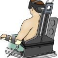
• Allen or candy cane stirrups are used to put the patient in lithotomy position.
• The Stamey procedure is a retropubic urethropexy approach in which sutures are used to raise the urethra and bladder neck and secure them to the surrounding tissue and bone.
Pyelolithotomy
• If the pleural cavity is to be entered, a chest tube may be necessary, because the 12th rib is removed to permit full visualization of the renal pelvis.
• The left kidney is larger than the right.
• The right kidney is located slightly lower than the left to accommodate the liver.
Ileal Conduit Procedure
• Be prepared for specimens to be taken for frozen sections.
• Seeding must be prevented in the case of tumor excision through isoslation of instruments that have come into contact with cancer cells.
• The patient will have a stoma (ileostomy).
Mapping
Here are a couple of example procedures for you:
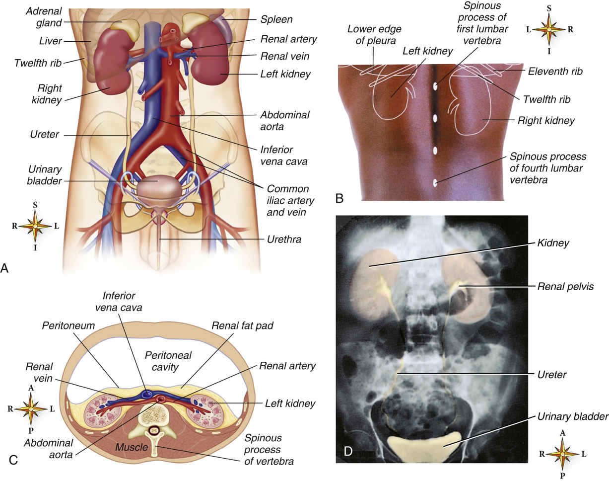
FIG. 7.15 Urinary system. A, Anterior view of urinary organs. B, Surface markings of the kidneys, 11th and 12th ribs, spinous processes of L1 to L4, and lower edge of pleura viewed from behind. C, Horizontal (transverse) section of the abdomen showing the retroperitoneal position of the kidneys. D, Colorized x-ray film of the urinary organs. (A, C, D, From Patton KT, Thibodeau GA: The human body in health & disease, ed 6, St Louis, 2014, Mosby. B, From Abrahams P, Hutchings RT, Marks SC: McMinn’s color atlas of human anatomy, ed 5, St Louis, 2003, Mosby.)
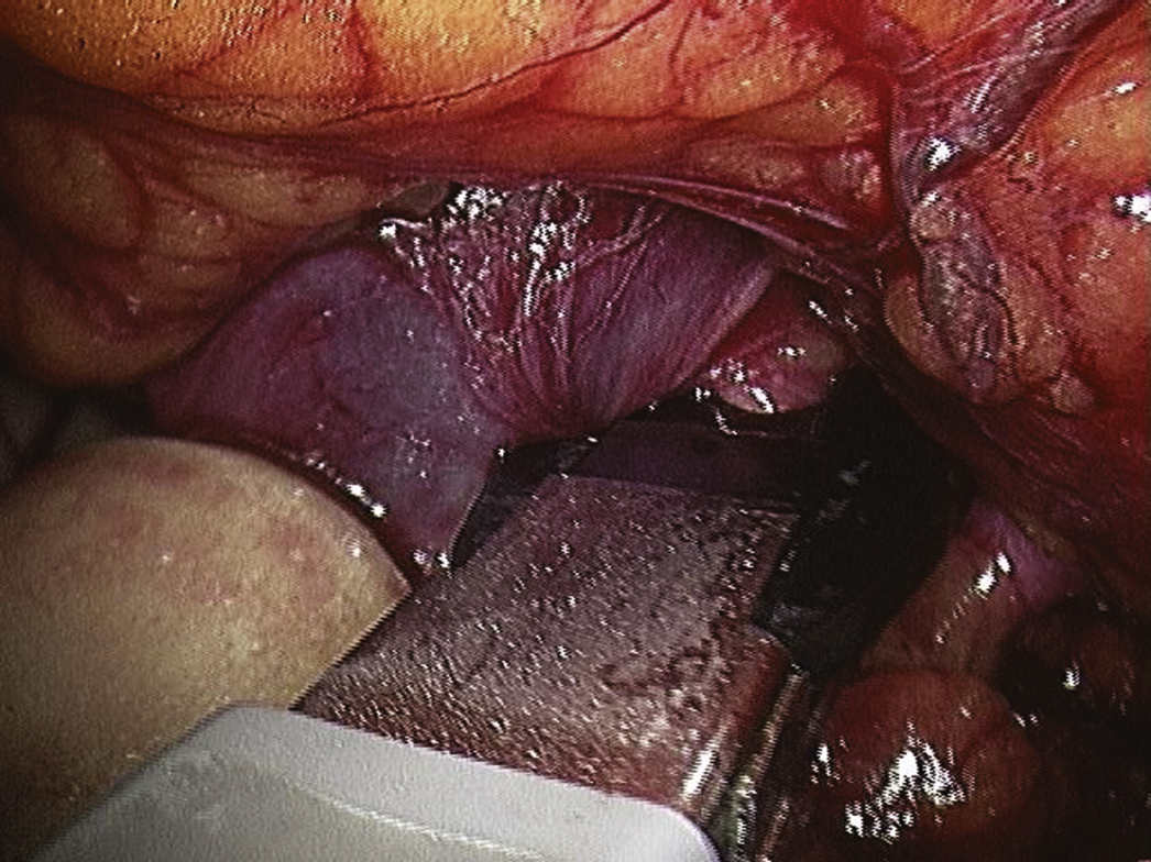
FIG. 7.16 Intraoperative photograph of a right radical nephrectomy. An automatic clip applier is being used to control the renal artery. Note the renal vein to the left of the instrument. (From Becker JM, Stucchi AF: Essentials of surgery, ed 1, Philadelphia, 2006, Saunders.)
Try mapping the remaining procedures using the template available on the Evolve Resources site:
• Wilms tumor excision
• Nephrectomy
• Kidney transplant
• Marshall-Marchetti-Krantz procedure
• Endoscopic suburethral sling
• Stamey procedure
Prostate Procedures
These surgeries may be performed endoscopically (laparoscopically) or as open procedures.
Procedures
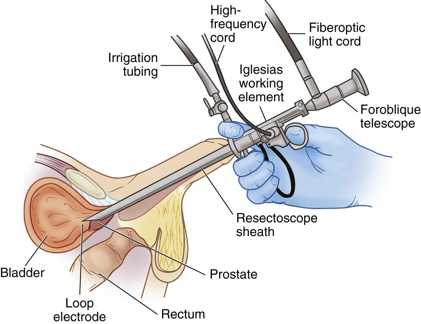
• Prostatectomy (Fig. 7.17)
• Suprapubic prostatectomy
• Laparoscopic robot-assisted prostatectomy
• Implantation of radioactive seeds into the prostate
Basic Laparoscopic Equipment Required
• Light cord
• Camera
• Insufflation tubing
• Trocars
• Laparoscopic instruments
• Minor soft Instruments
Additional Facts to Remember
• In endoscopic procedures, fluid is used as the medium for distention.
• Allen or candy cane stirrups are used to place the patient in the lithotomy position.
• A Foley catheter is inserted and maintained on the sterile field.
Mapping
Here’s how one of these procedures looks when mapped:
The remaining procedures are yours to map!
• Prostatectomy
• Laparoscopic robot-assisted prostatectomy
• Implantation of radioactive seeds into the prostate