FIG. 7.18 Human skeleton. The axial skeleton is distinguished by a blue tint. A, Anterior view. B, Posterior view. (From Patton KT, Thibodeau GA: The human body in health & disease, ed 6, St Louis, 2014, Mosby.)
• Arthroscopy of the shoulder (acromioplasty)
• Arthroscopy of the hip
• Arthroscopy of the ankle
Basic Equipment for Arthroscopic Procedures
• Basic orthopedic instrument tray
• Irrigation tubing and pump
• Arthroscopic tower
• Tourniquet
• Arthroscopic instrument tray
• Arthroscope with 30- and 70-degree lenses
• Arthroscopic shaver
• Camera
• Light source and cord
• 3000-mL bags of fluid (distention medium)
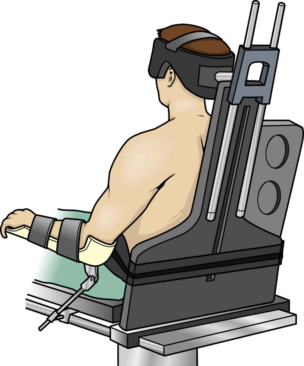
FIG. 7.19 Beach chair–style shoulder positioner allows distraction of the joint for visualization. (From Rothrock JC, Alexander SM: Alexander’s surgical procedures, ed 1, St. Louis, 2012, Mosby.)
• Suture anchors
• Beach chair positioner (for shoulder procedures; Fig. 7.19)
Additional Facts to Remember
• The patient is placed in the supine position/modified “beach chair” (Fowler) position for shoulder surgeries.
• For knee operations, the patient is placed in the supine/lateral position with the knee to be operated on placed below the break of the table.
• Before any arthroscopic procedure, you must check all videoscopic equipment, prime the pump, and white-balance the scope.
• Remember to monitor fluid input and output during procedure.
• Remember to monitor tourniquet time carefully:
• The surgeon should be informed once the cuff has been inflated for the first 60 minutes.
• The recommended inflation time for the arms is 60 minutes; it is 90 minutes for the legs.
• When the recommended time limit has been reached, the cuff should be deflated for 15 minutes to permit reperfusion of the extremity, after which the cuff may be reinflated for another recommended time period (i.e., 60 or 90 minutes).
• The limb should be re-exsanguinated before reinflation to help prevent the formation of venous thromboses.
Mapping
Here’s the map of one extremely common arthroscopic procedure:
Now it’s your turn: Use the template on the Evolve Resources site to try your hand at mapping the remaining procedures:
• Arthroscopy of the knee (diagnostic)
• Arthroscopy of the shoulder (diagnostic)
• Arthroscopy of the shoulder (Bankart procedure)
• Arthroscopy of the shoulder (acromioplasty)
• Arthroscopy of the hip
• Arthroscopy of the ankle