Superior Vena Cava Obstruction

Published on 18/07/2015 by admin

Filed under Radiology

Last modified 22/04/2025

Print this page

rate 1 star rate 2 star rate 3 star rate 4 star rate 5 star
Your rating: none, Average: 0 (0 votes)

This article have been viewed 2108 times

 Mediastinal mass, tumor, or lymphadenopathy

image Mediastinal calcification due to TB, histoplasmosis
image Thrombus in SVC
• Imaging findings suggesting SVC obstruction

image Dilated collateral veins (internal jugular veins, intercostal veins, azygous vein, lateral thoracic, etc.)
image Opacification of portions of liver parenchyma (usually medial segment, quadrate lobe) through collaterals
image Hot quadrate sign: Avid enhancement of left medial segment of liver on arterial phase (not seen on venous or delayed phases) due to intra- and perihepatic collaterals

PATHOLOGY

• > 75% of cases due to malignancy

image Lung cancer and lymphoma most common causes
• Other etiologies

image Thrombosis due to hypercoagulable state, long-term indwelling SVC catheter, or pacemaker
image Infectious lymphadenopathy from histoplasmosis, tuberculosis, coccidiomycosis
image Autoimmune or postradiation mediastinal fibrosis
image Fibrosing mediastinitis

CLINICAL ISSUES

• Symptoms vary based on rapidity of obstruction
• Patients with cerebral edema or airway obstruction (due to stridor) must be treated emergently

image SVC stent placement ± radiation therapy
• Remove catheter and anticoagulation for catheter-related thrombus ± catheter-directed thrombolysis

DIAGNOSTIC CHECKLIST

• Utilize coronal MIP imaging to identify collateral veins
image
(Left) Coronal CECT in an elderly woman who presented with a puffy face demonstrates obstruction of the superior vena cava (SVC) image
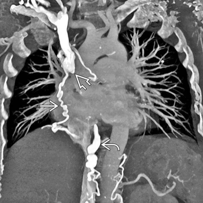
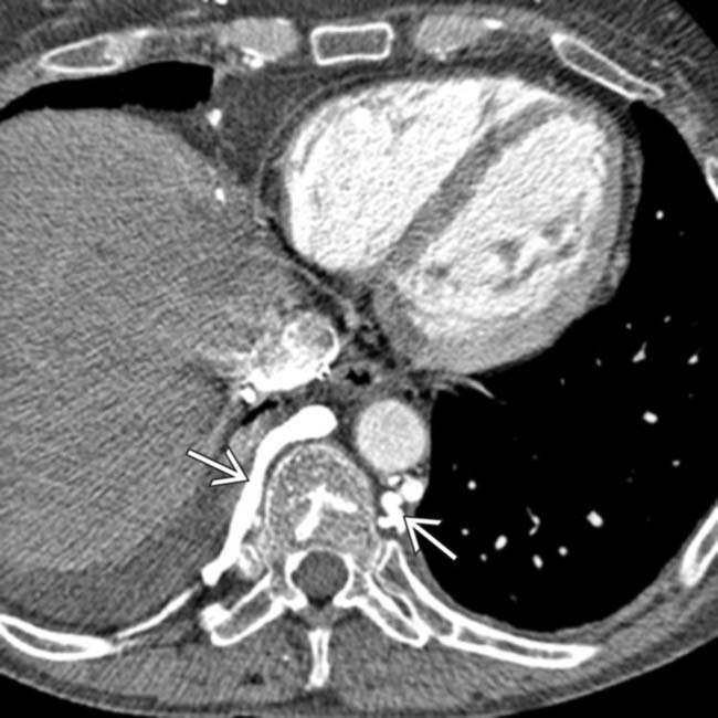
, with collateral flow through an enlarged azygous vein image as well as various mediastinal collateral veins image.

image
(Right) Axial CECT in the arterial phase in a patient with malignant SVC occlusion demonstrates a wedge-shaped perfusion abnormality image 
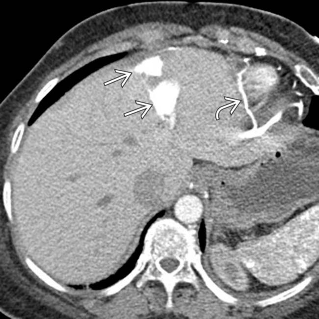
in the left medial segment of the liver (hot quadrate sign) and collateral veins image over the surface of the liver.
image
(Left) Axial CECT in a young woman evaluated for aortic dissection shows near-complete occlusion of the SVC image 
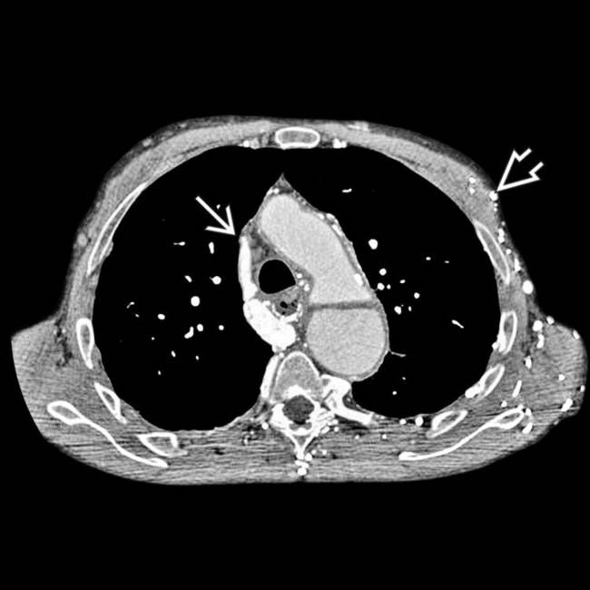
with extensive collateral veins image over the chest and abdominal walls.

image
(Right) Axial CECT in the same patient shows a segment of dense parenchymal enhancement in the medial segment of the liver image 
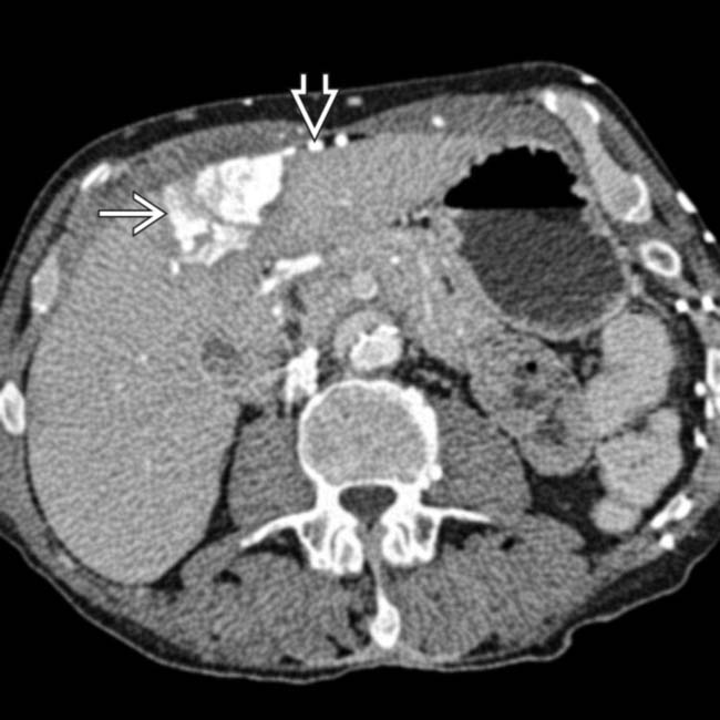
due to opacification of liver parenchyma alongside the intra- and perihepatic collaterals image. The liver perfusion abnormality was not seen on portal venous phase images (not shown).

TERMINOLOGY

Abbreviations

• Superior vena cava (SVC) obstruction

Synonyms

• Superior vena cava (SVC) syndrome

Definitions

• Occlusion of SVC by extrinsic compression, tumor invasion, or intrinsic thrombus

IMAGING

General Features

• Best diagnostic clue

image Mediastinal lymphadenopathy, masses, or calcifications with non-visualized SVC and multiple venous collaterals
• Location

image Superior mediastinum
• Size

image Mass 5-10 cm

Imaging Recommendations

• Best imaging tool

image CECT
• Protocol advice

image Bolus IV injection of contrast medium
image Multiplanar reformations (including maximum-intensity projection reconstructions) to display venous collaterals

CT Findings

• CECT

image Identify causes of SVC obstruction

– Thrombus in SVC
– Mediastinal mass, tumor, or lymphadenopathy
– Mediastinal calcification due to TB, histoplasmosis
image Imaging findings suggesting SVC obstruction

– Dilated collateral veins (dilated internal jugular veins, intercostal veins, azygous vein, lateral thoracic, etc.)
– Hyperperfusion of portions of liver parenchyma (usually medial segment) through collaterals
– Hot quadrate sign: Avid enhancement of left medial segment of liver on arterial phase (not on venous phase) due to intra-/perihepatic collaterals

Ultrasonographic Findings

• Grayscale ultrasound

image Dilated internal and external jugular veins
• Color Doppler

image Stagnant flow in jugular veins

Nuclear Medicine Findings

• Hot quadrate (medial segment of liver) following arm injection of Tc-99m sulfur colloid

image Due to collateral flow through liver (similar to CT finding)

Radiographic Findings

• Radiography

image Superior mediastinal widening or enlargement of right hilum due to lymphadenopathy or tumor
image Indwelling SVC catheter
image Pleural effusions

DIFFERENTIAL DIAGNOSIS

Fibrosing Mediastinitis

• Most commonly histoplasmosis or tuberculosis
• Autoimmune mediastinitis (± other processes, such as retroperitoneal fibrosis)
• Fibrotic soft tissue infiltration on CT, often with dense calcifications

Aortic Aneurysm or Dissection

• May mimic/cause SVC obstruction clinically and on imaging
• Ascending aortic aneurysm due to atherosclerosis, Marfan syndrome, or syphilis
• Internal flap if dissection present

Occlusion of Other Central Veins

• Subclavian/IJ vein occlusions can produce venous collaterals
• Often caused by venous catheters or pacemakers

PATHOLOGY

General Features

• Etiology

image > 75% of cases due to malignancy

– Lung cancer and lymphoma most common causes 

image Account for 95% of cases caused by malignancy
– Other malignancies include metastatic lymphadenopathy (esp. breast cancer), thymoma, germ cell tumors
image Other etiologies

– Thrombosis due to hypercoagulable state, long-term indwelling SVC catheter, or pacemaker

image 25% with pacemakers have central venous obstruction, but most asymptomatic
– Infectious lymphadenopathy from histoplasmosis, tuberculosis, coccidiomycosis
– Autoimmune or postradiation mediastinal fibrosis
– Fibrosing mediastinitis

image Most commonly histoplasmosis or tuberculosis
image Soft tissue infiltration with dense calcifications
image Venous compression due to thin endothelial wall of SVC

Gross Pathologic & Surgical Features

• Bulky mediastinal adenopathy from lung cancer or lymphoma

CLINICAL ISSUES

Presentation

• Most common signs/symptoms

image Facial/neck/arm swelling, dyspnea, cough
image Stridor, light headache, confusion, engorged neck veins
image Symptoms due to primary tumor
image Symptoms vary based on rapidity of obstruction

– Slow occlusions allow collaterals (often asymptomatic)
– Malignancies occlude SVC quickly, before collaterals have time to form; patients often very symptomatic
• Other signs/symptoms

image Other signs of lung cancer

– Horner syndrome, vocal cord paralysis, phrenic nerve paralysis

Demographics

• Age

image Obstruction in young patients often due to lymphoma
image Older patients: Lung cancer
• Epidemiology

image Mediastinal histoplasmosis in midwestern USA

Natural History & Prognosis

• If untreated, may progress to life-threatening cerebral edema and herniation
• If due to malignancy, poor prognosis

image 90% die within 2 years
image Average life expectancy with SVC syndrome: 6 months

Treatment

• Patients with cerebral edema or airway obstruction (due to stridor) must be treated emergently

image SVC stent placement ± radiation therapy
• Other symptoms may not need emergent treatment

image Emergent radiation might prevent histologic diagnosis
image In cases with unknown diagnosis and mild symptoms, stent placement first without radiation
• Radiation/chemo depending on sensitivity of tumor
• Remove catheter and anticoagulation for catheter-related thrombus ± catheter-directed thrombolysis
• Surgical bypass rare in malignant obstruction due to limited life expectancy, but possible for benign causes

image Sometimes used for malignant thymoma and thymic carcinoma (resistant to chemotherapy and radiation)

DIAGNOSTIC CHECKLIST

Consider

• Fibrosing mediastinitis, tuberculosis, or other granulomatous infections if calcified lymphadenopathy

Image Interpretation Pearls

• Coronal MIP reconstructions may help identify collateral veins
image
Axial CECT in the same patient reveals additional venous collaterals image. A subsequent CECT of the chest documented SVC obstruction related to the patient’s indwelling catheter.

image
Axial CECT in an asymptomatic 57-year-old non-Hodgkin lymphoma patient with an indwelling SVC catheter (in place for chemotherapy for over 6 months) presenting for routine surveillance. Note the numerous enhancing venous collaterals image 
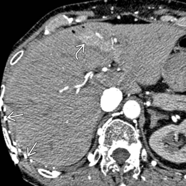
along the right chest wall, as well as the early enhancement of the liver in the region of the falciform ligament image from venous collaterals.
image
Axial CECT in the same patient again illustrates collateral veins image 
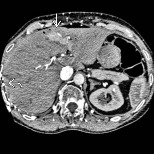
along the surface of the liver, with opacification of a portion of the medial segment image due to transhepatic collateral venous flow.
image
Coronal CECT demonstrates a characteristic wedge-shaped perfusion abnormality image 
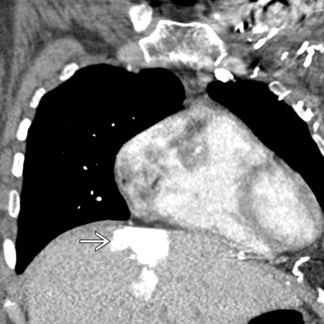
in the left medial segment of the liver in a patient with SVC obstruction.

Share this: