Subaxial Cervical and Upper Thoracic Spine Fractures in the Elderly

Published on 11/04/2015 by admin

Filed under Orthopaedics

Last modified 22/04/2025

Print this page

rate 1 star rate 2 star rate 3 star rate 4 star rate 5 star
Your rating: none, Average: 4 (1 votes)

This article have been viewed 3455 times

29 Subaxial Cervical and Upper Thoracic Spine Fractures in the Elderly

Introduction

The geriatric cervical spine is prone to injury. The susceptibility to bony, ligamentous, and neurological injury may be associated with age-related changes including osteoporotic bone, stiffened spinal articulations, preexisting stenosis, and altered spinal cord vasculature and morphology. Because of these factors, which influence injury susceptibility in the elderly patient, the majority of subaxial cervical and upper thoracic spine injuries occur secondary to low-energy mechanisms. Even within the geriatric population, age is an important predictor of injury location based on mechanism.

Although atlantoaxial fractures are more common than subaxial fractures in the elderly, there is considerable morbidity associated with subaxial cervical and upper thoracic spine fractures. These more caudal spine injuries are more likely to be associated with neurological deficits, in comparison to atlantoaxial injuries, and more likely to be associated with higher-energy mechanisms.1

Apart from the nearly ubiquitous osteoarthritic spondylosis seen in geriatric patients, other etiologies of severe cervical and thoracic spine ankylosis can alter the biomechanics of the cervical spine, causing increased susceptibility to fracture from minor traumatic events. These include ankylosing spondylitis (AS) and diffuse idiopathic skeletal hyperostosis (DISH). Both conditions result in a stiff and often osteoporotic spine. With injury, both the anterior and posterior columns may be completely disrupted, causing frank instability. Fractures of the ankylosed spine are associated with 50% morbidity and 30% mortality.2 For this reason, a high level of suspicion is required not to overlook potentially unstable fracture patterns.

The lateral cervical spine radiograph is widely used as a screening tool in the nongeriatric trauma patient. Because of the susceptibility of injury, the significant consequences of injury, and the potential for occult injury in the geriatric population, however, more extensive imaging may be warranted. This is particularly relevant in the spondylotic or ankylotic spine, to avoid missing injuries.

The treatment of subaxial cervical and upper thoracic spine fractures continues to be evaluated. The subaxial cervical spine injury classification system (SLIC) has been established to provide clinicians with standardization for making nonoperative versus operative decisions and, ultimately, how to surgically approach the injuries.3 The traditional thinking regarding the most optimal timing of surgical treatment of injuries is also changing. Specifically, there is now good evidence that early surgical treatment of central cord injuries is superior to late treatment for certain categories of patients. Geriatric surgical techniques are also evolving; the complex and overlapping pathologies of osteoporosis and ankylosis present challenges for which meticulous preoperative planning may prevent certain postoperative complications.

Basic Science

Cervical spine fractures occur in approximately 2% to 3% of blunt trauma patients. Subaxial fractures account for 40% to 60% of the cervical spine fractures. Of these, it has been found that nearly 20% involve the C7-T1 junction. Many subaxial cervical and upper thoracic spine fractures can be overlooked in the multiply-injured trauma patient. Geriatric patients, in particular, have characteristics that may make injury recognition difficult. These include preexisting spondylosis with or without degenerative deformities, as well as patient factors that make the physical examination difficult, including dementia, baseline weakness, and neuropathies. In the ankylosed spine, even minimally displaced segments can be unstable. The failure to recognize these sometimes subtle injuries can lead to devastating neurological consequences.

The radiographic and clinical evaluation of the cervical spine in the patient following trauma continues to be evaluated. There are ongoing modifications of recommendations regarding the role of radiographs, multiplanar CT, and MRI to rule out cervical spine injuries in the trauma patient. Multiplanar CT and MRI have been shown to have very high sensitivity for detecting cervical spine injury. Despite a high sensitivity, there are reports of cervical spine injury in obtunded patients with an unremarkable multiplanar CT.4 Brandenstein and colleagues recently reported on four patients with negative cervical CT scans and MRIs who later had evidence of cervical instability.5 They estimated that 0.2% to 0.4% of their patients would have cervical spine instability despite normal CT and MRI findings. Not surprisingly, three of the four patients with instability were geriatric. It is prudent to have a high degree of suspicion for cervical spine injuries in the geriatric patient despite seemingly normal imaging.

Ankylosing Spondylitis

Ankylosing spondylitis (AS) is a seronegative (RF-negative) spondyloarthropathy that predominantly affects the sacroiliac joints and spine. It typically, but not exclusively, affects HLA-B27–seropositive patients. The prevalence ranges from 0.1% in African and Eskimo populations to as high as 6% in Haida Native Americans in northern Canada. The white populations of the USA and UK have a prevalence of 0.5% to 1.0%. AS typically has its onset in the third decade of life, with a mean age of onset of 26. It rarely begins after the age of 40, although the diagnosis may be made at a later age because earlier symptoms are ignored or benign. A juvenile form of AS is described, but it does not affect the spine.

Clinical Case Examples

CASE 1

A 58-year-old male fell from standing and struck the back of his head. This resulted in temporary loss of consciousness and neck pain. He was transferred from an outside hospital when abnormal neurological findings were appreciated. On presentation at our institution, he was immobilized in a rigid cervical collar and was hemodynamically stable. His exam revealed weakness (4/5) of bilateral upper extremities muscle groups. He also had 4/5 strength in his quadriceps but other lower extremity strength was 5/5. His past medical history was significant only for hypertension.

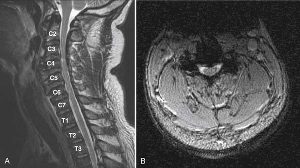
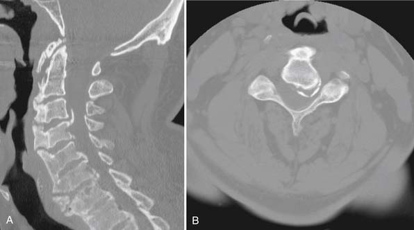
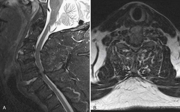
Imaging evaluation revealed extensive degenerative changes. Posterior osteophytes from C3 to C7 and calcification of the posterior longitudinal ligament were demonstrated on CT scanning. There was no fracture, malalignment, or prevertebral edema appreciated (Figure 29-1). An MRI revealed C3-4 disc osteophyte complex associated with spinal cord compression and T2 hyperintensity of the cord. Additional disc protrusions were seen at more caudal cervical levels (Figure 29-2).
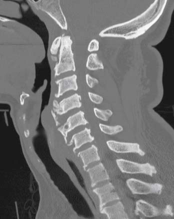
He was initially treated with cervical collar immobilization. His neurological examination was monitored. The patient showed no improvement in his neurological examination. The recommendation to decompress and stabilize the cervical spine was accepted by the patient. A posterior direct decompression with instrumented fusion was performed from C3 to T1 (Figure 29-3).

Postoperatively, his upper extremity weakness resolved. Two weeks later, however, he presented to the emergency department with recurrent weakness in elbow flexion bilaterally. His examination revealed 4/5 strength in bilateral deltoids and 3/5 strength in biceps and forearm supination. Otherwise his upper and lower extremity motor strength had improved to 5/5. He did not have any sensory deficits. Examination was consistent with C5 nerve palsy. He was treated with observation and analgesic medications. At latest follow-up, he is ambulatory with a fluid narrow-based gait and with resolved weakness in elbow flexion and supination.

Case 2

A 51-year-old male with known diffuse idiopathic skeletal hyperostosis (DISH) had a syncopal event and fell from the stands at Fenway Park, impacting his face and forehead. He had transient paralysis of bilateral upper extremities and severe neck pain. He was stabilized in a cervical collar at Fenway and transferred to our emergency department for evaluation.

On initial examination, he was found to have recovered motor function, and to have intact sensation to pain and light touch in bilateral upper extremities. He had persistent severe neck pain and severe burning pain and sensitivity to light touch in both hands, refractory to intravenous pain medications.

Imaging evaluation demonstrated several disc osteophyte complexes. The largest was observed at C3/C4, where there was 50% narrowing of the central canal. Extensive flowing nonmarginal osteophytes were also well characterized by CT scan (Figure 29-4). While no obvious unstable injuries were appreciated on CT, a subsequent MRI revealed extension distraction fractures with three column disruption at both C3-4 and C6-7. Each level of injury was associated with dissociation of the anterior longitudinal ligament and osteophytes (Figure 29-5). The spinal cord was compressed at C3-4 and C6-7.

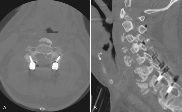
A C3 to C7 laminectomy and C3 to T1 instrumented fusion were performed. He tolerated the procedure well and was extubated in the operating room (Figure 29-6). Since the patient’s body habitus limited the adequacy of intraoperative radiographs, a CT scan was performed immediately postoperatively to evaluate the spinal alignment and instrumentation (Figure 29-7).
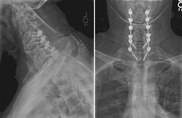
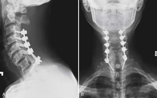
Postoperatively, the patient reported an immediate decrease in his burning hand pain. He was able to ambulate with PT and was discharged home on postoperative day 3. At his 6-week follow-up, he complained only of hyperesthesias of the right small and index fingers. He had no weakness and normal sensation, with resolution of his severe sensitivity to light touch.

Sacroiliitis is the most common presenting symptom, with bilateral or unilateral buttock pain being the chief complaint. Spinal stiffness and discomfort typically progress gradually and affect all joints in the spine. Extra-axial involvement includes plantar fasciitis, insertional Achilles tendinitis, eye lesions, enteritis, colitis, prostatitis, aortitis, and, rarely, fibrosis of the upper lung.

The hallmark spinal pathology seen in AS is due to enthesitis. The enthesis is the site of tendon and ligament attachment to bone. Local inflammation at the enthesis may lead to radiographic lysis of bone. AS affects the insertions and attachments of the discovertebral, costovertebral, and costotransverse joints, as well as the other interspinal ligaments. The reactive bone formation at the sites of inflammation and the remaining lysis result in a stiff and osteoporotic spine. This combination results in an increased susceptibility to spine fractures.6

Diffuse Idiopathic Skeletal Hyperostosis

Diffuse idiopathic skeletal hyperostosis (DISH) was first described by Forestier and Rotes-Querol in 1950, and it is often still referred to as Forestier disease. It has specific diagnostic criteria as outlined by Forestier. These include at least four contiguous vertebrae involved in ossification, without evidence of loss of disc height, and with relatively well-preserved facet joints and SI joints. The ossification is nonmarginal and flowing along the anterolateral vertebrae. Additionally, there are extraspinal manifestations such as increased heterotopic ossification after surgery.

DISH is not related to HLA-B27, and there has been no relationship found with other seronegative spondyloarthropathies such as AS. DISH has some relationship with HLA-8 and is relatively common, with prevalence as high as 28% in autopsy series. It is felt that 15% of women and 25% of men over the age of 50 have DISH, and the prevalence increases with age. There is no difference in prevalence between blacks and whites.

The thoracic spine is most commonly affected. The large syndesmophytes more often involve the right half of the vertebral body in the thoracic spine, contralateral to the aorta, whereas involvement is symmetric in the cervical or lumbar spine. DISH of the cervical spine usually involves the lower segments and can become large enough to exert a mass effect on the esophagus and cause dysphagia.

The bone morphology in DISH is different than in AS. Whereas vertebrae with inflammation-induced osteolysis adjacent to affected entheses are commonly seen in AS, in DISH, bone quality is relatively well preserved. Both conditions, however, are associated with increased risk of fracture through or adjacent to ankylosed vertebral segments. These patients can present a challenge in correctly identifying a cervical fracture.7

Biomechanics and Classification of Subaxial Spine Fractures

Ferguson and Allen reviewed 165 cases to develop a classification system for subaxial spine fractures based on the mechanism of injury. They developed six mechanisms with reproducible fracture patterns. The mechanisms described can be divided into three compression injuries (compression-flexion, compression, and compression-extension), two distraction injuries (distraction-flexion, distraction-extension), and lateral flexion. Each mechanism has varying degrees of severity based on radiographic findings. Although commonly used as a framework for fracture discussion, these results have never been validated in the current literature. Additionally, witnessed compression injuries have resulted in variable fracture morphology. This classification does not specifically grade the amount of ligamentous injury nor does it quantify the amount of neurological injury.

In addition to classification is the question of subaxial spine instability after injury. White and Panjabi published a biomechanic study evaluating clinical and radiographic markers of cervical spine instability. Their work focused on the ligamentous structures surrounding the vertebral bodies. They developed a checklist with point values, with a score of five or more indicating instability. The radiographic markers on plain film are sagittal plane translation of >3.5 mm, sagittal plane rotation >11 degrees, positive stretch test, and abnormal disc narrowing. Clinical criteria are cord damage, root damage, and if dangerous loading is anticipated. There are two additional criteria: anterior elements unable to function and posterior elements unable to function. While this checklist, published in 1976, is a tool for evaluating spine instability, the commonplace use of CT and MRI has provided a level of detail and sensitivity that has made the point system infrequently used.

More recently, a classification system has been developed using the injury morphology, discoligamentous complex, and neurological status to classify injuries and to determine the need for surgical intervention. This subaxial cervical spine injury classification system (SLIC) by the Spine Trauma Study Group (STSG) was developed not only to classify subaxial spine fractures but also to predict the need for surgical stabilization and/or reduction. The system is based on assigning points according to the severity of injury in three categories: proposed mechanism of injury, discoligamentous complex injury, and neurological injury. If a patient’s SLIC score is 1 to 3, then nonsurgical treatment is recommended. If the score is 5 or more, then surgical treatment is recommended (Table 29-1). To assess the reliability and validity of this classification system, 20 surgeons in two different settings used the system to classify 11 different spine fractures and clinical scenarios. They found the reliability to be slightly lower than that of the Ferguson and Allen classification. The validity was good in comparison to the Ferguson and Allen classification system. Of note is that 93% of the time, the surgical versus nonsurgical treatment recommended by SLIC was the treatment recommendation of the practicing clinician.

TABLE 29-1 The Subaxial Cervical Spine Injury Classification System (SLIC)

  Points
Morphology  
No abnormality 0
Compression + burst 1+1=2
Distraction (e.g., facet perch, hyperextension) 3
Rotation or translation (e.g., facet dislocation, unstable teardrop or advanced-stage flexion-compression injury) 4
Discoligamentous complex  
Intact 0
Indeterminate (e.g., isolated interspinous widening, MRI signal change only) 1
Disrupted (e.g., widening of anterior disc space, facet perch or dislocation) 2
Neurological status  
Intact 0
Root injury 1
Complete cord injury 2
Incomplete cord injury 3
Continuous cord compression (neuro modifier in the setting of a neurological deficit) +1

A score of 3 or less indicates nonoperative management. A score of 5 or greater indicates need for surgical intervention.

Instrumentation of Osteoporotic Lower Cervical And Upper Thoracic Spine

The mechanisms and fracture patterns of the cervical spine differ in the geriatric population as compared to the young trauma population. This is due in part to decreased bone mineral density, most typically related to osteoporosis. Osteoporosis is common and affects 55% of people older than 50 years old, with an increased prevalence in women. Osteoporosis is associated with preferential loss of trabecular (or cancellous) bone as compared to cortical bone. This pattern of bone loss has important bearing on spinal instrumentation and can preclude adequate screw purchase with traditional fixation methods. When considering surgical intervention in the geriatric population it is necessary to consider the diminished bone quality and utilize techniques that limit complications. Potential complications related to instrumentation of the geriatric cervical spine include loss of fixation, nonunion with subsequent hardware failure, loss of deformity correction, and adjacent segment fracture.

While not yet FDA approved, lateral mass screws are commonly used for reconstruction of the subaxial cervical spine. These short small-diameter screws are typically placed using unicortical technique and, as such, rely on sufficient trabecular bone to prevent loss of fixation. While it may be associated with other risks, bicortical placement of lateral mass screws may be considered when trabecular quantity is compromised. Additionally, an alternative technique has been described to place screws across three or four cortices by traversing the cervical facet articular cortex, as well as the posterior (and possibly the anterior) cortex of the lateral mass. Placement of transfacet screws can be technically demanding, and requires an approximately 40-degree caudal insertion angle in the sagittal plane and 20 degrees lateral in the coronal plane. For this reason, the occiput can interfere with placement in cephalad cervical segments. Additionally, transfacet placement cannot be used in the most caudal instrumented segment as it will violate the normal unfused facet joint. This is a technique that can certainly be helpful in salvage scenarios, such as when lateral mass fixation has been found to be inadequate intraoperatively.

One may also consider augmenting a construct that uses lateral mass instrumentation to ensure adequate fixation. For example, cranial or caudal extension of the construct is frequently useful in the geriatric spine. By extending the fusion, additional points of fixation may be included to reduce the stresses on any particular screw-bone interface. Furthermore, cranial extension may allow placement of a C2 pars screw, while caudal extension may allow placement of a pedicle screw in C7 or in the upper thoracic pedicles. These screws have the benefit of improved cortical contact throughout the length of the screw, which typically is longer than a lateral mass screw and, as such, offers improved fixation. A CT scan is useful to plan the placement of either a C2 pars screw or a cervical or thoracic pedicle screw. The CT scan will provide preoperative assessment of pedicle diameter, orientation, and bone quality. In the spondylotic spine, the pedicles can be entirely sclerosed and may be unable to accept a blunt pedicle probe; they may require alternative techniques for cannulation, such as drilling. When considering C2 pedicle fixation, the course of the vertebral artery must be known in detail. Intraoperative fluoroscopy can be employed to avoid injury, and to allow for the placement of a long and well-fixed screw along the medial cortex of the canal. It is also useful to access the canal in these cases, to establish the exact location and morphology of the medial wall cortex.

Other techniques that can be used to improve fixation in osteoporotic bone include the use of instrumentation that relies on the (relatively well-preserved) cortical bone. These include hooks, sublaminar wires, and translaminar screws. Placement of these does require at least part of the lamina to remain intact, which can limit their use in cases that require multilevel posterior decompression of the injured neural elements. Placement of hooks and wires can often be accomplished at the same vertebral level as a concomitant screw, enhancing fixation of that vertebra. This may be particularly useful at the construct’s terminal vertebra.

Until solid fusion has been achieved, stabilization can be augmented by use of rigid cervical orthosis. The duration of treatment should be based on the intraoperative assessment of the quality of fixation and the patient’s ability to tolerate a rigid orthosis. The most common intolerance is due to skin breakdown under the mandible, mastoid, occiput, or shoulders. Additionally, patients may complain of dysphagia or diminished respiratory capacity. These risks must be weighed against the need for supplemental cervical stabilization.

There are several techniques that may be used when planning instrumentation of the osteoporotic spine. To prevent intraoperative frustration, delays, and complications, a meticulous preoperative plan must be made and communicated to the OR staff.

Clinical Practice Guidelines

The importance of identifying cervical spine fracture, instability, or injury in the geriatric trauma patient cannot be overstated. Many characteristics of the geriatric population make identifying these injuries difficult. An accurate history of events surrounding the injury may be difficult, due to altered consciousness at the time of injury, associated head injuries, or baseline dementia. Additionally, a complete physical examination may be limited by altered mental status or by underlying medical conditions that alter the exam. Radiographic interpretation can, at times, be very difficult due to degenerative changes. Even when a diagnosis is made, there can be significant medical comorbidities that increase the morbidity and mortality related to these fractures.

Initial evaluation should consist of resuscitation as outlined by Advanced Trauma Life Support guidelines. Cervical spine precautions should be maintained even for perceived low-energy mechanisms. Patients with no complaints of neck pain but a history compatible with possible neck injury should have cervical collar stabilization until cleared. A thorough history and physical should be performed. An accurate history may require contacting family members or health care providers who can provide information regarding baseline function, impairments, and past medical history. Inspection of the face and cranium can give important clues to the mechanism of injury and energy transferred to the cervical spine. While maintaining cervical stability, the cervical orthosis should be removed and the posterior cervical midline palpated. With log roll, the entire spine should be palpated to assess for noncontiguous spine injuries that may be present in 10% to 15% of patients. Complete neurological examination must be performed.

Radiographic evaluation should be performed in all patients with suspected cervical spine injury. High-quality and complete images are required, and inadequate images cannot be accepted.. Standard radiographs that visualize from C1 to T1 can provide excellent information in regard to alignment, prevertebral swelling, disc height, and fractures. Standard radiographs, however, are often not adequate in the geriatric population because listhesis, spondylosis, ankylosis, and other degenerative changes make ruling out acute trauma difficult. As such, CT scan is often required to adequately evaluate the injured geriatric spine when radiographs are difficult to interpret. CT scans have much better sensitivity for detecting injuries and are also useful to aid in preoperative planning. MRI should be used in any patient with suspected ligamentous or neurological injury. MRI will provide the best information regarding neurological compression and is useful in determining if decompression is best performed from an anterior or posterior approach, or both.

Nonoperative treatment versus operative treatment of subaxial cervical and upper thoracic spine injuries is frequently based on instability and neurological injury. The SLIC score, although not a perfect tool, provides an excellent framework from which to build treatment strategies. An injured spine without neurological injury may be appropriate for nonoperative treatment in a rigid cervical orthosis, whereas the same injury with neurological injury may require decompression and stabilization to facilitate recovery or prevent further injury.

The timing of surgery, especially in regards to central cord syndrome, remains controversial. All patients with central cord syndrome should be treated aggressively with medical therapies. These therapies include rigid cervical orthosis, intensive care unit admission for monitoring, mean arterial pressure >85 mm Hg, intravenous pressors if needed to maintain blood pressure, and early involvement of physical and occuptional therapy. Surgical intervention in central cord injury is indicated if there is progressive neurological deficit or overt spinal instability requiring reduction and fixation. If surgery is to be performed, there is new evidence that early surgical decompression (<24 hours after injury) improves neurological recovery more than late surgical decompression (>24 hours after injury). However, there is no current consensus on the timing of surgical intervention for central cord syndrome.8

To decrease overall morbidity and mortality of the injured geriatric patient, it is imperative to optimize his or her medical management. This starts with an accurate past medical history and medication list as well as obtaining prior ancillary studies such as cardiac studies and pulmonary function tests. This information is critical for perioperative risk stratification. Early communication with anesthesia, internal medicine, geriatric medicine, and other consult services can prevent delays in surgical treatment and allow sufficient time for necessary interventions. Postoperatively, special attention should be paid to mobilizing the patient to prevent decubitus ulcers, deep vein thrombosis, and pulmonary complications. Additionally, the cervical orthosis should be checked to ensure adequate fit and appropriate distribution of contact pressure to prevent skin breakdown. Medical optimization and limited use of delirium-producing agents will allow the patient to rehabilitate from his or her injury. It must be communicated to patients and families that long-term care may be needed for patients with partial recovery from neurological injuries.

Clinical Case Examples: Treatment, Clinical Challenges, and Future Treatments

Case 1

This patient had a low-energy mechanism resulting in central cord syndrome with no evidence of fracture or ligamentous injury on initial CT imaging. Historically, central cord syndrome has been treated medically with a limited role for operative decompression. Hadley reviewed the historical trends of central cord syndrome and showed that historical treatments are being revisited.9 The age-old nonoperative treatment strategy was based on the case series reported by Schneider in the early 1950s. His initial series reported 50% mortality rate in two central cord patients surgically decompressed. The other patient had recovery similar to the nonoperatively treated patients. Four years later, in 1958, he published a second series in which 20 patients were reviewed. Three of the patients were treated surgically; one patient was treated early, and two patients weeks after their injury. The patient treated early had dramatic neurological improvement. Of the patients surgically decompressed later, one of the two late patients had neurological recovery similar to those managed medically. The other suffered neurological injury and was quadriplegic. Despite the fact that these patients underwent agressive retraction of the injured spinal cord, with transdural approaches to resect anterior osteophytes, and with sectioning of the dentate ligaments, the conclusion from this series was to treat central cord syndrome medically. This guided much of the thinking toward treatment of central cord syndrome from that time forward. It was not until Brodkey, in 1980, published his results of seven operatively treated patients with traumatic central cord syndrome that surgical management was again revisited. These patients were selected for surgery because their neurological recovery had plateaued and myelography showed anterior compression of the spinal cord. Of the seven patients, all patients’ trajectories of neurological recovery improved, and three patients had full neurological recovery. After reviewing the most recent data with regard to treatment of central cord syndrome, Hadley et al. concluded that early reduction of fracture-dislocation is recommended, and surgical decompression of the compressed spinal cord is warranted, especially if the compression is focal and anterior.

The operative technique highlights a well-reported complication of posterior cervical decompression. The incidence of C5 nerve palsy after posterior decompression is approximately 5%. Half of the patients have sensory deficits or pain, and the other 50% have motor weakness only. It is generally unilateral, but is seen bilaterally in about 10% of cases. A recent study showed the cervical spinal cord drifted posteriorly approximately 2.8 mm at 24 hours after surgery and then decreased to 1.9 mm, 2 weeks after surgery. Interestingly, the absolute posterior drift was at the C5-6 level. In their study of 19 patients, two patients developed C5 nerve palsy. These two patients had the largest degree of posterior drift seen at 24 hours and 2 weeks.10 This C5 nerve palsy associated with posterior decompression generally has a good prognosis. Nearly all patients with a C5 palsy with 3/5 or 4/5 strength regain their strength by 6 months, with half having full recovery by 3 months.

Case 2

The patient’s fall from a standing height resulted in a significant cervical spine injury. The ankylosed cervical spine was extended on impact and resulted in a fracture of the anterior column at two noncontiguous cervical levels. This case exemplifies the necessity of maintaining a high suspicion for unstable injuries in the anklyosed spine.

The degree of osteophyte formation made interpretation of the CT scan difficult. It was unclear to both the consulting spine surgeon and the interpreting radiologist whether there had been a bony injury to the cervical spine. An MRI demonstrated the fracture through the C3-4 and C6-7 disc spaces. The degree of injury is significant, and even after further review, difficult to see on CT scan. In patients with degenerative spines that preclude accurate assessment by x-ray or CT, it may be warranted to perform dynamic cervical spine x-rays or an MRI. Clinical clearance of the ankylosed cervical spine should be done with caution, as evaluation of neck pain in the DISH patient is not a reliable clinical tool; many DISH patients have neck pain at baseline.

This case also highlights the complexity of operative planning, as it is a two-level fracture involving the anterior spine. The severe overgrowth of osteophytes distorts the normal anatomy, makes anterior dissection more difficult, and potentially destabilizes the spine when removed. Although anterior stabilization of a spine with an anterior column fracture provides the most stable fixation, in this case it would be technically very difficult. Stabilization would require a 3-level corpectomy and extensive debridement of anterior osteophytes for plate positioning. The risks of bleeding, esophageal injury, and nonunion would be significant with the described procedure. Furthermore, such a multilevel corpectomy would require a posterior fusion to reduce the nonunion risk. Therefore a posterior approach was employed, as imaging demonstrated adequate cervical lordosis for posterior decompression. The goal was to allow posterior drift of the spinal cord and stabilize the spine with instrumentation and lateral mass fusion. Whereas AS is associated with osteoporotic bone, DISH patients generally have well-maintained bone mineral density. Excellent purchase was obtained in lateral masses and in the pedicles of T1. The construct was felt to have adequate stability without crossbars or supplemental techniques. There was no evidence of loss of fixation at follow-up.

Conclusion

The absolute number of geriatric cervical spine fractures and injuries will likely continue to increase as the population ages. The geriatric spine is prone to injury and to subaxial spine fractures, and more often has neurological injury with these in comparison to upper cervical spine fractures. The clinician should maintain a high degree of suspicion for lower cervical and upper thoracic spine injury, even with reportedly minor mechanisms of injury.

Two important disease processes that increase the susceptibility of the cervical spine to injury are AS and DISH. The stiffened and often osteoporotic vertebral segments can lead to devastating neurological injury. Central cord syndrome is often associated with trauma in the degenerative spine, and the indications and timing of surgery continue to be defined. Under specific circumstances, early decompression is associated with improved neurological recovery. The ankylotic and spondylotic changes in these cervical spines make diagnosis difficult with plain radiographs, and a very low threshold for ordering multiplanar imaging is indicated. It must be remembered that the most common severe complication of subaxial trauma is a missed injury.

Subaxial cervical spine fractures have had many classification systems developed over the past three decades. We have found the SLIC score to be very useful, in that it considers the neurological status, morphology of injury, and posterior ligamentous complex. It is our feeling that the reliability and validity of this scoring system will improve as more clinicians become familiar with its use. Surgical treatment of the injured geriatric spine requires meticulous planning and the use of techniques such as supplemental hooks and wires or screw techniques that rely more heavily on the screw and cortical bone interface. These techniques include C2 pedicle screws, laminar screws, and transfacet screws. A thorough understanding of the pathophysiology of subaxial and upper thoracic fractures in the geriatric patient, combined with conscientious treatment plans, will help prevent the morbidity and mortality so often associated with these injuries.

References

1. Sokolowski M.J., et al. Acute outcomes of cervical spine injuries in the elderly: atlantaxial vs subaxial injuries, J. Spinal Cord Med.. 2007;30(3):238-242.

2. Whang P., et al. The management of spinal injuries in patients with ankylosing spondylitis or diffuse idiopathic skeletal hyperostosis: a comparison of treatment methods and clinical outcomes. J Spinal Disord. Tech.. 2009;22(2):77-85.

3. Vaccaro A.R., et al. The subaxial cervical spine injury classification system. Spine. 2007;32:2365-2374.

4. Como J.J., et al. Practice management guidelines for identification of cervical spine injuries following trauma: update from the eastern association for the surgery of trauma practice management guidelines committee. J. Trauma. 2009;67(3):651-659.

5. Brandenstein D., et al. Unstable subaxial cervical spine injury with normal computed tomography and magnetic resonance initial imaging studies: a report of four cases and review of the literature. Spine. 2009;34(20):E743-E750.

6. Kubiak E.N., et al. Orthopedic management of ankylosing spondylitis. Journal of the American Academy of Orthopaedic Surgeons. 2005;13:267-278.

7. Belanger T.A., et al. Diffuse idiopathic skeletal hyperostosis: musculoskeletal manifestations. JAAOS. 2001;9:258-267.

8. Nowak D., et al. Central cord syndrome. Journal of the American Academy of Orthopaedic Surgeons. 2009;17(12):756-765.

9. Hadley M.N., et al. Management of acute central cervical spinal cord injuries. Neurosurgery. 2002;50(3):S166-S172.

10. Shiozaki T., et al. Spinal cord shift on magnetic resonance imaging at 24 hours after cervical laminoplasty. Spine. 2009;34(3):274-279.