Small Bowel NSAID Stricture

Published on 19/07/2015 by admin

Filed under Radiology

Last modified 22/04/2025

Print this page

rate 1 star rate 2 star rate 3 star rate 4 star rate 5 star
Your rating: none, Average: 0 (0 votes)

This article have been viewed 1408 times

 Multiple short-segment annular strictures ± partial SB obstruction

image Strictures may resemble normal plicae circulares on enteroclysis
• CT findings

image Strictures: Short segmental narrowing of lumen with dilation of bowel upstream
image Mucosal inflammation: Mucosal hyperenhancement and submucosal edema
• Capsule endoscopy can confirm diagnosis

TOP DIFFERENTIAL DIAGNOSES

• Crohn disease

image Longer segments of transmural involvement
• Celiac-sprue disease

image Jejunoileal fold pattern reversal
• Ischemic enteritis

image Bowel wall thickening; strictures can be late result
• Radiation enteritis

image Longer strictures; pelvic SB segments
• Small intestine vasculitis

image Long segments of submucosal edema

PATHOLOGY

• All NSAID (including aspirin) formulations can cause enterocolitis

image Slow-release formulations affect distal SB and colon

CLINICAL ISSUES

• Often asymptomatic; may have symptoms of bowel obstruction
• May require surgery or endoscopic balloon dilation for bowel obstruction
image
(Left) Axial NECT in a 40-year-old man shows a dilated proximal, mid small bowel image 
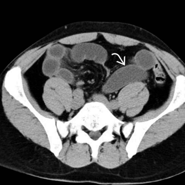
and collapsed colon.

image
(Right) Axial NECT in the same patient shows an abrupt transition from dilated to collapsed small bowel in the ileum image. There was no history of prior abdominal surgery, making adhesive bowel obstruction a less likely etiology.
image
(Left) Small bowel follow-through in the same patient shows dilation of the proximal small bowel and a short stricture in the ileum image 
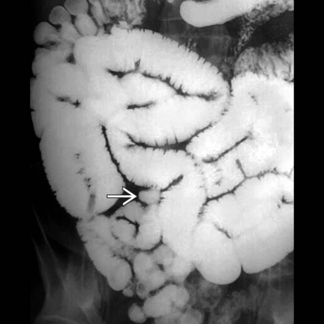
with collapsed bowel distal to this point.

image
(Right) Spot film from the small bowel study in the same patient shows a short stricture image 
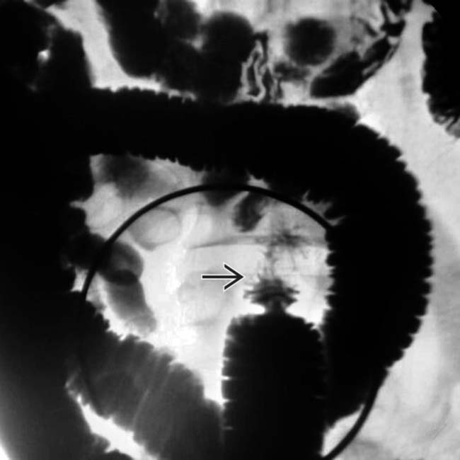
in the ileum. At surgery, the stricture was confirmed, resected, and determined to be due to injury from chronic use of NSAIDs.

TERMINOLOGY

Abbreviations

• Nonsteroidal anti-inflammatory drugs (NSAIDs)

Synonyms

• Diaphragm disease

Definitions

• Focal strictures in small bowel (SB) secondary to NSAID use

IMAGING

General Features

• Best diagnostic clue

image Multiple short-segment annular strictures ± partial SB obstruction
• Location

image Middle and distal small bowel
image May also be present in ascending colon

– Associated with slow-release NSAID formulations
• Size

image 2-4 mm thick septa

– Can reduce lumen to pinhole size
• Morphology

image Circumferential, ring-like
image Patient may have 20-50+ strictures
image Long tapered strictures have been reported as well

Fluoroscopic Findings

• Enteroclysis

image Strictures may resemble normal plicae circulares

– May be easily missed unless they cause SB obstruction
image Mucosal erosions or ulcerations

– Fluoroscopy may identify ulcers and bowel spasm

CT Findings

• Stricture(s): Short segmental narrowing of lumen

image Dilation of SB upstream from stricture
• Mucosal inflammation: Mucosal hyperenhancement and submucosal edema
• Bowel perforation: Free intraperitoneal air and fluid (rare)

Imaging Recommendations

• Best imaging tool

image Enteroclysis or CT enterography
image Capsule endoscopy

– Capsule may fail to pass through tight stricture
image Enteroscopy and colonoscopy can directly visualize strictures within reach of scope

– Double-balloon enteroscopy may reach more distal SB

Radiographic Findings

DIFFERENTIAL DIAGNOSIS

Crohn Disease

• Segmental areas of ileocolonic ulceration on barium study

image Cobblestoning: Longitudinal and transverse ulcers
• CT: Mucosal hyperenhancement, submucosal edema, mesenteric adenopathy

Celiac-Sprue Disease

• Enteroclysis: ↓ number of jejunal folds (< 3 inches) and ↑ number of ileal folds (4-6 inches)

image Jejunoileal fold pattern reversal: Sensitivity in diagnosing disease increases to 83%

Ischemic Enteritis

• Occlusion of superior mesenteric artery or vein
• Bowel wall thickening; strictures can be late result

Radiation Enteritis

• Pelvic SB segments affected most often
• Mural thickening and luminal narrowing
• Single or multiple strictures of varying length

Small Intestine Vasculitis

• Long segments of submucosal edema

PATHOLOGY

General Features

• Etiology

image Chronic NSAIDs use

– All NSAID (including aspirin) formulations can cause enterocolitis

image Slow-release formulations affect distal small bowel or colon
– Also associated with celecoxib (selective COX-2 inhibitor)

Gross Pathologic & Surgical Features

• Multiple strictures with small central openings
• Superficial or deep ulcerations may be seen

Microscopic Features

• Transmural inflammation
• Submucosal deposition of collagen fiber

CLINICAL ISSUES

Presentation

• Most common signs/symptoms

image Asymptomatic or pain due to bowel obstruction
• Other signs/symptoms

image Occult blood loss, anemia

Treatment

• Cessation of NSAIDs use
• May require surgery or endoscopic balloon dilation for bowel obstruction

DIAGNOSTIC CHECKLIST

Consider

• Consider NSAID enteritis for patients with focal strictures of bowel

Image Interpretation Pearls

• Strictures may resemble normal anatomy on enteroclysis

SELECTED REFERENCES

1. Gill, RS, et al. Small bowel stricture characterization and outcomes of dilatation by double-balloon enteroscopy: a single-centre experience. Therap Adv Gastroenterol. 2014; 7(3):108–114.

2. Kishi, T, et al. CT enteroclysis/enterography findings in drug-induced small-bowel damage. Br J Radiol. 2014; 87(1044):20140367.

3. Lim, YJ, et al. Recent advances in NSAIDs-induced enteropathy therapeutics: new options, new challenges. Gastroenterol Res Pract. 2013; 2013:761060.

4. Scholz, FJ, et al. Diaphragmlike strictures of the small bowel associated with use of nonsteroidal antiinflammatory drugs. AJR Am J Roentgenol. 1994; 162(1):49–50.

Fortun, PJ, et al. Nonsteroidal antiinflammatory drugs and the small intestine. Curr Opin Gastroenterol. 2005; 21(2):169–175.