• Antiperistaltic flow of barium proximal to obstruction
• Relief of obstruction in prone, knee-chest, or left lateral decubitus positions
TOP DIFFERENTIAL DIAGNOSES
• Duodenal obstruction (other causes)
• Intestinal scleroderma
• Duodenal stricture
PATHOLOGY
• Predisposing conditions
Weight loss → depletion of retroperitoneal fat, leading to narrowed aorto-mesenteric angle
Anatomical and congenital anomalies
Postoperative states (e.g., scoliosis)
CLINICAL ISSUES
• Postprandial epigastric pain, nausea, vomiting
Pain relieved in prone, knee-chest, or left lateral decubitus position
• Surgery (bypassing duodenum) indicated when conservative therapy fails
DIAGNOSTIC CHECKLIST
• Can be mimicked by or made worse by other causes of duodenal dilation (e.g., scleroderma)
(Left) Supine film from an upper GI series in a woman with recent weight loss and early satiety shows an abrupt, straight-line cut-off of the 3rd portion of duodenum as it crosses over the midline, with dilation and slow emptying of the proximal duodenum. There is also a duodenal diverticulum .
(Right) Axial CECT shows marked distention of the 2nd portion of the duodenum and stomach. The 3rd portion of the duodenum is compressed as it passes between the aorta and the superior mesenteric artery (SMA).
(Left) Coronal reformatted CT in the same case shows dilation of the second portion of duodenum , while the remaining bowel is collapsed. Note this patient’s thin body habitus.
(Right) Sagittal-reformatted CT in the same case shows a very narrow angle between the superior mesenteric artery and the aorta, with compression of the 3rd portion of duodenum as it passes between these vessels.
TERMINOLOGY
Definitions
• Vascular compression of 3rd portion of duodenum between aorta and superior mesenteric artery (SMA)
IMAGING
General Features
• Best diagnostic clue
Dilated 1st and 2nd portions of duodenum with abrupt, straight-line transition to collapsed duodenum as it crosses spine
Imaging Recommendations
• Best imaging tool
Barium upper GI series with CECT
• Protocol advice
Obtain thin slice CECT with good contrast bolus
– Reformat in sagittal plane to see aorta and SMA
Fluoroscopic Findings
• Dilatation of 1st and 2nd portions of duodenum ± gastric dilatation


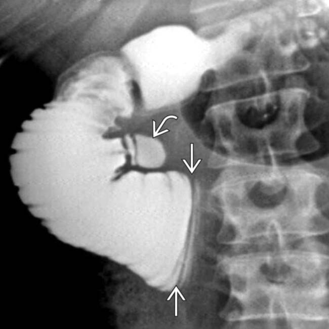
 of the 3rd portion of duodenum as it crosses over the midline, with dilation and slow emptying of the proximal duodenum. There is also a duodenal diverticulum
  .
.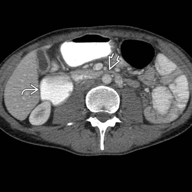
 and stomach. The 3rd portion of the duodenum
  is compressed as it passes between the aorta and the superior mesenteric artery (SMA).
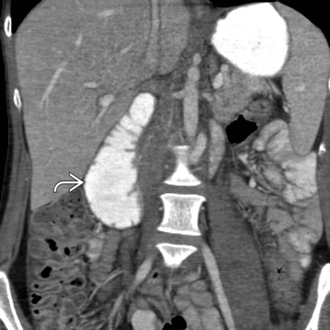
 , while the remaining bowel is collapsed. Note this patient’s thin body habitus.
 and the aorta, with compression of the 3rd portion of duodenum
  as it passes between these vessels.


