Chapter 411 Skeletal Diseases Influencing Pulmonary Function
Pulmonary function is influenced by the structure of the chest wall (Chapter 365). Chest wall abnormalities can lead to restrictive or obstructive pulmonary disease, impaired respiratory muscle strength, and decreased ventilatory performance in response to physical stress. The congenital chest wall deformities include pectus excavatum, pectus carinatum, sternal clefts, Poland syndrome, and skeletal and cartilage dysplasias. Vertebral anomalies such as kyphoscoliosis can alter pulmonary function in children and adolescents.
411.1 Pectus Excavatum (Funnel Chest)
Etiology
Midline narrowing of the thoracic cavity is usually an isolated skeletal abnormality. The cause is unknown. Pectus excavatum can occur in isolation or it may be associated with a connective tissue disorder (Marfan [Chapter 693] or Ehlers-Danlos syndrome [Chapter 651]). It may be acquired secondarily to chronic lung disease, neuromuscular disease, or trauma.
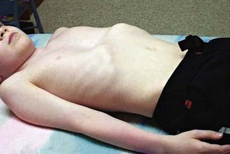
Clinical Manifestations
The deformity is present at or shortly after birth but is usually not associated with any symptoms at that time. In time, decreased exercise tolerance, fatigue, chest pain, palpitations, recurrent respiratory infections, wheezing, stridor, and cough may be present. Because of the cosmetic nature of this deformity, children may experience significant psychologic stress. Physical examination may reveal sternal depression, protracted shoulders, kyphoscoliosis, inferior rib flares, rib cage rigidity, forward head tilt, scapular winging, and loss of vertebral contours (Fig. 411-1). Patients exhibit paroxysmal sternal motion and a shift of point of maximal impulse to the left. Innocent systolic murmurs may be heard.
Laboratory Findings
Lateral chest radiograms demonstrate the sternal depression. Use of the Haller index on chest CT (maximal internal transverse diameter of the chest divided by the minimal anteroposterior diameter at the same level) in comparison with age- and gender-appropriate normative values for determining the extent of depression of the chest wall anomaly has become useful in determining the extent of the anatomic abnormality. An electrocardiogram may show a right-axis deviation or Wolff-Parkinson-White syndrome (Chapter 429); an echocardiogram may demonstrate mitral valve prolapse (Chapter 422.3) and ventricular compression. Results of static pulmonary function tests may be normal but commonly show an obstructive defect in the lower airways and, less commonly, a restrictive defect due to abnormal chest wall mechanics. Exercise testing may demonstrate either normal tolerance or limitations from underlying cardiopulmonary dysfunction that appear associated with the severity of the defect. Ventilatory limitations are commonly seen in younger children and adolescents, whereas cardiac limitations secondary to stroke volume impairments are more commonly seen in older adolescents and young adults.
Brigato RR, Campos JR, Jatene FB, et al. Pectus excavatum: evaluation of Nuss technique by objective methods. Interact Cardiovasc Thorac Surg. 2008;7:1084-1088.
Borowitz D, Cerny F, Zallen G, et al. Pulmonary function and exercise response in patients with pectus excavatum after Nuss repair. J Pediatr Surg. 2003;38:544-547.
Daunt SW, Cohen JH, Miller SF. Age-related normal ranges for the Haller index in children. Pediatr Radiol. 2004;34:326-330.
Haller JAJr, Loughlin GM. Cardiorespiratory function is significantly improved following corrective surgery for severe pectus excavatum: proposed treatment guidelines. J Cardiovasc Surg. 2000;41:125-130.
Koumbourlis AC, Stolar CJ. Lung growth and function in children and adolescents with idiopathic pectus excavatum. Pediatr Pulmonol. 2004;38:339-343.
Malek MH, Fonkalsrud EW, Cooper CB. Ventilatory and cardiovascular responses to exercise in patients with pectus excavatum. Chest. 2003;124:870-882.
Nuss D, Kelly REJr. Minimally invasive surgical correction of chest wall deformities in children (Nuss procedure). Adv Pediatr. 2008;55:395-410.
Ohno K, Morotomi Y, Nakahira M, et al. Indications for surgical repair of funnel chest based on indices of chest wall deformity and psychologic state. Surg Today. 2003;33:662-665.
Protopapas AD, Athanasiou T. Peri-operative data on the Nuss procedure in children with pectus excavatum: independent survey of the first 20 years’ data. J Cardiothorac Surg. 2008 Jul 4;3:40.
Rowland T, Moriarty K, Banever G. Effect of pectus excavatum deformity on cardiorespiratory fitness in adolescent boys. Arch Pediatr Adolesc Med. 2005;159:1069-1073.
Shamberger RC, Welch KJ, Sanders SP. Mitral valve prolapse associated with pectus excavatum. J Pediatr. 1987;111:404-407.
Zhao L, Feinberg MS, Gaides M, et al. Why is exercise capacity reduced in subjects with pectus excavatum? J Pediatr. 2000;136:163-167.
411.2 Pectus Carinatum and Sternal Clefts
Abramson H, D’Agostino J, Wuscovi S. A 5-year experience with a minimally invasive technique for pectus carinatum repair. J Pediatr Surg. 2009;44:118-123.
Coelho Mde S, Guimarães Pde S. Pectus carinatum. J Bras Pneumol. 2007;33:463-474.
Fonkalsrud EW, Anselmo DM. Less extensive techniques for repair of pectus carinatum: the undertreated chest deformity. J Am Coll Surg. 2004;198:898-905.
Goretsky MJ, Kelly RE, Croitoru D, et al. Chest wall anomalies: pectus excavatum and pectus carinatum. Adolesc Med Clin. 2004;15:455-471.
Ohye RG, Rutherford JA, Bove EL. Congenital sternal clefts. Pediatr Cardiol. 2002;23:472-473.
Waters P, Welch K, Micheli LJ, et al. Scoliosis in children with pectus excavatum and pectus carinatum. J Pediatr Orthop. 1989;9:551-556.
Williams AM, Crabbe DC. Pectus deformities of the anterior chest wall. Paediatr Respir Rev. 2003;4:237-242.
411.3 Asphyxiating Thoracic Dystrophy (Thoracic-Pelvic-Phalangeal Dystrophy)
Davis JT, Long FR, Adler BH, et al. Lateral thoracic expansion for Jeune syndrome: evidence of rib healing and new bone formation. Ann Thorac Surg. 2004;77:445-448.
Kajantic E, Anderson S, Kaitila I. Familial asphyxiating thoracic dysplasia: clinical variability and impact of improved neonatal intensive care. J Pediatr. 2001;139:130-133.
Phillips JD, van Aalst JA. Jeune’s syndrome (asphyxiating thoracic dystrophy): congenital and acquired. Semin Pediatr Surg. 2008;17:167-172.
Sharoni E, Erez E, Chorev G, et al. Chest reconstruction in asphyxiating thoracic dystrophy. J Pediatr Surg. 1998;33:1578-1581.
Tahernia AC, Stamps P. “Jeune syndrome” (asphyxiating thoracic dystrophy): report of a case, a review of the literature, and an editor’s commentary. Clin Pediatr. 1977;16:903-908.
Wiebicke W, Pasterkamp H. Long-term continuous positive pressure in a child with asphyxiating thoracic dystrophy. Pediatr Pulmonol. 1988;4:54-58.
411.4 Achondroplasia
Achondroplasia is the most common condition characterized by disproportionate short stature. This condition is inherited as an autosomal dominant disorder that results in disordered growth (Chapter 687). Much has been learned about this disorder, including its genetic origins and how to minimize its serious complications.
Treatment
Treatment of sleep apnea, if present, is supportive (Chapter 17). Physiotherapy and bracing may minimize the complications of both kyphosis and severe lordosis. Aggressive treatment of respiratory infections and scoliosis is warranted.
Boulet S, Althuser M, Nugues F, Schaal JP, et al. Prenatal diagnosis of achondroplasia: new specific signs. Prenat Diagn. 2009;29:697-702.
Hunter AG, Reid CS, Pauli RM, et al. Standard curves of chest circumference in achondroplasia and the relationship of chest circumference to respiratory problems. Am J Med Genet. 1996;62:91-97.
Mogayzel PJJr, Carroll JL, Loughlin GM, et al. Sleep-disordered breathing in children with achondroplasia. J Pediatr. 1998;132:667-671.
Stokes DC, Phillips JA, Leonard CO, et al. Respiratory complications of achondroplasia. J Pediatr. 1983;102:534-541.
Stokes DC, Wohl ME, Wise RA, et al. The lungs and airways in achondroplasia: do little people have little lungs? Chest. 1990;98:145-152.
Tasker RC, Dundas I, Laverty A, et al. Distinct patterns of respiratory difficulty in young children with achondroplasia: a clinical, sleep, and lung function study. Arch Dis Child. 1998;79:99-108.
Trotter TL, Hall JG, American Academy of Pediatrics Committee on Genetics. Health supervision for children with achondroplasia. Pediatrics. 2005;116:771-783.
411.5 Kyphoscoliosis: Adolescent Idiopathic Scoliosis and Congenital Scoliosis
Pathogenesis
Adolescent idiopathic scoliosis (AIS) is characterized by lateral bending of the spine (Chapter 671). It commonly affects children during their teen years as well as during periods of rapid growth. The cause is unknown. Congenital scoliosis is uncommon, affecting girls more than boys, and is apparent in the 1st year of life (Chapter 671.2).
Chen S, Huang T, Lee Y, Hsu RW. Pulmonary function after thoracoplasty in adolescent idiopathic scoliosis. Clin Orthop Related Res. 2002;399:152-161.
Hedequist D, Emans J. Congenital scoliosis. J Am Acad Orthop Surg. 2004;12:266-275.
Kearon C, Viviani GR, Killian KJ. Factors influencing work capacity in adolescent idiopathic thoracic scoliosis. Am Rev Respir Dis. 1993;148:295-303.
Leech JA, Ernst P, Rogala EJ, et al. Cardiorespiratory status in relation to mild deformity in adolescent idiopathic scoliosis. J Pediatr. 1985;106:143-149.
McNicholas WT. Impact of sleep in respiratory failure. Eur Respir J. 1997;10:920-933.
Mezon BL, West P, Israels J, et al. Sleep breathing abnormalities in kyphoscoliosis. Am Rev Respir Dis. 1980;122:617-621.
Yuan N, Skaggs DL, Dorey F, et al. Preoperative predictors of prolonged postoperative mechanical ventilation in children following scoliosis repair. Pediatr Pulmonol. 2005;40:414-419.
411.6 Congenital Rib Anomalies
Bronsther B, Coryllos E, Epstein B, et al. Lung hernias in children. J Pediatr Surg. 1968;3:544-550.
Campbell RMJr, Smith MD, Mayes TC, et al. The characteristics of thoracic insufficiency syndrome associated with fused ribs and congenital scoliosis. J Bone Joint Surg Am. 2003;85-A:399-408.
Mehta MH, Patel RV, Mehta LV, et al. Congenital absence of ribs. Indian Pediatr. 1992;29:1149-1152.
Rickham PP. Lung hernia secondary to congenital absence of ribs. Arch Dis Child. 1959;34:14-17.


