Sigmoid Volvulus

Published on 19/07/2015 by admin

Filed under Radiology

Last modified 22/04/2025

Print this page

rate 1 star rate 2 star rate 3 star rate 4 star rate 5 star
Your rating: none, Average: 0 (0 votes)

This article have been viewed 2829 times

 Midline; directed toward RUQ or LUQ; elevation of hemidiaphragm

• Diagnosis: Abdominal radiography, water-soluble contrast enema, CT

image Coronal reformatted CT is especially useful in diagnosis
image Abdominal radiographs: Supine, upright, prone, and decubitus views

PATHOLOGY

• Major predisposing factors

image Diet: Fiber increase → increased bulk of stool, elongation and dilatation of colon
image Chronic constipation and obtundation from medications → gaseous distension
• Comorbid disease: 30% with psychiatric disease, 13% are institutionalized at time of diagnosis

CLINICAL ISSUES

• Most common signs/symptoms

image Acute or insidious onset
image Abdominal pain (< 33%), vomiting, distension, obstipation
• Treatment: Sigmoidoscopic decompression of obstruction ± stabilization via rectal tube insertion

image Usually followed by surgical resection of sigmoid colon

DIAGNOSTIC CHECKLIST

• Rule out other causes of distal colonic obstruction
• Dilated sigmoid colon in inverted “U” shape with absent haustra; “beaking,” whirl sign, northern exposure sign
image
(Left) Supine film of the abdomen shows marked dilation of the sigmoid colon. The sigmoid is folded back upon itself, and the apposed walls of the redundant sigmoid colon image 
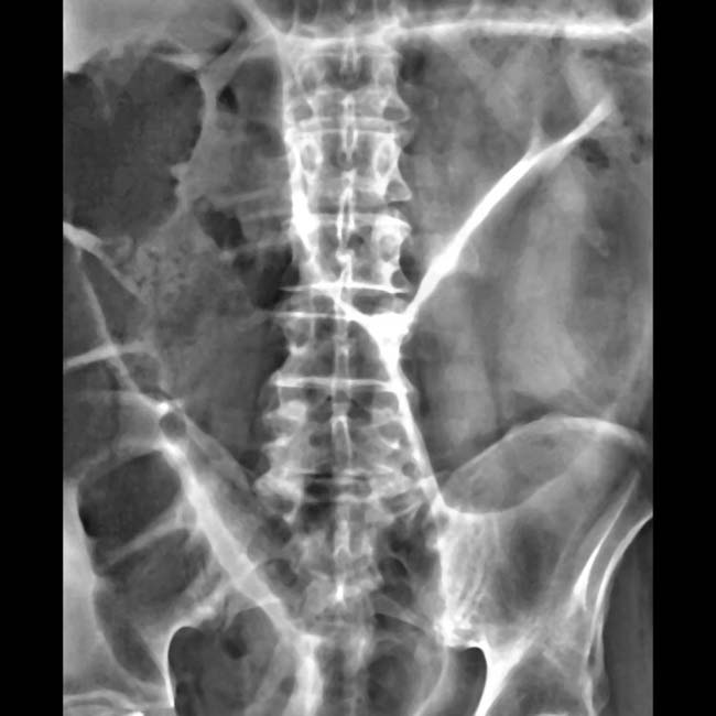
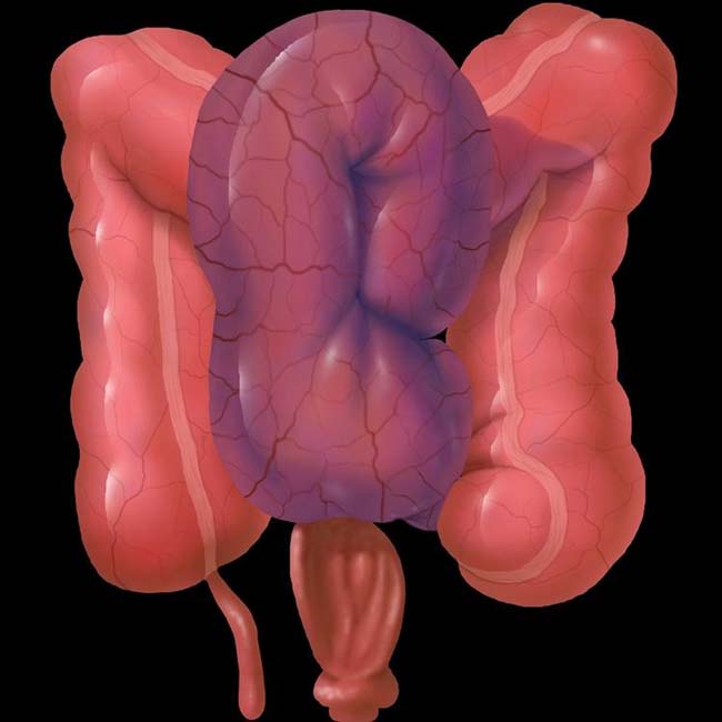
form the “seam” of the football (or coffee bean) shape. The sigmoid extends into the upper abdomen above the transverse colon image.

image
(Right) Axial CECT in the same case shows the dilated sigmoid lumen image 
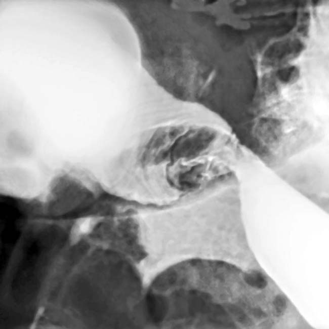
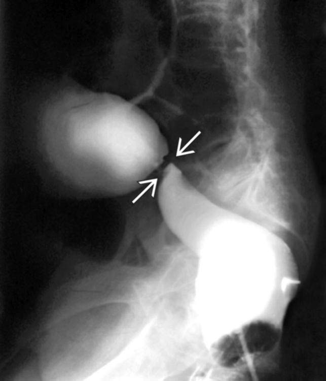
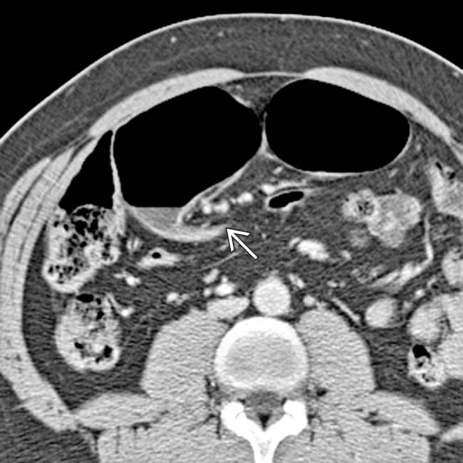
with abrupt narrowing at its base image.
image
(Left) Coronal reformatted CT in the same patient shows twisting and displacement of the base of the sigmoid colon and its mesentery image. The dilated colonic segments upstream from the volvulus may be easier to distinguish on coronal sections.

image
(Right) Another CT section in this case shows the whirl sign image 
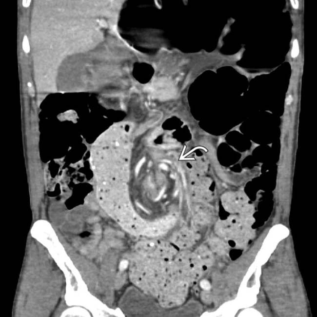
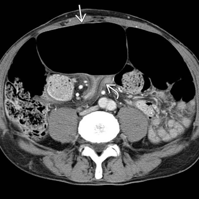
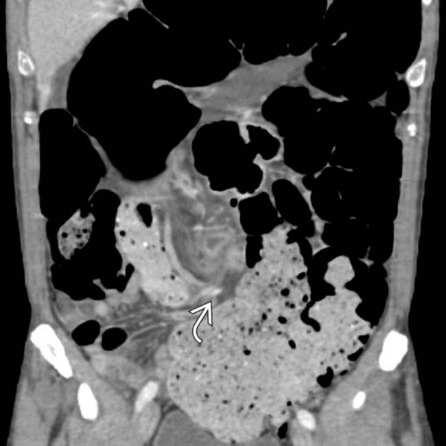
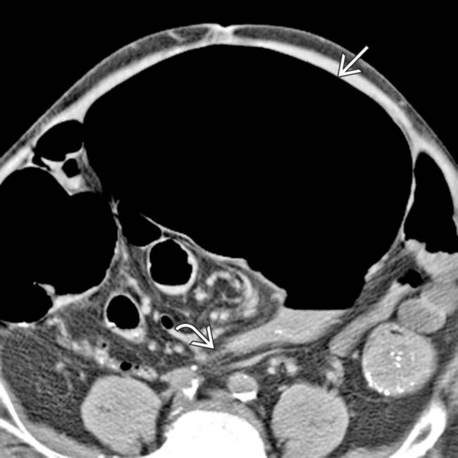
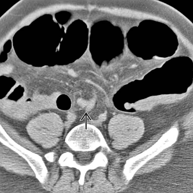
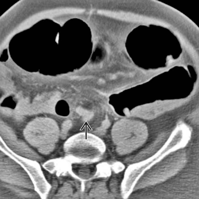
of twisted colon and vessels at the base of the sigmoid mesentery.

TERMINOLOGY

Definitions

• Torsion or twisting of sigmoid colon around its mesenteric axis

IMAGING

General Features

• Best diagnostic clue

image Dilated sigmoid colon with inverted “U” configuration and absent haustra
• Location

image Midline; directed toward RUQ or LUQ; elevation of hemidiaphragm

Radiographic Findings

• Radiography

image Sigmoid volvulus

– Diagnostic in 75% of cases
– Vertical dense white line: Apposed inner walls of sigmoid colon pointing toward pelvis
– Closed loop obstruction: Segment of bowel obstructed at 2 points
– Gas in proximal small intestine and colon; absence of gas in rectum
– Absent rectal gas in spite of prone or decubitus views
– Inverted “U” shape with absent haustra
– Northern exposure sign: Dilated, twisted sigmoid colon projects above transverse colon on supine radiograph
– Apex above T10 vertebra and under left hemidiaphragm; directed toward right shoulder
image Compound volvulus

– Dilated sigmoid loop in mid abdomen extending to RLQ with distended small bowel
– Medially deviated distal left colon

Fluoroscopic Findings

• Water-soluble contrast enema

image Can use low-pressure barium enema without balloon inflation
image “Beaking”: Smooth, tapered narrowing or point of torsion at rectosigmoid junction
image Mucosal folds often show corkscrew pattern at point of torsion
image Shouldering: Localized wall thickening at site of twist (in chronic or recurrent volvulus)

CT Findings

• CECT

image “Beaking”: Progressive tapering of afferent and efferent limbs leading into twist
image Whirl sign: Tightly twisted mesentery and bowel near base of volvulus
image Compound volvulus: Medial deviation of distal left colon with pointed appearance of medial border

Imaging Recommendations

• Best imaging tool

image Abdominal radiography, water-soluble contrast enema, CT

– Supine, upright, prone, and decubitus views of abdomen
– Coronal reformatted CT is especially useful in diagnosis

DIFFERENTIAL DIAGNOSIS

Acute Ileus

• Postop, medication, post-traumatic injury, ischemia
• Dilated large and small bowel with no transition point
• Air-fluid levels without peristalsis
• No colonic obstruction

Functional Megacolon

• Gross constipation without organic cause
• Markedly dilated, ahaustral, air- or stool-filled colon
• Ogilvie syndrome: Nonobstructive dilation of colon

Toxic Megacolon

• Dilated ahaustral transverse colon in patient with known ulcerative or infectious colitis
• “Thumbprinting” due to edematous mucosa
• Mucosal surface is ulcerated or sloughed

Distal Colon Obstruction

• Change in stool caliber over several months
• Gas-filled intestinal loops proximal to obstruction; no distal gas
• Abrupt transition at site of obstruction
• Malignancy

image Most common cause of colonic obstruction (55%)
image Insidious onset
image Weakness, weight loss, anorexia, rectal bleeding
image “Apple core” configuration, mucosal destruction
image Positive fecal occult blood test highly suggestive of colon cancer
• Stricture secondary to diverticulitis

image 2nd most common cause of colonic obstruction (12%)
image History of recurrent diverticulitis
image Other diverticula present
image Signs of diverticulitis (e.g., infiltrated mesocolic fat, extraluminal gas or fluid)

PATHOLOGY

General Features

• Etiology

image Major predisposing factors

– Diet: Fiber increase → increased bulk of stool, elongation and dilatation of colon
– Chronic constipation and obtundation from medications → gaseous distension
image Compound volvulus (ileosigmoid knot)

– Hyperactive ileum winding around narrow pedicle of passive sigmoid colon
image Etiology in children

– Malrotation and other mesenteric attachment abnormalities
– Constipation (mental retardation, Hirschsprung disease, cystic fibrosis, aerophagia)
• Associated abnormalities

image Comorbid disease: 30% with psychiatric disease, 13% are institutionalized at time of diagnosis

Gross Pathologic & Surgical Features

• Twisted narrow segment with markedly dilated sigmoid loop

Microscopic Features

• Localized thickening of mucosal folds; ischemic and necrotic changes

CLINICAL ISSUES

Presentation

• Most common signs/symptoms

image Acute or insidious onset
image Abdominal pain (< 33%), vomiting, distension, obstipation
image Compound volvulus

– Rapid deterioration (more than with other colonic volvulus types)
– Pain disproportionate to physical findings; absolute constipation
image Diagnosis: Radiography is definitive in 75% of cases

Demographics

• Age

image 60-80 years
• Epidemiology

image 3rd most common cause of colonic obstruction (10%)
image 60-75% of colonic volvulus cases involve sigmoid colon
image Causes 1-2% of intestinal obstructions in USA
image Increased incidence in elderly men and residents of nursing homes &/or mental hospitals (constipation and obtundation)
image Significant increase in South America and Africa (increased fiber in diet)

Natural History & Prognosis

• Complications

image Closed loop obstruction → strangulation
image Ischemia, necrosis (15-20%), and perforation
image Ileosigmoid knot → strangulation and gangrene of small bowel within hours
• Prognosis

image Uncomplicated: Good
image Complicated: Poor
image 40-50% recurrence after nonoperative reduction
image Degree of rotation relative to chance of nonsurgical decompression: 180° (35%), 360° (50%), 540° (10%)

– Twist > 360° does not resolve spontaneously
image 3% recurrence after nonoperative and operative reduction

Treatment

• Nonoperative

image Sigmoidoscopic decompression of obstruction ± stabilization via rectal tube insertion
image 70-80% success rate
• Nonoperative and operative

image Decompression, mechanical cleansing, and elective sigmoid resection
• Complicated cases

image Surgical emergency; colonic resection
• Follow-up

image Water-soluble contrast enema to rule out underlying colon cancer

DIAGNOSTIC CHECKLIST

Consider

• Acute abdomen; rule out other causes of obstruction

Image Interpretation Pearls

• Dilated sigmoid colon in inverted “U” shape with absent haustra; “beaking,” whirl sign, northern exposure sign
image
(Left) Axial CECT in a 47-year-old man presenting with a 6-day history of abdominal distension and pain, constipation, and absence of flatus shows the sigmoid volvulus evident in the LLQ with the beaked appearance of the descending loop image of the twisted sigmoid colon.

image
(Right) Coronal CECT in the same patient illustrates a swirled appearance of the mesenteric pedicle at the site of the sigmoid volvulus image
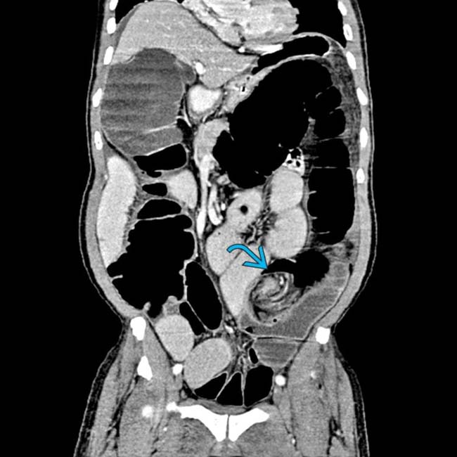
, characteristic findings in this setting.
image
(Left) Radiograph in a 65-year-old man presenting with abdominal pain and distention demonstrates a dilated redundant sigmoid colon appearing as an inverted “U” loop arising from the pelvis image 
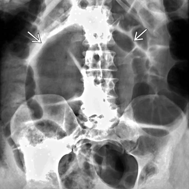
in this typical presentation of a sigmoid volvulus.

image
(Right) Supine frontal image from a contrast enema in the same patient demonstrates luminal tapering at the site of stenosis, a.k.a. the bird’s beak sign image
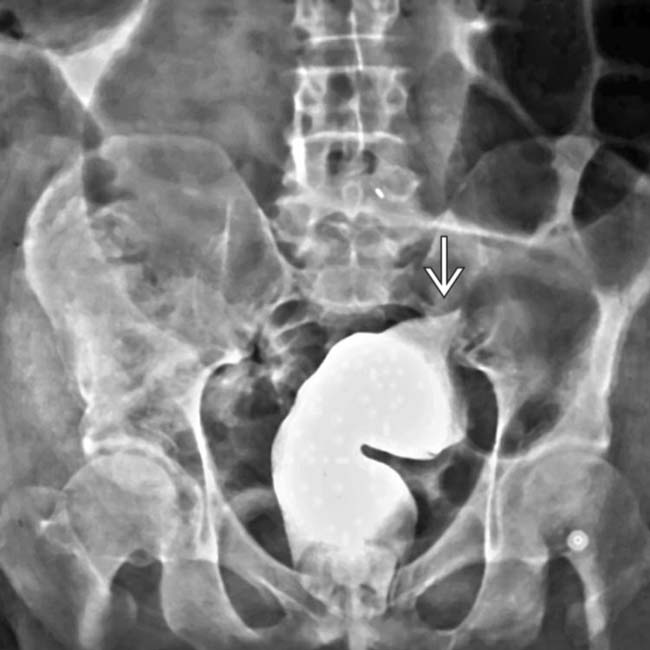
. This configuration is pathognomonic for sigmoid volvulus.
image
(Left) Supine digital scout radiograph in an 89-year-old man who is a long-time resident of a nursing home, presenting with a 2-day history of severe abdominal pain and marked abdominal distension, reveals marked dilation of the sigmoid colon image 
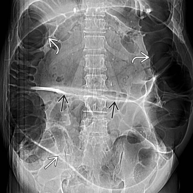
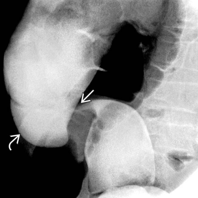
as well as the ascending and descending colon image. Note the apposed walls image of the sigmoid colon.

image
(Right) Axial CECT in the same patient illustrates the bird’s beak sign image 
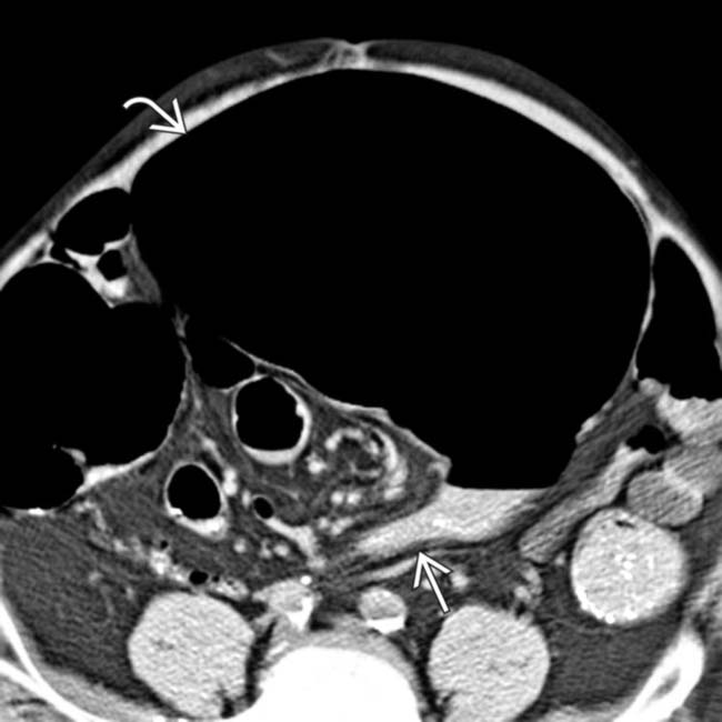
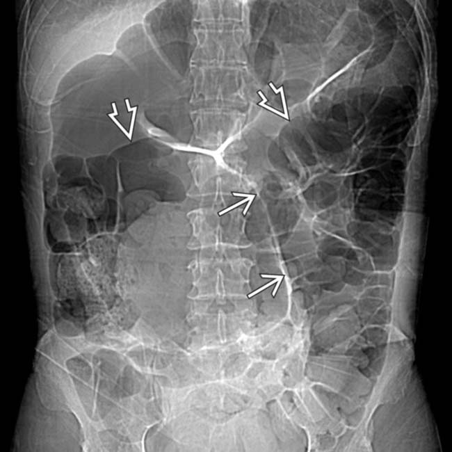
from the volvulus obstructing the massively dilated sigmoid colon image.
image
Graphic shows a dilated, twisted, elongated sigmoid colon with venous engorgement and colonic obstruction.

image
Axial NECT demonstrates sigmoid volvulus. Note the massive dilatation of the sigmoid colon with beaking image 
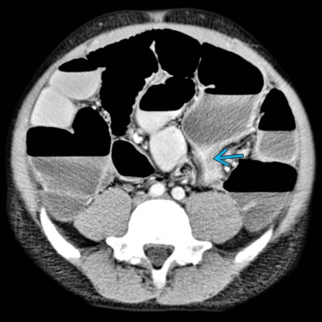
at the site of volvulus image.
image
Frontal radiograph shows a classic case of sigmoid volvulus. Note the classic inverted, U-shaped, massively dilated sigmoid colon directed toward the right hemidiaphragm image.
image
Lateral contrast enema in the same patient shows a contrast-filled rectum and “bird’s beak” sign image corresponding to luminal narrowing at the site of sigmoid obstruction.
image
Axial CECT shows massive colonic redundancy and distention of the large and small bowel. Sigmoid volvulus is evident in LLQ by the beaked appearance of the descending loop image 
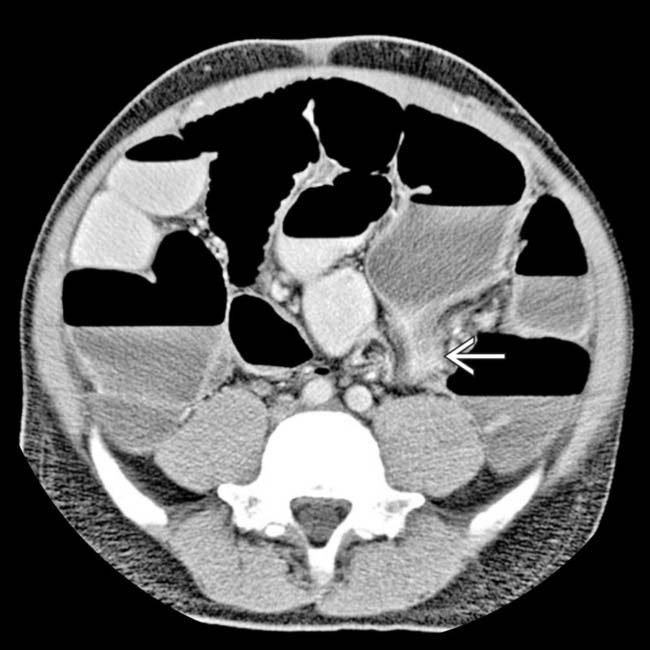
of twisted sigmoid colon.
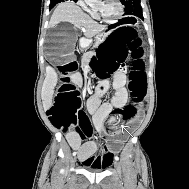
image
Coronal CECT in the same patient demonstrates the classic swirled appearance of the mesenteric pedicle at the site of sigmoid volvulus image.
image
Axial NECT shows a partially obstructing sigmoid volvulus. Note the “beaking” of the sigmoid at the site of the twist image.
image
Oblique water-soluble contrast enema of the sigmoid colon in the same patient reveals “beaking” at the site of the volvulus image 
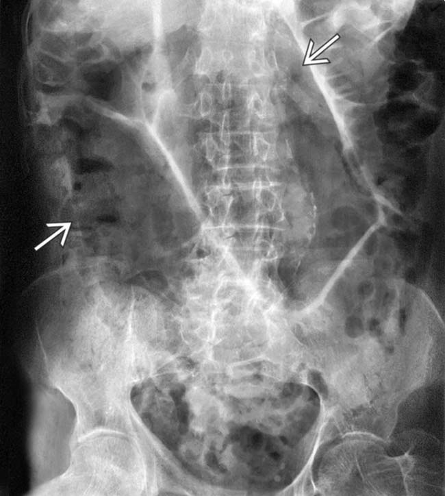
and distal filling of the dilated colon image due to partial obstruction.
image
Supine radiograph shows dilation of the entire colon. The vertical white line image 
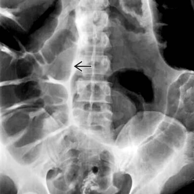
represents the apposed walls of the dilated, inverted sigmoid colon and points toward the mesenteric volvulus.
image
Supine radiograph shows dilated colon. The apex of the sigmoid colon image 
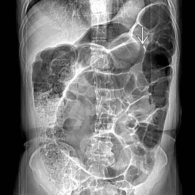
can be seen above the transverse colon, the “northern exposure” sign of sigmoid volvulus.
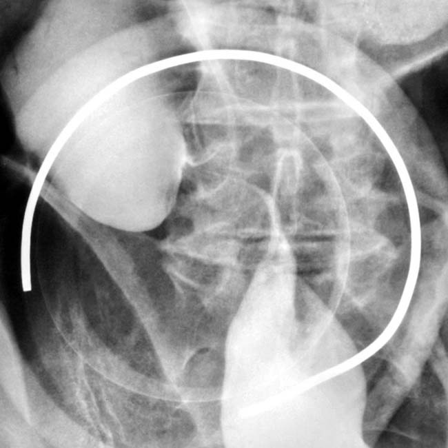
image
Single contrast BE shows a smooth tapered beak obstructing the lumen of the sigmoid colon.
image
Axial CECT of sigmoid volvulus shows diffuse dilation of the colon. There is a swirl of sigmoid mesocolic blood vessels that converge at the site of the volvulus image.
image
Axial CECT shows a swirl of mesocolic vessels at the base of the volvulus image.
image
Supine radiograph shows a dilated, inverted, U-shaped sigmoid colon.
image
Single contrast BE shows twist and beak at the point of the volvulus with dilated colon beyond the twist.

SELECTED REFERENCES

1. Maddah, G, et al. Management of sigmoid volvulus: options and prognosis. J Coll Physicians Surg Pak. 2014; 24(1):13–17.

2. Selçuk Atamanalp, S. Treatment for ileosigmoid knotting: a single-center experience of 74 patients. Tech Coloproctol. 2014; 18(3):233–237.

3. Meyer, F, et al. Unusual “twister”-like appearance of a sigmoid volvulus on computed tomography. Endoscopy. 2007; 39(Suppl 1):E295.

4. Tiah, L, et al. Sigmoid volvulus: diagnostic twists and turns. Eur J Emerg Med. 2006; 13(2):84–87.

5. Agaoglu, N, et al. Surgical treatment of the sigmoid volvulus. Acta Chir Belg. 2005; 105(4):365–368.

6. Chandrasekaran, TV, et al. Minimally invasive stapled surgical approach to the management of sigmoid volvulus. Ann R Coll Surg Engl. 2005; 87(5):381–382.

7. Matsumoto, S, et al. Computed tomographic imaging of abdominal volvulus: pictorial essay. Can Assoc Radiol J. 2004; 55(5):297–303.

8. Chiu, HH, et al. Recurrent sigmoid volvulus. Gastrointest Endosc. 2002; 56(3):419–420.

9. Kuzu, MA, et al. Emergent resection for acute sigmoid volvulus: results of 106 consecutive cases. Dis Colon Rectum. 2002; 45(8):1085–1090.

10. Chirdan, LB, et al. Sigmoid volvulus and ileosigmoid knotting in children. Pediatr Surg Int. 2001; 17(8):636–637.

11. Moore, CJ, et al. CT of cecal volvulus: unraveling the image. AJR Am J Roentgenol. 2001; 177(1):95–98.

12. Daniels, IR, et al. Recurrent sigmoid volvulus treated by percutaneous endoscopic colostomy. Br J Surg. 2000; 87(10):1419.

13. Dulger, M, et al. Management of sigmoid colon volvulus. Hepatogastroenterology. 2000; 47(35):1280–1283.

14. Lee, SH, et al. The ileosigmoid knot: CT findings. AJR Am J Roentgenol. 2000; 174(3):685–687.

15. Madiba, TE, et al. The management of sigmoid volvulus. J R Coll Surg Edinb. 2000; 45(2):74–80.

16. Chung, YF, et al. Minimizing recurrence after sigmoid volvulus. Br J Surg. 1999; 86(2):231–233.

17. Javors, BR, et al. The northern exposure sign: a newly described finding in sigmoid volvulus. AJR Am J Roentgenol. 1999; 173(3):571–574.

18. Choi, D, et al. Endoscopic sigmoidopexy: a safer way to treat sigmoid volvulus? J R Coll Surg Edinb. 1998; 43(1):64.

19. Catalano, O. Computed tomographic appearance of sigmoid volvulus. Abdom Imaging. 1996; 21(4):314–317.

20. Frizelle, FA, et al. Colonic volvulus. Adv Surg. 1996; 29:131–139.

21. Forde, KA. Therapeutic colonoscopy. World J Surg. 1992; 16(6):1048–1053.

22. Gibney, EJ. Volvulus of the sigmoid colon. Surg Gynecol Obstet. 1991; 173(3):243–255.