CHAPTER 4 Shock and trauma
Shock
Hypovolaemic shock
Symptoms and signs
Treatment
Shock is a surgical emergency and needs rapid treatment.
Septic shock
Treatment
Cardiogenic shock
Causes
Investigations
Urgent investigations include portable CXR, FBC, U&E, cardiac enzymes, D-dimers, ABGs, ECG, CXR.
Neurogenic shock
Trauma
Initial assessment of the trauma patient
Primary survey
 ABCDE of emergency management:
Secondary survey
The secondary survey is a head-to-toe evaluation of the trauma patient, i.e. a complete history and physical examination, including a reassessment of all vital signs. Each area of the body should be completely examined. A full neurological examination is carried out including a GCS (Glasgow Coma Score) determination (Table 4.1).
| Responses | Score |
|---|---|
| Eye-opening response | |
| Spontaneous | 4 |
| To voice | 3 |
| To pain | 2 |
| None | 1 |
| Best verbal response | |
| Orientated | 5 |
| Confused | 4 |
| Inappropriate speech | 3 |
| Incomprehensible speech | 2 |
| None | 1 |
| Best motor response | |
| Obeys commands | 6 |
| Localizes pain | 5 |
| Withdraws to pain | 4 |
| Flexion to pain | 3 |
| Extension to pain | 2 |
| None | 1 |
| Total | 3–15 |
A score of 3 indicates a severe injury with a poor prognosis. A score of 13–15 indicates minor injury with a good prognosis.
Examination
Head injury (→ Ch. 18)
Management
The management of specific head injury is dealt with in the section on Neurosurgery (→ Ch. 18) but the basic principles are outlined here as far as trauma management is concerned.
Hypotension in adults is not due to intracranial blood loss. However, in children, significant blood loss can occur in head injuries and can be responsible for hypotension. The scalp should be examined for lacerations and boggy wounds. Observation should be made for bleeding and CSF leakage from the ear and nose. The cranial nerves should be checked and the limbs examined. Assessment of head injured patients include skull X-rays and CT scan; indications for these are detailed in Chapter 18.
Thoracic trauma
Airway obstruction
Aortic disruption (traumatic rupture of the aorta – TRA)
Tracheobronchial injury
Larynx
Pneumothorax (→ Ch. 9)
Miscellaneous thoracic injuries
Rib fractures
Common injury following blunt trauma. Rib fractures can lead to:
Sternal fractures
Abdominal trauma
Organs in the retroperitoneum include:
Penetrating trauma
Management
Initial management is via ATLS protocols, however, note specific points to remember:
Blunt trauma
Investigations in abdominal trauma
Specific organ injuries
Duodenum
Symptoms and signs
Bloody NG aspirate, retroperitoneal air, raised amylase (with associated pancreatic injury).
Liver
Management
Spleen
Most commonly injured in blunt trauma.
Management
Radiological
As with liver injury, certain splenic injuries can be managed via angiography and embolization.
Complications
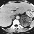
These include LUQ haematoma (may progress to abscess), pleural effusion, pseudoaneurysm of the splenic artery, arteriovenous fistula between the artery and vein, pancreatic injury/fistula and overwhelming post-splenectomy sepsis (OPSI, → Ch. 14). With splenic haematomas that were treated non-operatively, there is a small risk of delayed rupture, which can occur days or weeks later. It is prudent to monitor these patients with a repeat USS to identify an enlarging haematoma.
Small bowel
Commonly injured in penetrating trauma. In blunt trauma, injuries may occur due to:
Urinary trauma
Lower urinary tract
Urethra
Management
Limb trauma
Limb trauma involves injury to: soft tissues; blood vessels (→ Ch. 15); nerves; bones (→ Ch. 17). It can be life- or limb-threatening.
General principles of management:
Vascular trauma (→ Ch. 15)
Skeletal trauma (→ Ch. 17)
Nerve injuries
Symptoms and signs
These depend upon the site of injury to the nerve.

 The importance of an adequate drug and sensitivity history cannot be overemphasized. Always make sure before giving parenteral injections that resuscitation equipment and drugs are available.
 Immediate management depends on severity. The presence of abnormal pupillary reflexes, asymmetrical motor signs or deteriorating level of consciousness is an immediate indication for treatment.


