Published on 18/03/2015 by admin
Filed under Dermatology
Last modified 22/04/2025
This article have been viewed 5706 times
Richard J. Motley
Evidence Levels: A Double-blind study B Clinical trial ≥ 20 subjects C Clinical trial < 20 subjects D Series ≥ 5 subjects E Anecdotal case reports
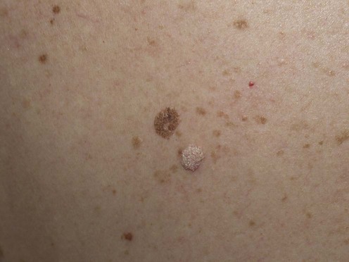
Seborrheic keratosis is a benign, exophytic, warty, lightly pigmented growth of the skin surface that becomes increasingly common with age. Found mainly on the trunk, often at sites of pressure, it is a cosmetic nuisance and rarely a cause for diagnostic confusion. Several variants exist and are described below.
Many patients present with seborrheic keratoses because of concern about possible melanoma, and reassurance may be all that is required. Occasionally the lesion can become ‘irritated’ and show erythema, crusting, and itching (lower lesion in photograph). In this case the appearance may resemble a pyogenic granuloma or squamous cell carcinoma.
Where treatment is requested there are several options and the choice will depend on patient and physician preference. Surgical excision, although effective, is never the treatment of choice and usually indicates that the physician failed to make the correct clinical diagnosis or is unfamiliar with alternative treatments. When the diagnosis is in doubt then material should be taken for histologic examination, preferably by a ‘shave’ or tangential biopsy technique or by sharp curettage. Blunt curettage provides poor material for histologic assessment. Cautery is very effective at softening the lesion and often allows it to be removed with minimal effort, the heat separating the lesion at the dermoepidermal junction. The ‘melting’ of the lesion observed after the application of heat is almost diagnostic. Smaller lesions can be ‘flicked off’ the skin using a traditional curette, often without local anesthesia. Heat can be applied to the surface of the lesion followed by curettage, and for superficial lesions ‘curettage’ can be achieved using a cotton gauze swab to wipe away the softened lesion. Cautery can also be used after shave excision to treat any tissue remnants. With all these treatments the aim is to remove the lesion, but little of the underlying skin surface.
Cryotherapy using a liquid nitrogen spray is an alternative method of treatment. Liquid nitrogen is sprayed onto the lesion until it is frozen, and then continued for 5 to 10 seconds. It can be undertaken without local anesthesia because freezing reduces sensation of pain, and this can be an advantage when treating multiple lesions. After a day or two the treated lesion blisters and crumbles away. The underlying wound heals over after several days and is often quite exudative, requiring daily cleansing by the patient. Overall the recovery period is longer and the wound slower to heal than following curettage and cautery. Whereas following cautery hyperpigmentation is common, following cryotherapy, hypopigmentation occurs. For this reason it is not recommended in black people.
Senile or ‘solar’ lentigines can be considered to be flat versions of the seborrheic keratosis. Sometimes referred to as ‘age’ or ‘liver’ spots, these small pigmented papules and plaques are more commonly seen on areas of frequent sun exposure, such as the face and dorsa of the hands. Their true nature can be recognized by the slight velvety texture to the lesional surface, which is best seen with tangential lighting. This indicates that the lesion is not a true lentigo but a superficial keratosis. These lesions are amenable to very minor treatments. Topical tretinoin cream can be effective. Other treatments include light abrasion with an exfoliating cream, light dermabrasion or laser resurfacing, cryotherapy, or chemical peels using trichloroacetic acid or phenol. A favorite treatment is minimal cautery followed by ‘curettage’ with a cotton gauze swab. This leaves an erythematous superficial wound that heals rapidly.
Dermatosis papulosa nigra is commonly seen on the cheeks of black adults. These small seborrheic warts are easily treated by light cautery or diathermy followed by cotton gauze curettage, but patients should be warned about the possibility of hyperpigmentation.
Stucco keratoses are small grayish-white seborrheic keratoses which are typically found on the forearms and lower legs and are easily removed with curettage without bleeding. The edge of these lesions is often curled up away from the skin surface.
Giant seborrheic keratoses are large lesions, usually found on the scalp, and are often several centimeters in diameter.
Seborrheic keratoses may have a familial tendency, especially when multiple. It is these patients who are often the most challenging to treat and also the most affected by their condition. Many middle-aged to elderly patients will not undress for sporting activities such as swimming because they are embarrassed by the appearance of their skin. Patients not infrequently present with multiple lesions and request their complete removal. In these circumstances it is reasonable to offer several different types of treatment so that the patient can determine their preference.
The sudden onset of multiple seborrheic keratoses may be associated with underlying malignancy (the sign of Leser–Trelat) and should prompt a full clinical examination for underlying malignancy.
Consider the sign of Leser–Trelat and investigate for underlying malignancy
Schwartz RA. J Am Acad Dermatol 1996; 35: 88–95.
The sign of Leser–Trelat is rare. The sudden eruption of multiple seborrheic keratoses or their rapid increase in size is caused by a malignancy. Its association with malignant acanthosis nigricans, seen in 35% of patients, is one of several of its features that support its legitimacy as a true paraneoplastic disorder.
Long CC, Motley RJ, Holt PJ. Br J Dermatol 1994; 131: 732–3.
Ring curettage followed by aluminum chloride 25% in 70% isopropyl alcohol gave a superior cosmetic result compared to using a traditional curette and light cautery.
Motley R. Dermatol Pract 1997; 5: 6–7.
A practical review of treatment options.
Dawber R, Colver G, Jackson A, eds. London: Martin Dunitz, 1997.
A well-illustrated practical guide to all that is cutaneous cryosurgery, including seborrheic warts!
Goetze S, Ziemer M, Lipman RD, Elsner P. Dermatol Surg 2006; 32: 661–8.
Hydrocolloid dressings produced superior healing of superficial wounds following curettage of seborrhoeic keratoses.
Chun EY, Lee JB, Lee KH. Dermatol Surg 2004; 30; 512–16.
After cleansing the skin with alcohol, 65% trichloroacetic acid was applied focally to seborrheic keratoses using a sharpened wooden applicator, to create evenly frosted spots on each lesion. Crusts separated from the skin with gentle washing after 4 to 7 days, and healing was complete 10 to 14 days later. Twenty-three patients required a mean of 1.5 treatments, and in 57% of these the results were rated as excellent.
Mehrabi D, Brodell RT. Dermatol Surg 2002; 28: 437–9.
A patient with multiple seborrheic keratoses was treated in four sessions with a normal-mode alexandrite laser (755 nm wavelength) spot size 8 mm, fluence 100 J/cm2. The entire surface of each lesion was treated in a checkerboard fashion. Twelve-day follow-up revealed excellent cosmetic resolution of the lesions with minimal scarring and hypopigmentation, especially compared to areas previously treated with liquid nitrogen cryotherapy.
Trafeli JP, Kwan JM, Meehan KJ, Domankevitz Y, Gilbert S, Malomo K, et al. Dermatol Surg 2007; 33: 1477–82.
Patients with solar lentigines were treated with a long-pulse alexandrite laser (755 nm wavelength). Optimal settings were spot size 10 mm, pulse duration 3 ms, fluence 22 J/cm2 (without skin cooling). The authors advocate undertaking some initial test spots and observing the result after 10 minutes before proceeding to treat an entire area. The desired end-point is a slight darkening of each lesion and/or perilesional erythema. Treated lesions darken and crust and fall off within about 10 days.
Fitzpatrick RE, Goldman MP, Ruiz-Esparza J. J Dermatol Surg Oncol 1994; 20: 449–56.
Continuous and superpulsed CO2 lasers were compared in their effect on a variety of lesions, including seborrheic keratoses. Ideal parameters prevent unwanted thermal damage.
Khatri KA. J Cosmet Laser Ther 2003; 5: 150–3.
Erbium : YAG laser was used to remove multiple benign skin lesions and found to be safe and effective for this purpose.
Gulbertson GR. Dermatol Surg 2008; 34: 525–8.
A red color marker was applied to seborrheic keratoses to enhance the laser absorption prior to treatment with a 532 nm diode laser; 93% of lesions responded completely.
Tsuji T, Morita A. J Dermatol 1995; 22: 74–5.
An unusual giant scalp seborrheic keratosis was successfully treated with topical fluorouracil.
Herron MD, Bowen AR, Krueger GG. Int J Dermatol 2006; 43: 300–2.
One treatment with cryotherapy was effective and acceptable to all patients. In seven out of 15 patients tazarotene 0.1% applied twice daily produced clinical improvement.
Burkhart CG, Burkhart CN. Skinmed 2008; 1: 15–18.
A topical 50% urea-containing product applied under occlusion was combined with superficial scraping to remove hyperkeratotic seborrheic keratoses on the trunk and extremities.
Treatment of Skin Disease Comprehensive Therapeutic Strategies 4e
WhatsApp us