Sclerosing Mesenteritis

Published on 19/07/2015 by admin

Filed under Radiology

Last modified 22/04/2025

Print this page

rate 1 star rate 2 star rate 3 star rate 4 star rate 5 star
Your rating: none, Average: 0 (0 votes)

This article have been viewed 2617 times

 Misty mesentery: Increased attenuation of mesentery with fat stranding and induration

image Thin (usually < 3 mm) pseudocapsule encasing inflamed mesentery (tumoral pseudocapsule sign)
image Cluster of mildly enlarged mesenteric nodes
image Usually located in left upper quadrant mesentery
image Mesenteric vessels and nodes have halo of spared fat (fat ring or fat halo sign)
• Chronic phase

image Chronic fibrosis results in discrete fibrotic soft tissue mass with desmoplastic reaction
image “Stellate” appearance with calcification within mass
image Encasement of mesenteric vessels (especially portomesenteric veins) with resultant collaterals

– Small bowel wall edema and mucosal hyperemia as result of lymphatic/venous obstruction and ischemia
image Tethering of bowel loops can lead to bowel obstruction

TOP DIFFERENTIAL DIAGNOSES

• Retroperitoneal and mesenteric lymphoma
• Carcinoid tumor
• Mesenteric edema
• Desmoid tumor (fibromatosis)
• Carcinomatosis (mesenteric metastases)
• Primary visceral malignancy
• Inflammatory pseudotumor

PATHOLOGY

• Unknown etiology, but associations with prior surgery, trauma, infection, autoimmune diseases, and malignancies
• Classified histologically into 3 types or stages based on predominant tissue type in mass

image Mesenteric lipodystrophy: Fat necrosis > inflammation or fibrosis
image Mesenteric panniculitis: Acute inflammation ± fat necrosis > fibrosis
image Retractile mesenteritis: Fibrosis/retraction > inflammation or fat necrosis

CLINICAL ISSUES

• Acute mesenteritis can be cause of acute abdominal pain
• Natural history or frequency of progression to chronic fibrotic phase not well understood

image Most patients have stable or slowly progressing disease without symptoms
• Common complications in chronic phase include bowel obstruction, urinary tract obstruction, and bowel ischemia
• Treatment: Immunosuppressive therapy should be attempted initially

image Limited role for surgery, as surgical excision is very difficult in advanced cases due to vascular involvement
image
(Left) Initial CECT evaluation in an elderly man with abdominal pain shows infiltration of the jejunal mesentery marked by a pseudocapsule image.

image
(Right) Axial CECT in the same patient shows clusters of mildly enlarged mesenteric nodes image. No diagnosis was made at this time, and the patient was not given treatment.
image
(Left) The same patient returned for evaluation 5 years later, having had chronic pain intermittently for the entire time without a diagnosis being made. Axial CECT shows a soft tissue mass image 
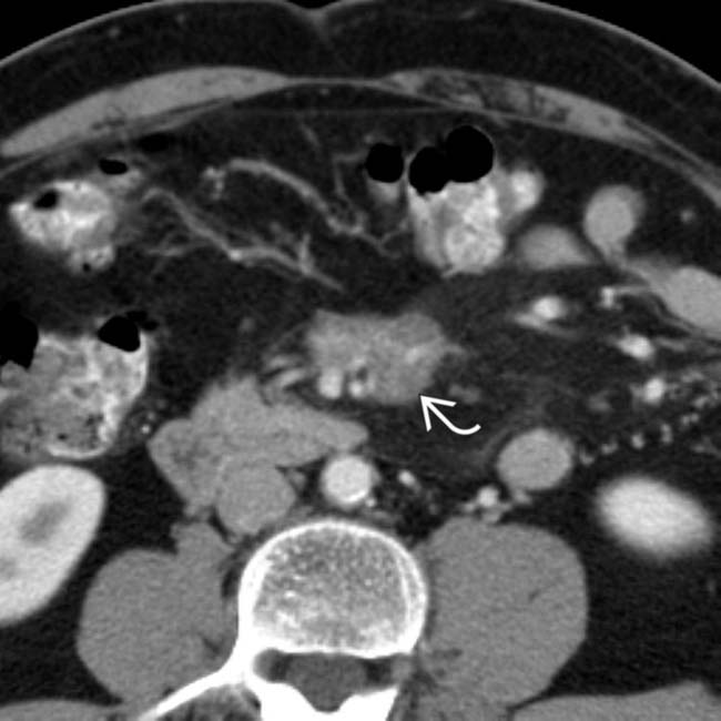
in the mesentery that encases and narrows the mesenteric vessels.

image
(Right) Axial CECT in a 2nd study of the same patient shows a focus of calcification image 
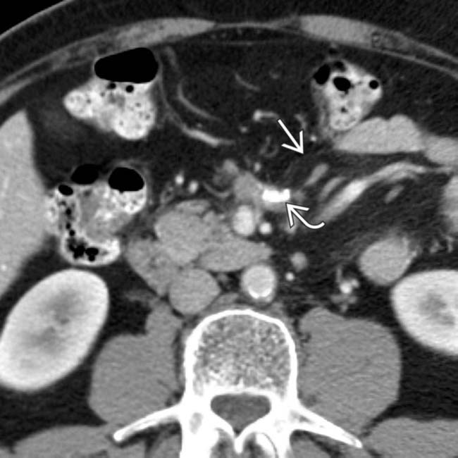
within the fibrotic mesenteric mass. The infiltrated mesentery and pseudocapsule image are still evident. This case illustrates progression of the disease over time.

TERMINOLOGY

Synonyms

• Retractile mesenteritis, fibrosing mesenteritis, mesenteric panniculitis, mesenteric lipodystrophy, liposclerotic mesenteritis, systemic nodular panniculitis, xanthogranulomatous mesenteritis

Definitions

• Idiopathic inflammatory and fibrotic disorder affecting mesentery of unknown etiology

IMAGING

General Features

• Best diagnostic clue

image “Misty mesentery” with surrounding pseudocapsule, clustered prominent mesenteric lymph nodes, and halo of spared fat surrounding nodes and vessels
• Location

image Most common site: Root of jejunal mesentery

– 90% involve small bowel mesentery and primarily to left of midline (jejunal mesentery)
image Occasionally: Colon (transverse or rectosigmoid)
image Rarely: Peripancreatic, omentum, and retroperitoneum
• Morphology

image Mostly characterized by mixture of mesenteric inflammation, fat necrosis, and fibrosis
• Key concepts

image Uncommon, benign, inflammatory process involving mesenteric fat
image Classified histologically into 3 types or stages based on predominant tissue type in mass

– Mesenteric panniculitis: Acute inflammation ± fat necrosis > fibrosis
– Mesenteric lipodystrophy: Fat necrosis > inflammation or fibrosis
– Retractile mesenteritis: Fibrosis/retraction > inflammation or fat necrosis
image Retractile mesenteritis

– Considered as final, chronic form, with collagen deposition, fat necrosis, fibrosis, and tissue retraction
image Often associated with other idiopathic inflammatory disorders (> 1 condition may be present)
image May coexist with malignancy (e.g., lymphoma, breast, lung, colon cancer, and melanoma)

CT Findings

• Acute mesenteritis (mesenteric panniculitis and lipodystrophy)

image Often associated with misty mesentery: Increased attenuation of mesentery with fat stranding and induration

– Nonspecific finding that can be seen with other etiologies
– Usually located in left upper quadrant mesentery
– Often discrete fat-attenuation “mass” with increasing soft tissue component as disease progresses
image Thin (usually < 3 mm) pseudocapsule encasing inflamed portion of mesentery (tumoral pseudocapsule sign)
image Cluster of mesenteric nodes (only rarely enlarged > 1 cm) within misty mesentery

– Mesenteric vessels and nodes have halo of surrounding spared fat (fat ring or fat halo sign)
• Chronic phase (retractile mesenteritis)

image Chronic fibrosis results in discrete fibrotic soft tissue mass with desmoplastic reaction

– Usually located in root of mesentery (left upper quadrant)
– Mass often has “stellate” appearance with internal calcification (and within adjacent lymph nodes)

image Can rarely show internal cystic or necrotic components
– Encasement of mesenteric and collateral vessels with frequent narrowing/occlusion of portomesenteric veins

image Collaterals or engorged vessels may be present
image Small bowel wall edema and mucosal hyperemia as result of lymphatic/venous obstruction and ischemia
– Tethering of bowel loops can lead to bowel obstruction

MR Findings

• Variable signal intensity due to varying elements of inflammation, fat necrosis, calcification, and fibrosis
• Acute mesenteritis (panniculitis and lipodystrophy)

image T1WI: Mixed signal intensity
image T2WI: Mixed signal intensity (usually mildly hyperintense)
• Chronic phase (retractile mesenteritis): Imaging findings of mature fibrotic reaction

image T1WI: Decreased signal intensity
image T2WI: Very low signal intensity
image Narrowing/occlusion of flow in mesenteric vessels

– Collateral or engorged vessels may be seen

Nuclear Medicine Findings

• Mesenteritis usually not FDG avid (unlike malignancy), although multiple reports suggests mesenteritis can be FDG avid

Radiographic Findings

• Fluoroscopic-guided barium study

image Dilation, fixed narrowing, and tethering of jejunum with fold thickening and luminal narrowing in advanced cases

Imaging Recommendations

• CT with 3D volume rendering is optimal study

DIFFERENTIAL DIAGNOSIS

Retroperitoneal and Mesenteric Lymphoma

• Early-stage lymphoma appears identical to mesenteritis with misty mesentery, mildly enlarged nodes, and rarely, fat halo sign
• Large discrete/confluent lymph nodes in advanced cases

image Calcification within lymph nodes very uncommon prior to treatment, but can be seen with treated lymphoma
• Treated mesenteric lymphoma can look identical to mesenteritis (with chronic misty mesentery)
• Lack of fat halo and lymphadenopathy outside of mesentery favors lymphoma

Carcinoid Tumor

• Can appear identical to retractile mesenteritis, with calcified mesenteric mass, desmoplastic reaction, occlusion of vessels, and bowel obstruction
• Usually involves ileal mesentery in right lower quadrant
• May be associated with hypervascular primary tumor (usually in ileum) and liver metastases
• Increased urinary 5-HIAA
• Somatostatin-receptor scintigraphy usually detects carcinoid
• Preservation of fat ring sign favors diagnosis of sclerosing mesenteritis

Mesenteric Edema

• Fluid infiltrates mesentery and increases attenuation of mesenteric fat, simulating acute mesenteritis
• Misty mesentery can be seen in multiple other conditions, such as cirrhosis, heart failure, portal or mesenteric vein thrombosis, vasculitis, acute pancreatitis, and other abdominal inflammatory conditions

Desmoid Tumor (Fibromatosis)

• Benign proliferation of fibrous tissue that usually presents as discrete solid mass (isodense to muscle)
• Displaces but does not usually encase mesenteric vessels
• Associated with Gardner syndrome or prior surgery
• Diagnosis: Biopsy and histologic analysis

Carcinomatosis (Mesenteric Metastases)

• Dominant tumor implant in left upper quadrant mesentery can mimic retractile mesenteritis
• Additional implants usually found elsewhere, such as omentum, surface of liver, spleen, or bowel
• Calcification uncommon with most tumors, but can be seen with mucinous adenocarcinoma
• Carcinomatosis is more frequently associated with ascites

Amyloidosis, Abdominal Manifestations

• Rare disease but often involves GI tract
• Mesentery may have multifocal of diffuse infiltration
• Typically has coarse calcifications

Inflammatory Pseudotumor

• a.k.a. myofibroblastic tumor
• Rare, usually affects younger individuals
• Can affect mesentery and almost any organ

Liposarcoma

• Mass with varying soft tissue and fat components (depending on degree of tumor differentiation) and discrete surrounding capsule
• More likely to present with mass effect on adjacent structures than mesenteritis

Primary Visceral Malignancy

• Can mimic primary malignancy when mesenteritis abuts a visceral organ (such as bowel or pancreas)
• Can resemble pancreatic cancer when it occurs near or within pancreatic parenchyma

PATHOLOGY

General Features

• Etiology

image Exact etiology remains unknown
image Several different theories have been suggested

– Prior abdominal surgery or trauma
– Possible associations with autoimmune disease, infections, and ischemic insults
– Possible result of paraneoplastic syndrome 

image Possible association with underlying malignancy (particularly lymphoma) in 1-70% of patients
• Associated abnormalities

image Reported association with different idiopathic or IgG4-related inflammatory and fibrotic disorders

– Retroperitoneal fibrosis, sclerosing cholangitis, Riedel thyroiditis, orbital pseudotumor, and autoimmune pancreatitis
• Fibrosis → thickened mesentery contracts → adhesions → nodular changes

Gross Pathologic & Surgical Features

• Encapsulated firm/hard mass(es)
• Nodules of fat, areas of necrosis, and fibrosis
• Thickened mesentery, adhesions, displaced bowel loops

Microscopic Features

• Fat with lipid-laden macrophages and fibrous septa
• Lymphocytes, plasma cells, eosinophils, ± calcifications
• Invasion of bowel muscle/submucosa

CLINICAL ISSUES

Presentation

• Most common signs/symptoms

image Often incidental finding in asymptomatic patient
image Abdominal pain (most common symptom), fever, nausea, vomiting, weight loss, and palpable mass
image Bowel obstruction in chronic phase
image Chylous ascites
• Other signs/symptoms

image Lab data: Elevated ESR and C-reactive protein may be used to gauge response to therapy
image Diagnosis: Usually presumptive diagnosis in acute phase based on imaging and elevated inflammatory markers

– Percutaneous or surgical excisional biopsy often required for diagnosis in chronic phase

Demographics

• Age

image 2nd-8th decades of life; average age: 60-70 years
image Very rare in pediatric age group
• Gender

image M:F = 2:1
• Epidemiology

image Mayo Clinic reports ∼ 5 cases per year (pathologically proven)
image Seems much more commonly recognized on CT (perhaps up to 0.6% of all patients undergoing abdominal CT)
image Disease is probably underreported and underdiagnosed, as few patients go on to biopsy or specific treatment

Natural History & Prognosis

• Natural history not clearly understood due to rarity of disorder and lack of follow-up in many patients

image Frequency of progression from acute mesenteritis to chronic stage is unknown
image Most patients have stable or slowly progressing disease without symptoms
• Complications most common in chronic setting

image Bowel or urinary tract obstruction
image Ischemia due to occlusion of mesenteric vasculature
image Questionable risk of developing malignancy
• Prognosis

image Partial or complete resolution in some cases (with some patients spontaneously resolving)
image Most will respond to medical therapy
image Some progress to bowel obstruction/ischemia and death

Treatment

• Immunosuppressive therapy should be attempted initially

image Tamoxifen + prednisone is first-line regimen

– Most effective before fibrotic change
image Other options include cyclophosphamide, thalidomide, colchicine, and progesterone
• Relatively limited role for surgery

image Surgical excision very difficult in advanced cases due to vascular involvement
image Surgery may be palliative, such as bypass to relieve bowel obstruction
image Questionable role for radiation therapy

DIAGNOSTIC CHECKLIST

Consider

• Differentiate sclerosing mesenteritis from other more common mimics, including other idiopathic inflammatory disorders and malignancies

Image Interpretation Pearls

• CT appearances vary depending on predominant tissue component (fat, inflammation, or fibrosis)
• Fat ring sign: Differentiates sclerosing mesenteritis from lymphoma, carcinoid, and mesenteric metastases
• Check for other idiopathic inflammatory disorders and malignancies

image
(Left) Axial CECT in a patient with pain and fever shows infiltration of the jejunal mesentery, demarcated by a pseudocapsule image
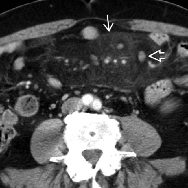
. Multiple prominent mesenteric nodes are present image with a fat halo. PET showed no increased activity, and symptoms improved with steroids.
image
(Right) Axial NECT in a patient with chronic pain shows infiltration of the jejunal mesentery with a pseudocapsule image
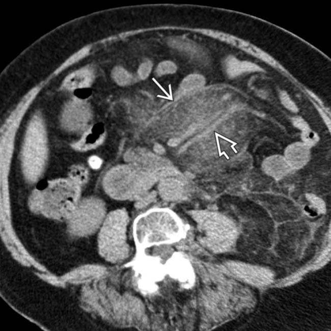
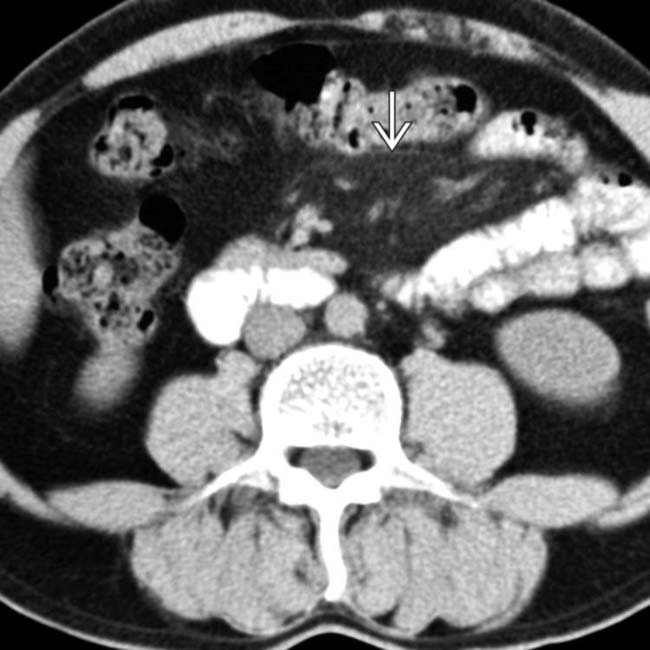
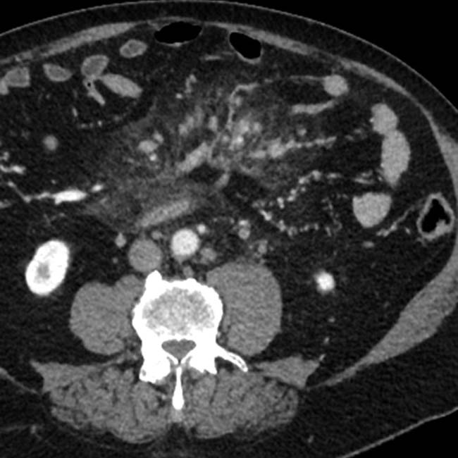
. The mesenteric vessels image are encased but not obstructed. This was found to represent sclerosing mesenteritis.
image
(Left) Axial CECT shows an infiltrative mesenteric mass image 
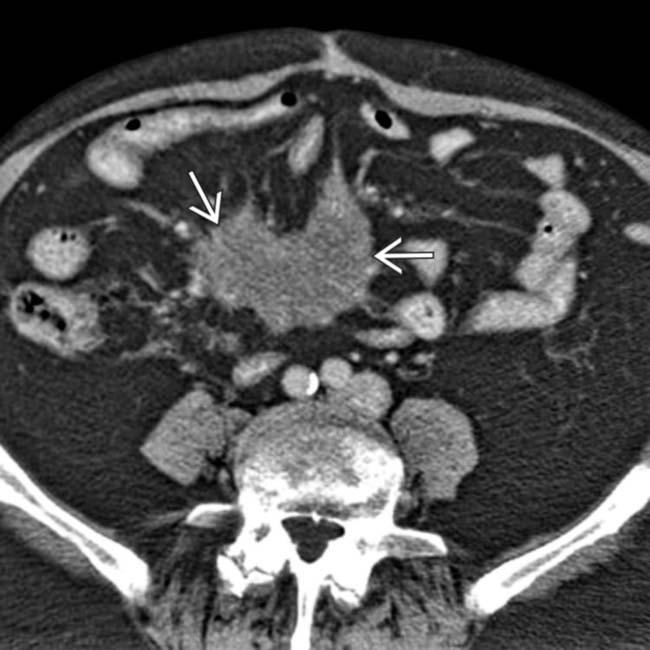
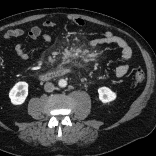
that encases blood vessels. Note the engorgement of the mesenteric veins. This was found to represent sclerosing mesenteritis.

image
(Right) Axial CECT in an elderly woman with chronic pain and diarrhea shows a soft tissue mass image 
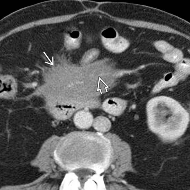
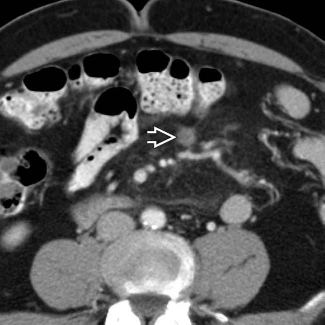
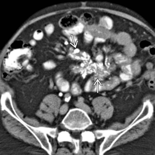
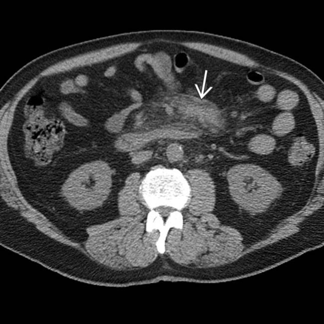
at the base of the small bowel mesentery, encasing and narrowing the mesenteric vessels image. This was found to represent fibrosing mesenteritis at biopsy.
image
(Left) Axial CECT demonstrates a calcified mass image 
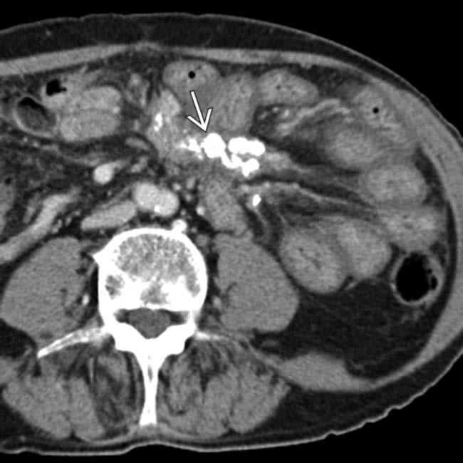
in the left upper quadrant mesentery with tethering of surrounding bowel loops.

image
(Right) Coronal CECT in the same patient demonstrates the calcified mass image
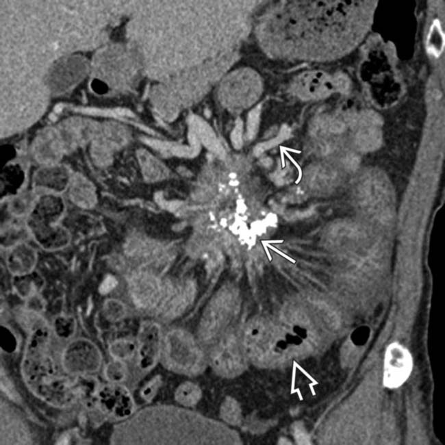
. Note the venous collaterals image due to obstruction of the superior mesenteric vein, as well as diffuse small bowel wall thickening image due to venous/lymphatic obstruction. This was biopsy-proven retractile mesenteritis.
image
Axial CECT shows a mesenteric mass image 
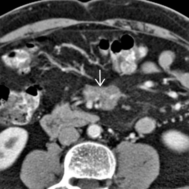
encasing vessels, causing mesenteric venous distention. This was found to represent sclerosing mesenteritis.

image
Axial CECT shows focal calcification within a mesenteric mass image 
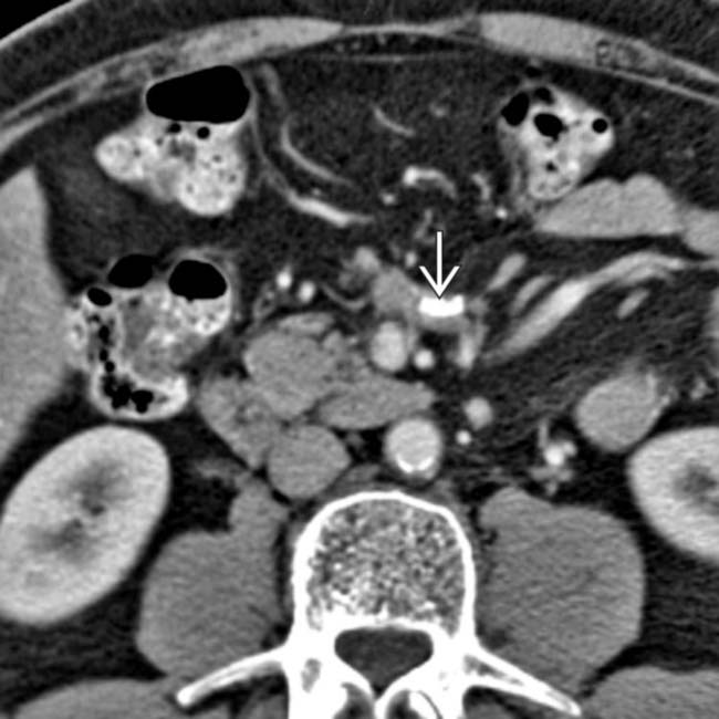
and vascular engorgement. This was found to represent sclerosing mesenteritis.
image
Axial CECT in a 59-year-old woman shows a misty mesentery image 
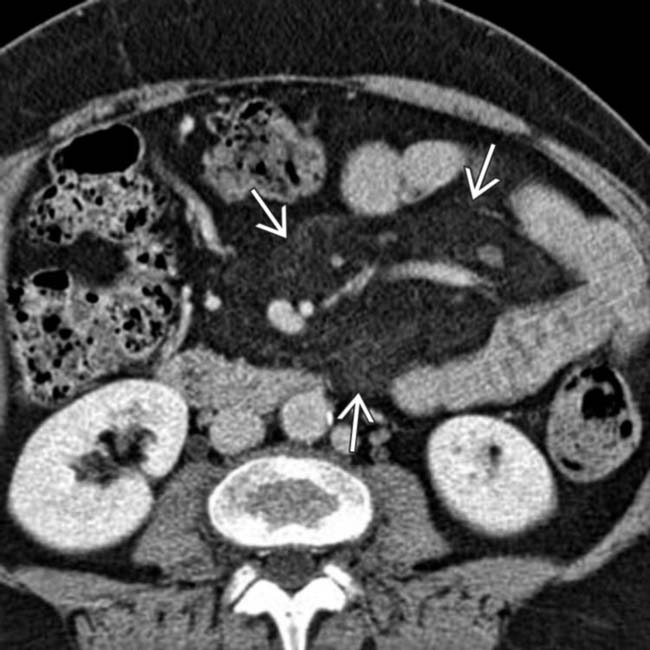
and mesenteric adenopathy due to mesenteritis.
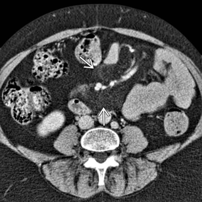
image
Axial CECT in a 59-year-old woman demonstrates mesenteritis with subtle infiltration of mesenteric fat image.
image
Axial CECT of an 84-year-old patient with retractile mesenteritis demonstrates a calcified, spiculated mass image that encases vessels and distorts the bowel.
image
Axial CECT in a patient with dermatomyositis demonstrates sclerosing mesenteritis image 
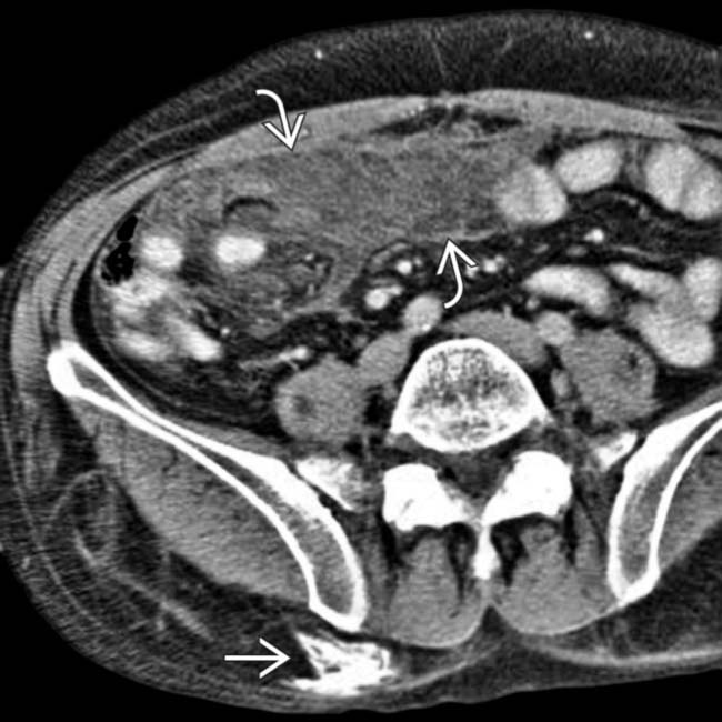
along with subcutaneous calcification image.
image
Axial CECT demonstrates an upper abdominal misty mesentery with multiple prominent internal lymph nodes.
image
Axial CECT in the same patient again demonstrates increased mesenteric attenuation with several prominent internal lymph nodes. This is a nonspecific finding but was found to represent sclerosing mesenteritis in this case.
image
Axial NECT demonstrates infiltration and mass effect image within the jejunal mesentery.
image
Fused axial PET/CT from the same patient shows that the lesion image 
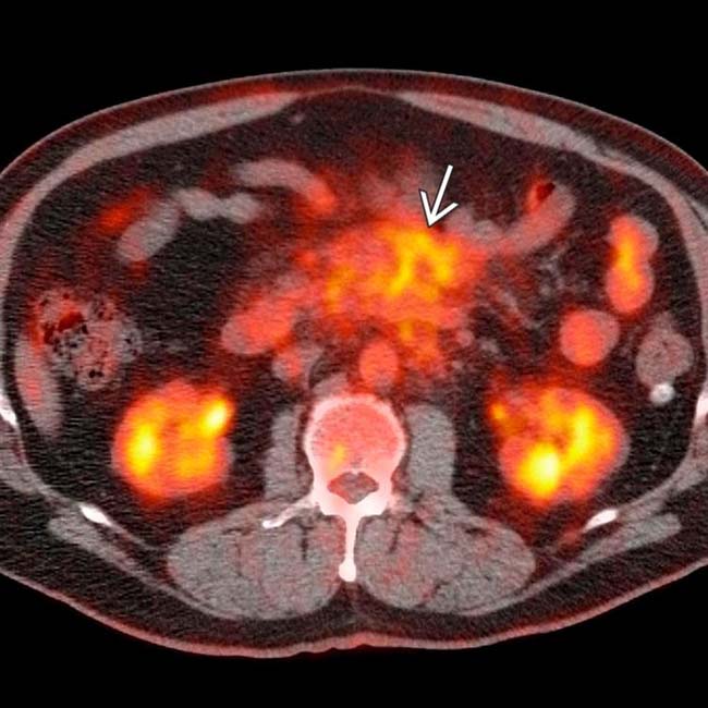
was moderately FDG avid, raising concern for malignancy, either metastatic from the patient’s known esophageal carcinoma (considered unlikely) or from a 2nd malignancy, such as non-Hodgkin lymphoma. Laparoscopic biopsy of the mass demonstrated only inflammation and fibrosis, interpreted as sclerosing mesenteritis. Acute and chronic inflammatory processes, such as sclerosing mesenteritis and IgG4-related sclerosing disease, often demonstrate moderate to marked FDG avidity on PET, constituting a potential pitfall in misdiagnosis as malignancy.
image
Axial CT section shows mesenteric inflammation, nodes image 
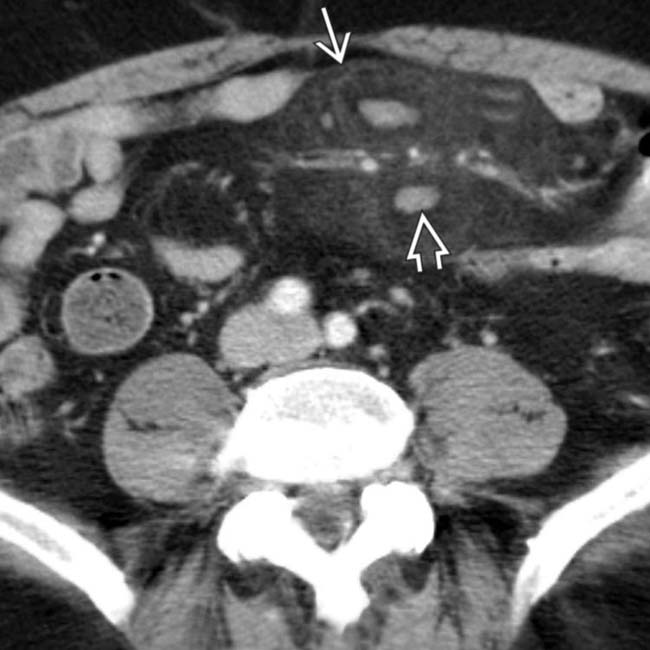
with a surrounding halo and a pseudocapsule image. A positron emission tomogram (PET scan) showed no increased metabolic activity within the mass, helping to exclude the diagnosis of lymphoma. Symptoms improved with oral steroid medication.

SELECTED REFERENCES

1. McLaughlin, PD, et al. The “misty mesentery”: mesenteric panniculitis and its mimics. AJR Am J Roentgenol. 2013; 200(2):W116–W123.

Canyigit, M, et al. Multidetector-row computed tomography findings of sclerosing mesenteritis with associated diseases and its prevalence. Jpn J Radiol. 2011; 29(7):495–502.

Coulier, B. Mesenteric panniculitis. Part 1: MDCT—pictorial review. JBR-BTR. 2011; 94(5):229–240.

Scudiere, JR, et al. Sclerosing mesenteritis involving the pancreas: a mimicker of pancreatic cancer. Am J Surg Pathol. 2010; 34(4):447–453.

Arora, M, et al. A clinical case study: sclerosing mesenteritis presenting as chylous ascites. Medscape J Med. 2008; 10(2):30.

Chen, TS, et al. Are tumefactive lesions classified as sclerosing mesenteritis a subset of IgG4-related sclerosing disorders? J Clin Pathol. 2008; 61(10):1093–1097.

Grieser, C, et al. Sclerosing mesenteritis as a rare cause of upper abdominal pain and digestive disorders. Acta Radiol. 2008; 49(7):744–746.

Preda, M, et al. Sclerosing mesenteritis presenting with abdominal pain and fever in a young woman: computed tomography findings. ANZ J Surg. 2008; 78(8):709–710.

Akram, S, et al. Sclerosing mesenteritis: clinical features, treatment, and outcome in ninety-two patients. Clin Gastroenterol Hepatol. 2007; 5(5):589–596. .

Levy, AD, et al. From the archives of the AFIP: benign fibrous tumors and tumorlike lesions of the mesentery: radiologic-pathologic correlation. Radiographics. 2006; 26(1):245–264.

Matsuki, M, et al. CT and MR features of sclerosing mesenteritis mimicking a mesenteric metastasis from the carcinoid tumor. Radiat Med. 2006; 24(3):220–223.

Zissin, R, et al. Mesenteric panniculitis in oncologic patients: PET-CT findings. Br J Radiol. 2006; 79(937):37–43.

Horton, KM, et al. CT findings in sclerosing mesenteritis (panniculitis): spectrum of disease. Radiographics. 2003; 23(6):1561–1567.

Nguyen, BD. F-18 FDG PET demonstration of sclerosing mesenteritis. Clin Nucl Med. 2003; 28(8):670–671.

Seo, BK, et al. Segmental misty mesentery: analysis of CT features and primary causes. Radiology. 2003; 226(1):86–94.

Lawler, LP, et al. Sclerosing mesenteritis: depiction by multidetector CT and three-dimensional volume rendering. AJR Am J Roentgenol. 2002; 178(1):97–99.

Daskalogiannaki, M, et al. CT evaluation of mesenteric panniculitis: prevalence and associated diseases. AJR Am J Roentgenol. 2000; 174(2):427–431.

Sabate, JM, et al. Sclerosing mesenteritis: imaging findings in 17 patients. AJR Am J Roentgenol. 1999; 172(3):625–629.

Fujiyoshi, F, et al. Retractile mesenteritis: small-bowel radiography, CT, and MR imaging. AJR Am J Roentgenol. 1997; 169(3):791–793.

Mindelzun, RE, et al. The misty mesentery on CT: differential diagnosis. AJR Am J Roentgenol. 1996; 167(1):61–65.

Kronthal, AJ, et al. MR imaging in sclerosing mesenteritis. AJR Am J Roentgenol. 1991; 156(3):517–519.