Salmonella

Published on 27/03/2015 by admin

Filed under Pediatrics

Last modified 22/04/2025

Print this page

rate 1 star rate 2 star rate 3 star rate 4 star rate 5 star
Your rating: none, Average: 0 (0 votes)

This article have been viewed 4320 times

Chapter 190 Salmonella

Salmonellosis is a common and widely distributed food-borne disease that is a global major public health problem that affects millions of individuals and results in significant mortality. Salmonellae live in the intestinal tracts of warm- and cold-blooded animals. Some species are ubiquitous, whereas others are specifically adapted to a particular host.

The sequencing of the Salmonella enterica serovar Typhi (previously called Salmonella typhi) and Salmonella typhimurium genomes has indicated an almost 95% genetic homology between the organisms. However, the clinical diseases caused by the 2 organisms differ considerably. Orally ingested salmonellae survive at the low pH of the stomach and evade the multiple defenses of the small intestine in order to gain access to the epithelium. Salmonellae preferentially enter M cells, which transport them to the lymphoid cells (T and B) in the underlying Peyer patches. Once across the epithelium, Salmonella serotypes that are associated with systemic illness enter intestinal macrophages and disseminate throughout the reticuloendothelial system. By contrast, non-typhoidal Salmonella (NTS) serovars induce an early local inflammatory response, which results in the infiltration of polymorphonuclear leukocytes into the intestinal lumen and diarrhea. The NTS serovars cause a gastroenteritis of rapid onset and brief duration, in contrast to typhoid, which has a considerably longer incubation period and duration of illness and in which systemic illness predominates and only a small proportion of children get diarrhea. These differences in the manifestations of infection by the two groups of pathogens, one predominantly causing intestinal inflammation and the other leading to systemic disease, may be related to specific genetic pathogenicity islands on the organisms. NTS serovars are unable to overcome defense mechanisms that limit bacterial dissemination from the intestine to systemic circulation in immunocompetent individuals and produce a self-limiting gastroenteritis. In contrast, S. typhi may possess unique virulence traits that allow it to overcome mucosal barrier functions in immunocompetent hosts, resulting in a severe systemic illness. Interestingly, the frequencies of typhoid in immunocompetent and immunocompromised individuals do not differ.

The nomenclature of Salmonella reflects the species name Salmonella enterica with a number of serovars. Salmonella nomenclature has undergone considerable alterations. The original taxonomy was based on clinical syndromes (S. typhi, S. choleraesuis, S. paratyphi). With adoption of serologic analysis, a Salmonella species was defined subsequently as “a group of related fermentation phage-type,” with the result that each Salmonella serovar was regarded as a species in itself. Although this classification is simplistic, its use until 2004 resulted in identification of 2,501 serovars of Salmonella, which led to the need for further categorization to aid communication among scientists, public health officials, and the public.

All Salmonella serovars form a single DNA hybridization group, a single species called S. enterica composed of several subspecies (Table 190-1). Each subspecies contains various serotypes defined by the O and H antigens. To further simplify the nomenclature for physicians and epidemiologists, the names for the common serovars are kept for subspecies I strains, which represent >99.5% of the Salmonella strains isolated from humans and other warm-blooded animals.

Table 190-1 SALMONELLA NOMENCLATURE

TRADITIONAL USAGE FORMAL NAME CDC DESIGNATION
S. typhi S. enterica* subsp. enterica ser. Typhi S. ser. Typhi
S. dublin S. enterica subsp. enterica ser. Dublin S. ser. Dublin
S. typhimurium S. enterica subsp. enterica ser. Typhimurium S. ser. Typhimurium
S. choleraesuis S. enterica subsp. enterica ser. Choleraesuis S. ser. Choleraesuis
S. marina S. enterica subsp. houtenae ser. Marina S. ser. Marina

CDC, U.S. Centers for Disease Control and Prevention; subsp, subspecies; ser., serovar.

* Some authorities prefer S. choleraesuis or S. enteritidis rather than S. enterica to describe the species.

190.1 Nontyphoidal Salmonellosis

Epidemiology

Salmonellosis constitutes a major public health burden and represents a significant cost to society in many countries. It is estimated that in the USA alone an estimated 1.4 million nontyphoidal Salmonella infections occurred in 2007, with an estimated $2.5 billion cost due to lost productivity and medical treatment. Although there is little information on the epidemiology and the burden of Salmonella gastroenteritis from developing countries, Salmonella infections are recognized as major causes of childhood diarrheal illness. With the growing burden of HIV infections and malnutrition in Africa, nontyphoidal Salmonella bacteremic infections have emerged as a major cause of morbidity and mortality among children and adults.

Nontyphoidal Salmonella infections have a worldwide distribution, with an incidence proportional to the standards of hygiene, sanitation, availability of safe water, and food preparation practices. In the developed world, the incidence of Salmonella infections and outbreaks has increased several-fold over the past few decades, which may be related to modern practices of mass food production that increase the potential for epidemics. Salmonella gastroenteritis accounts for over half of all episodes of bacterial diarrhea in the USA, with incidence peaks at the extremes of ages, among young infants and the elderly. Most human infections have been caused by S. Enteritidis; the prevalence of this organism has decreased over the past decade, with S. Typhimurium overtaking it in some countries.

The rise in Salmonella infections in many parts of the world over the past 3 decades may also be related to intensive animal husbandry practices, which selectively promote the rise of certain strains, especially drug-resistant varieties that emerge in response to the use of antimicrobials in food animals. Poultry products were traditionally regarded as a common source of salmonellosis, but consumption of a range of foods has now been associated with outbreaks, including fruits and vegetables. Although this change in epidemiology may be related to selective pressure from the use of antimicrobials, there may be other factors, such as the rise of strains with a selective propensity to develop resistance and virulence. It appears that multidrug-resistant strains of Salmonella are more virulent than susceptible strains and that poorer outcome does not simply relate to the delay in treatment response due to empirical choice of an ineffective antibiotic. Strains of multidrug-resistant Salmonella such as S. Typhimurium phage type DT104 harbor a genomic island that contains many of the drug resistance genes. It is possible that these integrons also contain genes that express virulence factors. The global spread of multidrug-resistant S. Typhimurium phage type DT104 in animals and humans may be related to the growing use of antimicrobials and may be facilitated by international and national trade of infected animals.

Several risk factors are associated with outbreaks of Salmonella infections. Animals constitute the principal source of human nontyphoidal Salmonella disease, and cases have occurred in which individuals have had contact with infected animals, including domestic animals such as cats, dogs, reptiles, pet rodents, and amphibians. Specific serotypes may be associated with particular animal hosts; children with S. enterica serovar Marina typically have exposure to pet lizards. In 1996 more than 50,000 cases of salmonellosis related to domestic lizards were reported to the U.S. Centers for Disease Control and Prevention (CDC). Domestic animals probably acquire the infection in the same way that humans do, through consumption of contaminated raw meat, poultry, or poultry-derived products. Animal feeds containing fishmeal or bone meal contaminated with Salmonella are an important source of infection for animals. Moreover, subtherapeutic concentrations of antibiotics are often added to animal feed to promote growth. Such practices promote the emergence of antibiotic-resistant bacteria, including Salmonella, in the gut flora of the animals, with subsequent contamination of their meat. There is strong evidence to link resistance of S. Typhimurium to fluoroquinolones with the use of this group of antimicrobials in animal feeds. Animal-to-animal transmission can occur, but most infected animals are asymptomatic.

An increasing number of produce-associated foodborne outbreaks in the USA associated with bacterial contamination are primarily from Salmonella. Although almost 80% of Salmonella infections are discrete, outbreaks can pose an inordinate burden on public health systems. In an evaluation of 604 outbreaks of foodborne disease in schools in the USA, Salmonella was the most commonly identified pathogen, accounting for 36% of outbreak reports with a known etiology. Salmonella infections in chickens increase the risk for contamination of eggs, and both poultry and eggs have been regarded as a dominant cause of common-source outbreaks. However, a growing proportion of Salmonella outbreaks are also associated with other food sources. The CDC reports that between 2002 and 2003, 31 food produce–associated Salmonella outbreaks were reported, compared with only 29 poultry-related outbreaks. The food sources included many fruits and vegetables, such as tomatoes, sprouts, watermelon, cantaloupe, lettuce, and mangoes.

In addition to the effect of antibiotic use in animal feeds, the relationship of Salmonella infections to prior antibiotic use among children in the previous month is well recognized. This increased risk for infection in people who have received antibiotics for an unrelated reason may be related to alterations in gut microbial ecology, which predispose them to colonization and infection with antibiotic-resistant Salmonella isolates. These resistant strains of Salmonella are also more virulent. It is estimated that antimicrobial resistance in Salmonella may result in about 30,000 additional Salmonella infections annually, leading to about 300 hospitalizations and 10 deaths.

Given the ubiquitous nature of the organism, nosocomial infections with nontyphoidal Salmonella strains can also occur through contaminated equipment and diagnostic or pharmacologic preparations, particularly those of animal origin (pancreatic extracts, pituitary extracts, bile salts). Hospitalized children are at increased risk for severe and complicated Salmonella infections, especially with drug-resistant organisms.

Pathogenesis

The estimated number of bacteria that must be ingested to cause symptomatic disease in healthy adults is 106 to 108 Salmonella organisms. The gastric acidity inhibits multiplication of the salmonellae, and most organisms are rapidly killed at gastric pH ≤2.0. Achlorhydria, buffering medications, rapid gastric emptying after gastrectomy or gastroenterostomy, and a large inoculum enable viable organisms to reach the small intestine. Neonates and young infants have hypochlorhydria and rapid gastric emptying, which contribute to their increased vulnerability to symptomatic salmonellosis. In infants who typically take fluids, the inoculum size required to produce disease is also comparatively smaller because of faster transit through the stomach.

Once they reach the small and large intestines, the ability of Salmonella organisms to multiply and cause infection depends on the infecting dose as well as competition with normal flora. Prior antibiotic therapy may alter this relationship, as might factors such as co-administration of antimotility agents. The typical intestinal mucosal response to nontyphoidal Salmonella infection is an enterocolitis with diffuse mucosal inflammation and edema, sometimes with erosions and microabscesses. Salmonella organisms are capable of penetrating the intestinal mucosa, although destruction of epithelial cells and ulcers are usually not found. Intestinal inflammation with polymorphonuclear leukocytes and macrophages usually involves the lamina propria. Underlying intestinal lymphoid tissue and mesenteric lymph nodes enlarge and may demonstrate small areas of necrosis. Such lymphoid hypertrophy may cause interference with the blood supply to the gut mucosa. Hyperplasia of the reticuloendothelial system (RES) is also found within the liver and spleen. If bacteremia develops, it may lead to localized infection and suppuration in almost any organ.

Although S. Typhimurium can cause systemic disease in humans, intestinal infection usually results in a localized enteritis that is associated with a secretory response in the intestinal epithelium. Intestinal infection also induces secretion of interleukin-8 (IL-8) from the basolateral surface and other chemoattractants from the apical surface, directing recruitment and transmigration of neutrophils into the gut lumen and thus preventing the systemic spread of the bacteria (Fig. 190-1).

image

Figure 190-1 On contact with the epithelial cell, salmonellae assemble the Salmonella pathogenicity island 1–encoded type III secretion system (TTSS-1) and translocate effectors (yellow spheres) into the eukaryotic cytoplasm. Effectors such as SopE, SopE2 and SopB then activate host Rho guanosine triphosphatase (GTPases), resulting in the rearrangement of the actin cytoskeleton into membrane ruffles, induction of mitogen-activated protein kinase (MAPK) pathways, and destabilization of tight junctions. Changes in the actin cytoskeleton, which are further modulated by the actin-binding proteins SipA and SipC, lead to bacterial uptake. MAPK signaling activates the transcription factors activator protein-1 (AP-1) and nuclear factor-κB (NF-κB), which turn on production of the proinflammatory polymorphonuclear leukocyte (PMN) chemokine interleukin (IL)-8. SipB induces caspase-1 activation in macrophages, with the release of IL-1β and IL-18, so augmenting the inflammatory response. In addition, SopB stimulates Cl secretion by its inositol phosphatase activity. The destabilization of tight junctions allows the transmigration of PMNs from the basolateral to the apical surface, paracellular fluid leakage, and access of bacteria to the basolateral surface. However, the transmigration of PMNs also occurs in the absence of tight-junction disruption and is further promoted by SopA. The actin cytoskeleton is restored, and MAPK signaling is turned off by the enzymatic activities of SptP. This also results in the down-modulation of inflammatory responses, to which SspH1 and AvrA also contribute by inhibiting activation of NF-κB.

(From Haraga A, Ohlson MB, Miller SI: Salmonellae interplay with host cells, Nat Rev Microbiol 6:53–66, 2008.)

Interestingly, virulence traits that contribute to the host response are common to all nontyphoidal Salmonella serovars. These include (1) the type III secretion system (TTSS-1) encoded on Salmonella pathogenicity island-1 (SP1), which mediates invasion of the intestinal epithelium; (2) the TTSS encoded on SP2 (TTSS-2), which is required for survival within macrophages; and (3) expression of strong agonists of innate pattern recognition receptors (lipopolysaccharide and flagellin), which are important for triggering an TLR-mediated inflammatory response mediated by Toll-like receptors (TLRs). These observations suggest that S. Typhimurium must have acquired additional factors that further modulate the host response during infection.

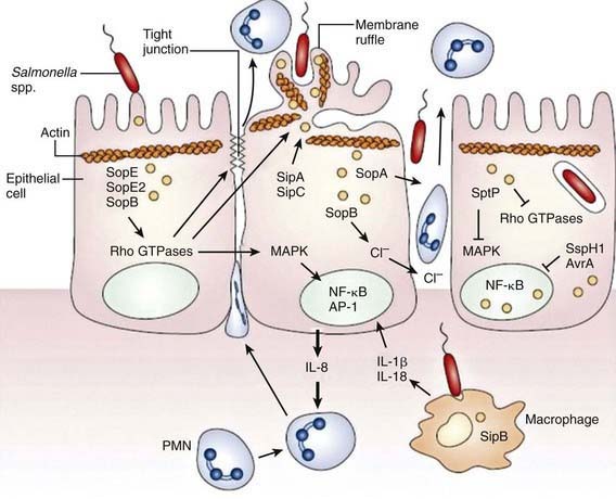
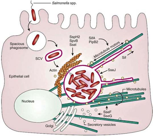
Salmonella species invade epithelial cells in vitro by a process of bacteria-mediated endocytosis involving cytoskeletal rearrangement, disruption of the epithelial cell brush border, and the subsequent formation of membrane ruffles (Fig. 190-2). An adherent and invasive phenotype of S. enterica is activated under conditions similar to those found in the human small intestine (high osmolarity, low oxygen). The invasive phenotype is mediated in part by Salmonella pathogenicity island 1, a 40-kb region that encodes regulator proteins such as HilA, the type 3 secretory system involved in invasion of epithelial cells, and a variety of other products. In humans the TLR-dependent interleukin-12/interferon-λ (IL-12/IFN-λ) is a major immunoregulatory system that bridges innate and adaptive immunity and is responsible for restricting the systemic spread of nontyphoidal Salmonella.

Shortly following invasion of the gut epithelium, invasive Salmonella organisms encounter macrophages within the gut-associated lymphoid tissue. The interaction between Salmonella and macrophages results in alteration in the expression of a number of host genes, including those encoding proinflammatory mediators (inducible nitric oxide synthase [iNOS], chemokines, IL-1β), receptors or adhesion molecules (tumor necrosis factor-α receptor [TNF-αR], CD40, intercellular adhesion molecule 1 [ICAM-1]), and anti-inflammatory mediators (transforming growth factor-β1 and -β2 [TGF-β1] and TGF-β2). Other upregulated genes include those involved in cell death or apoptosis (intestinal epithelial cell protease, TNF-R1, Fas) and transcription factors (early growth response 1[Egr-1], IFN regulatory factor 1 [IRF-1]). S. Typhimurium can induce rapid macrophage death in vitro, which depends on the host cell protein caspase-1 and is mediated by the effector protein SipB (Salmonella invasion protein B). Intracellular S. Typhimurium is found within specialized Salmonella organisms containing vacuoles that have diverged from the normal endocytic pathway. This ability to survive within monocytes/macrophages is essential for S. Typhimurium to establish a systemic infection in the mouse. The mucosal proinflammatory response to S. Typhimurium infection and the subsequent recruitment of phagocytic cells to the site may also facilitate systemic spread of the bacteria.

Some virulence traits are shared by all salmonellae, but others are serotype restricted. These virulence traits have been defined in tissue culture and murine models, and it is likely that clinical features of human Salmonella infection will eventually be related to specific DNA sequences. With most diarrhea-associated nontyphoidal salmonelloses, the infection does not extend beyond the lamina propria and the local lymphatics. Specific virulence genes are related to the ability to cause bacteremia. These genes are found significantly more often in strains of S. Typhimurium isolated from the blood than in strains recovered from stool. Although both S. dublin and S. choleraesuis have a greater propensity to rapidly invade the bloodstream with little or no intestinal involvement, the development of disease after infection with Salmonella depends on the number of infecting organisms, their virulence traits, and several host defense factors. Various host factors may also affect the development of specific complications or clinical syndromes (Table 190-2) and of these, HIV infections are assuming greater importance in Africa in all age groups.

Table 190-2 HOST FACTORS AND CONDITIONS PREDISPOSING TO THE DEVELOPMENT OF SYSTEMIC DISEASE WITH NONTYPHOIDAL SALMONELLA STRAINS

Neonates and young infants (≤3 mo of age)

HIV/AIDS

Other immunodeficiencies and chronic granulomatous disease

Immunosuppressive and corticosteroid therapies

Malignancies, especially leukemia and lymphoma

Hemolytic anemia, including sickle cell disease, malaria, and bartonellosis

Collagen vascular disease

Inflammatory bowel disease

Achlorhydria or use of antacid medications

Impaired intestinal motility

Schistosomiasis, malaria

Malnutrition

Bacteremia is possible with any Salmonella serotype, especially in individuals with reduced host defenses and especially in those with altered reticuloendothelial or cellular immune function. Thus, children with HIV infection, chronic granulomatous disease, and leukemia are more likely to demonstrate bacteremia after Salmonella infection, although the majority of children with Salmonella bacteremia in Africa are HIV negative. Children with Schistosoma mansoni infection and hepatosplenic involvement as well as chronic malarial anemia are also at a greater risk for development of chronic salmonellosis. Children with sickle cell disease are at increased risk for Salmonella septicemia and osteomyelitis. This risk may be related to the presence of numerous infarcted areas in the gastrointestinal tract, bones, and RES as well as reduced phagocytic and opsonizing capacity of patients, which allow the organism to flourish.

Some inherited defects, such as IL-12 deficiency (IL-12 β1 chain deficiency, IL-12 p40 subunit deletion) are associated with increased risk for Salmonella infections, suggesting a key role for IL-12 in the clearance of Salmonella. IL-12 is produced by activated macrophages and is a potent inducer of IFN-γ by natural killer cells and T lymphocytes. Given the putative protective role of IL-12 against malarial infection, Salmonella infection of phagocytes may secondarily affect IL-12 production and thus produce a vicious circle of chronic malaria and salmonella co-infection.

Clinical Manifestations

Complications

Salmonella gastroenteritis can be associated with acute dehydration and complications that result from delayed presentation and inadequate treatment. Bacteremia in younger infants and immunocompromised individuals can have serious consequences and potentially fatal outcomes. Salmonella organisms can seed many organ systems, leading to osteomyelitis in children with sickle cell disease, among other infections. Reactive arthritis may follow Salmonella gastroenteritis, usually in adolescents with the HLA-B27 antigen.

In certain high-risk groups, especially those with impaired immunity, the course of Salmonella gastroenteritis may be more complicated. Neonates, infants younger than 6 mo, and children with primary or secondary immunodeficiency may have symptoms that persist for several weeks. The course of illness and complications may also be affected by coexisting pathologies. In children with AIDS, Salmonella infection frequently becomes widespread and overwhelming, causing multisystem involvement, septic shock, and death. In patients with inflammatory bowel disease, especially active ulcerative colitis, Salmonella gastroenteritis may lead to rapid development of toxic megacolon, bacterial translocation, and sepsis. In children with schistosomiasis, the Salmonella may persist and multiply within schistosomes, leading to chronic infection unless the schistosomiasis is effectively treated. Prolonged or intermittent bacteremia is associated with low-grade fever, anorexia, weight loss, diaphoresis, and myalgias and may occur in children with underlying problems and an RES dysfunction such as hemolytic anemia or malaria.

Treatment

Appropriate therapy relates to the specific clinical presentation of Salmonella infection. In children with gastroenteritis, rapid clinical assessment, correction of dehydration and electrolyte disturbances, and supportive care, are key (Chapter 332). Antibiotics are not generally recommended for the treatment of isolated uncomplicated Salmonella gastroenteritis because they may suppress normal intestinal flora and prolong both the excretion of Salmonella and the remote risk for creating the chronic carrier state (usually in adults). However, given the risk for bacteremia in infants (<3 mo of age) and that of disseminated infection in high-risk groups with immune compromise (HIV, malignancies, immunosuppressive therapy, sickle cell anemia, immunodeficiency states), these children must receive an appropriate empirically chosen antibiotic until culture results are available (Table 190-3). The S. Typhimurium phage type DT104 strain is usually resistant to the following 5 drugs: ampicillin, chloramphenicol, streptomycin, sulfonamides, and tetracycline. An increasing proportion of S. Typhimurium phage type DT104 isolates also have reduced susceptibility to fluoroquinolones. Given the higher mortality associated with multidrug-resistant Salmonella infections, it is necessary to perform susceptibility tests on all human isolates. Infections with suspected drug-resistant Salmonella should be closely monitored and treated with appropriate antimicrobial therapy.

Table 190-3 TREATMENT OF SALMONELLA GASTROENTERITIS

ORGANISM AND INDICATION DOSE AND DURATION OF TREATMENT
Salmonella infections in infants <3 mo of age or immunocompromised persons (in addition to appropriate treatment for underlying disorder) Cefotaxime 100-200 mg/kg/day every 6 hr for 5-14 days
or
Ceftriaxone 75 mg/kg/day once daily for 7 days
or
Ampicillin 100 mg/kg/day every 6 hr for 7 days
or
Cefixime 15 mg/kg/day for 7-10 days

Prevention

Control of the transmission of Salmonella infections to humans requires control of the infection in the animal reservoir, judicious use of antibiotics in dairy and livestock farming, prevention of contamination of foodstuffs prepared from animals, and use of appropriate standards in food processing in commercial and private kitchens (Table 190-4). Because large outbreaks are often related to mass food production, it should be recognized that contamination of just one piece of machinery used in food processing may cause an outbreak; meticulous cleaning of equipment is essential. Clean water supply and education in handwashing and food preparation and storage are critical to reducing person-to-person transmission. Salmonella may remain viable when cooking practices prevent food from reaching a temperature greater than 150°F (65.5°C) for >12 min. Parents should be advised of the risk of reptiles as pets in households with young infants.

Table 190-4 RECOMMENDATIONS FOR PREVENTING TRANSMISSION OF SALMONELLA FROM REPTILES AND AMPHIBIANS TO HUMANS

Pet store owners, health care providers, and veterinarians should provide information to owners and potential purchasers of reptiles and amphibians about the risks for and prevention of salmonellosis from these pets.

Persons at increased risk for infection or serious complications from salmonellosis (e.g., children aged <5 yr and immunocompromised persons) should avoid contact with reptiles and amphibians and any items that have been in contact with reptiles and amphibians.

Reptiles and amphibians should be kept out of households that include children aged <5 yr or immunocompromised persons. A family expecting a child should remove any pet reptile or amphibian from the home before the infant arrives.

Reptiles and amphibians should not be allowed in child-care centers.

Persons should always wash their hands thoroughly with soap and water after handling reptiles and amphibians or their cages.

Reptiles and amphibians should not be allowed to roam freely throughout a home or living area.

Pet reptiles and amphibians should be kept out of kitchens and other food preparation areas. Kitchen sinks should not be used to bathe reptiles and amphibians or to wash their dishes, cages, or aquariums. If bathtubs are used for these purposes, they should be cleaned thoroughly and disinfected with bleach.

Reptiles and amphibians in public settings (e.g., zoos and exhibits) should be kept from direct or indirect contact with patrons except in designated animal contact areas equipped with adequate handwashing facilities. Food and drink should not be allowed in animal contact areas.

From the Centers for Disease Control and Prevention: Reptile-associated salmonellosis—selected states, 1998-2002, MMWR Morbid Mortal Wkly Rep 52:1206–1210, 2003.

In contrast to developed countries, relatively little is known about the transmission of nontyphoidal Salmonella infections in developing countries, and it is likely that person-to-person transmission may be relatively more important in some settings. Although some vaccines have been used in animals, no human vaccine against nontyphoidal Salmonella infections is currently available. Infections should be reported to public health authorities so that outbreaks can be recognized and investigated. Given the rapid rise of antimicrobial resistance among Salmonella isolates, it is imperative that there is rigorous regulation of the use of antimicrobials in animal feeds.

Bibliography

Centers for Disease Control and Prevention. Human salmonellosis associated with animal-derived pet treats—United States and Canada, 2005. MMWR Morbid Mortal Wkly Rep. 2006;55:702-705.

Centers for Disease Control and Prevention. Outbreak of multidrug-resistant Salmonella typhimurium associated with rodents purchased at retail pet stores—United States, December 2003-October 2004. MMWR Morbid Mortal Wkly Rep. 2005;54:429-434.

Centers for Disease Control and Prevention. Reptile-associated salmonellosis—selected states, 1998–2002. MMWR. 2003;52:1206-1210.

Chiu CH, Chuang CH, Chiu S, et al. Salmonella enterica serotype choleraesuis infections in pediatric patients. Pediatrics. 2006;117:e1193-e1196.

Coburn B, Grassl GA, Finlay BB. Salmonella, the host and disease: a brief review. Immunology Cell Biology. 2007;85:112-118.

Graham SM. Salmonellosis in children in developing and developed countries and populations. Curr Opin Infect Dis. 2000;15:507-512.

Hanning IB, Nutt JD, Ricke SC. Salmonellosis outbreaks in the United States due to fresh produce: sources and potential intervention measures. Foodborne Pathogens Dis. 2009;6:635-648.

Haraga A, Ohlson MB, Miller SI. Salmonellae interplay with host cells. Nat Rev Microbiol. 2008;6:53-66.

Helms M, Simonsen J, Molbak K. Quinolone resistance is associated with increased risk of invasive illness or death during infection with Salmonella serotype Typhimurium. J Infect Dis. 2004;190:1652-1654.

Helms M, Vastrup P, Gerner-Smidt P, et al. Excess mortality associated with antimicrobial drug-resistant Salmonella typhimurium. Emerg Infect Dis. 2002;8:490-495.

Hohmann EL. Nontyphoidal salmonellosis. Clin Infect Dis. 2001;32:263-269.

International Food Safety Authorities Network. Antimicrobial-resistant Salmonella (pdf). www.who.int/foodsafety/fs_management/en/No_03_Salmonella_Apr05_en.pdf, 2010. Accessed September 16

Jones TF, Ingram LA, Fullerton KE, et al. A case-control study of the epidemiology of sporadic salmonella infection in infants. Pediatrics. 2006;118:2380-2387.

McEwen SA, Fedorka-Cray PJ. Antimicrobial use and resistance in animals. Clin Infect Dis. 2002;34(Suppl 3):S93-S106.

Morpeth SC, Ramadhani HO, Crump JA. Invasive non-Typhi Salmonella disease in Africa. Clin Infect Dis. 2009;49:606-611.

Santos RL, Tsolis RM, Baumler AJ, et al. Pathogenesis of Salmonella-induced enteritis. Braz J Med Biol Res. 2003;36:3-12.

Sirinavin S, Chiemchanya S, Vorachit M. Systemic nontyphoidal Salmonella infection in normal infants in Thailand. Pediatr Infect Dis J. 2001;20:581-587.

Stevens MP, Humphrey TJ, Maskell DJ. Molecular insights into farm animal and zoonotic Salmonella infections. Philos Trans R Soc Lond B Biol Sci. 2009;364:2709-2723.

Walsh AL, Phiri AJ, Graham SM, et al. Bacteremia in febrile Malawian children: clinical and microbiologic features. Pediatr Infect Dis J. 2000;19:312-318.

190.2 Enteric Fever (Typhoid Fever)

Enteric fever (more commonly termed typhoid fever) remains endemic in many developing countries. Given the ease of modern travel, cases are regularly reported from most developed countries, usually from returning travelers.

Epidemiology

It is estimated that more than 21.7 million typhoid cases and more than 200,000 deaths occur annually, the vast majority in Asia. Additionally, an estimated 5.4 million cases due to paratyphoid occur each year. Given the paucity of microbiologic facilities in developing countries, these figures may be more representative of the clinical syndrome rather than of culture-proven disease. In most developed countries, the incidence of typhoid fever is <15 cases/100,000 population, with most cases occurring in travelers. In contrast, the incidence may vary considerably in the developing world, with estimated rates ranging from 100 to 1,000 cases/100,000 population. There are significant differences in the age distribution and population at risk. Population-based studies from South Asia also indicate that the age-specific incidence of typhoid may be highest in children <5 yr of age, in association with comparatively higher rates of complications and hospitalization.

Typhoid fever has been notable for the emergence of drug resistance. Following sporadic outbreaks of chloramphenicol-resistant typhoid, many strains of S. Typhi have developed plasmid-mediated multidrug resistance to all 3 of the primary antimicrobials: ampicillin, chloramphenicol, and trimethoprim-sulfamethoxazole. There has also been considerable increase in nalidixic acid–resistant isolates of S. Typhi as well as the emergence of fluoroquinolone-resistant isolates. Nalidixic acid–resistant isolates first emerged in Southeast Asia and India and now account for the majority of travel-associated cases of typhoid fever in the USA.

S. Typhi is highly adapted to infection of humans to the point that it has lost the ability to cause transmissible disease in other animals. The discovery of the large number of pseudogenes in S. Typhi suggests that the genome of this pathogen has undergone degeneration to facilitate a specialized association with the human host. Thus, direct or indirect contact with an infected person (sick or chronic carrier) is a prerequisite for infection. Ingestion of foods or water contaminated with S. Typhi from human feces is the most common mode of transmission, although water-borne outbreaks due to poor sanitation or contamination have been described in developing countries. In other parts of the world, oysters and other shellfish cultivated in water contaminated by sewage and the use of night soil as fertilizer may also cause infection.

Pathogenesis

Enteric fever occurs through the ingestion of the organism, and a variety of sources of fecal contamination have been reported, including street foods and contamination of water reservoirs.

Human volunteer experiments established an infecting dose of about 105-109 organisms, with an incubation period ranging from 4 to 14 days, depending on the inoculating dose of viable bacteria. After ingestion, S. Typhi organisms are thought to invade the body through the gut mucosa in the terminal ileum, possibly through specialized antigen-sampling cells known as M cells that overlie gut-associated lymphoid tissues, through enterocytes, or via a paracellular route. S. Typhi crosses the intestinal mucosal barrier after attachment to the microvilli by an intricate mechanism involving membrane ruffling, actin rearrangement, and internalization in an intracellular vacuole. In contrast to nontyphoidal Salmonella, S. Typhi expresses virulence factors that allow it to downregulate the pathogen recognition receptor–mediated host inflammatory response. Within the Peyer patches in the terminal ileum, S. Typhi can traverse the intestinal barrier through several mechanisms, including the M cells in the follicle-associated epithelium, epithelial cells, and dendritic cells. At the villi, Salmonella can enter through the M cells or by passage through or between compromised epithelial cells.

On contact with the epithelial cell, S. typhi assembles TTSS-1 and translocates effectors into the cytoplasm. These effectors activate host Rho guanosine triphosphatases (GTPases), resulting in the rearrangement of the actin cytoskeleton into membrane ruffles, induction of mitogen-activated protein kinase (MAPK) pathways, and destabilization of tight junctions. Changes in the actin cytoskeleton are further modulated by the actin-binding proteins SipA and SipC and lead to bacterial uptake. MAPK signaling activates the transcription factors activator protein-1 (AP-1) and nuclear factor-κB (NF-κB), which turn on production of IL-8. The destabilization of tight junctions allows the transmigration of polymorphonuclear leukocytes (PMNs) from the basolateral surface to the apical surface, paracellular fluid leakage, and access of bacteria to the basolateral surface. Shortly after internalization of S. Typhi by macropinocytosis, salmonellae are enclosed in a spacious phagosome that is formed by membrane ruffles. Later, the phagosome fuses with lysosomes, acidifies, and shrinks to become adherent around the bacterium, forming the Salmonella-containing vacuole (SCV). TTSS-2 is induced within the SCV and translocates effector proteins SifA and PipB2, which contribute to Salmonella-induced filament (Sif) formation along microtubules (see Fig. 190-2).

After passing through the intestinal mucosa, S. Typhi organisms enter the mesenteric lymphoid system and then pass into the bloodstream via the lymphatics. This primary bacteremia is usually asymptomatic, and blood culture results are frequently negative at this stage of the disease. The blood-borne bacteria are disseminated throughout the body and are thought to colonize the organs of the RES, where they may replicate within macrophages. After a period of bacterial replication, S. Typhi organisms are shed back into the blood, causing a secondary bacteremia that coincides with the onset of clinical symptoms and marks the end of the incubation period (Fig. 190-3).

image

Figure 190-3 Pathogenesis of typhoid fever. RES, reticuloendothelial system.

(Adapted from Richens J: Typhoid fever. In Cohen J, Powderly WG, Opal SM, editors: Infectious diseases, ed 2, London, 2004, Mosby, pp 1561–1566.)

In vitro studies with human cell lines have shown qualitative and quantitative differences in the epithelial cell response to S. Typhi and S. Typhimurium with regard to cytokine and chemokine secretion. Thus, by avoiding the triggering of an early inflammatory response in the gut, S. Typhi could instead colonize deeper tissues and organ systems. Infection with S. Typhi produces an inflammatory response in the deeper mucosal layers and underlying lymphoid tissue, with hyperplasia of Peyer patches and subsequent necrosis and sloughing of overlying epithelium. The resulting ulcers can bleed but usually heal without scarring or stricture formation. The inflammatory lesion may occasionally penetrate the muscularis and serosa of the intestine and produce perforation. The mesenteric lymph nodes, liver, and spleen are hyperemic and generally have areas of focal necrosis as well. A mononuclear response may be seen in the bone marrow in association with areas of focal necrosis. The morphologic changes of S. Typhi infection are less prominent in infants than in older children and adults.

It is thought that several virulence factors, including TTSS-2, may be necessary for the virulence properties and ability to cause systemic infection. The surface Vi polysaccharide capsular antigen found in S. Typhi interferes with phagocytosis by preventing the binding of C3 to the surface of the bacterium. The ability of organisms to survive within macrophages after phagocytosis is an important virulence trait encoded by the PhoP regulon and may be related to metabolic effects on host cells. The occasional occurrence of diarrhea may be explained by the presence of a toxin related to cholera toxin and E. coli heat-labile enterotoxin. The clinical syndrome of fever and systemic symptoms is produced by a release of proinflammatory cytokines (IL-6, IL-1β, and TNF-α) from the infected cells.

In addition to the virulence of the infecting organisms, host factors and immunity may also play an important role in predisposition to infection. There is an association between susceptibility to typhoid fever and human genes within the major histocompatibility complex class II and class III loci. Patients who are infected with HIV are at significantly higher risk for clinical infection with S. Typhi and S. Paratyphi. Similarly, patients with Helicobacter pylori infection have an increased risk of acquiring typhoid fever.

Clinical Features

The incubation period of typhoid fever is usually 7-14 days but depends on the infecting dose and ranges between 3 and 30 days. The clinical presentation varies from a mild illness with low-grade fever, malaise, and slight, dry cough to a severe clinical picture with abdominal discomfort and multiple complications.

Many factors influence the severity and overall clinical outcome of the infection. They include the duration of illness before the initiation of appropriate therapy, choice of antimicrobial treatment, age, previous exposure or vaccination history, virulence of the bacterial strain, quantity of inoculum ingested, and several host factors affecting immune status.

The presentation of typhoid fever may also differ according to age. Although data from South America and other parts of Africa suggest that typhoid may manifest as a mild illness in young children, presentation may vary in different parts of the world. There is emerging evidence from south Asia that the presentation of typhoid may be more dramatic in children <5 yr of age, with comparatively higher rates of complications and hospitalization. Diarrhea, toxicity, and complications such as disseminated intravascular coagulopathy (DIC) are also more common in infancy, resulting in higher case fatality rates. However, some of the other features and complications of typhoid fever seen in adults, such as relative bradycardia, neurologic manifestations, and gastrointestinal bleeding, are rare in children.

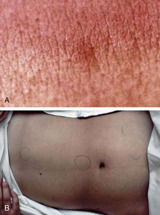
Typhoid fever usually manifests as high-grade fever with a wide variety of associated features, such as generalized myalgia, abdominal pain, hepatosplenomegaly, abdominal pain, and anorexia (Table 190-5). In children, diarrhea may occur in the earlier stages of the illness and may be followed by constipation. In the absence of localizing signs, the early stage of the disease may be difficult to differentiate from other endemic diseases such as malaria and dengue fever. The fever may rise gradually, but the classic stepladder rise of fever is relatively rare. In about 25% of cases, a macular or maculopapular rash (rose spots) may be visible around the 7th-10th day of the illness, and lesions may appear in crops of 10-15 on the lower chest and abdomen and last 2-3 days (Fig. 190-4). These lesions may be difficult to see in dark-skinned children. Patients managed as outpatients present with fever (99%) but have less emesis, diarrhea, hepatomegaly, splenomegaly, and myalgias than patients who require admission to the hospital.

Table 190-5 COMMON CLINICAL FEATURES OF TYPHOID FEVER IN CHILDREN*

FEATURE RATE (%)
High-grade fever 95
Coated tongue 76
Anorexia 70
Vomiting 39
Hepatomegaly 37
Diarrhea 36
Toxicity 29
Abdominal pain 21
Pallor 20
Splenomegaly 17
Constipation 7
Headache 4
Jaundice 2
Obtundation 2
Ileus 1
Intestinal perforation 0.5

* Data collected in Karachi, Pakistan, from 2,000 children.

image

Figure 190-4 A, A rose spot in a volunteer with experimental typhoid fever. B, A small cluster of rose spots is usually located on the abdomen. These lesions may be difficult to identify, especially in dark-skinned people.

(From Huang DB, DuPont HL: Problem pathogens: Extra-intestinal complications of Salmonella enterica serotype Typhi infection, Lancet Infect Dis 5:341–348, 2005.)

The presentation of typhoid fever may be tempered by coexisting morbidities and early diagnosis and administration of antibiotics. In malaria-endemic areas and in parts of the world where schistosomiasis is common, the presentation of typhoid may also be atypical. It is also recognized that multidrug-resistant S. Typhi infection is a more severe clinical illness with higher rates of toxicity, complications, and case fatality rates, which may be related to the greater virulence as well as higher numbers of circulating bacteria. The emergence of typhoid infections resistant to nalidixic acid and fluorquinolones has been associated with higher rates of morbidity and treatment failure. These findings may have implications for treatment algorithms, especially in endemic areas with high rates of multidrug-resistant and nalidixic acid– or fluoroquinolone-resistant typhoid.

If no complications occur, the symptoms and physical findings gradually resolve within 2-4 wk; however, the illness may be associated with malnutrition in a number of affected children. Although enteric fever caused by S. Paratyphi organisms has been classically regarded as a milder illness, there have been several outbreaks of infection with drug-resistant S. Paratyphi A, suggesting that paratyphoid fever may also be severe, with significant morbidity and complications.

Complications

Although altered liver function is found in many patients with enteric fever, clinically significant hepatitis, jaundice, and cholecystitis are relatively rare and may be associated with higher rates of adverse outcome. Intestinal hemorrhage (<1%) and perforation (0.5-1%) are infrequent among children. Intestinal perforation may be preceded by a marked increase in abdominal pain (usually in the right lower quadrant), tenderness, vomiting, and features of peritonitis. Intestinal perforation and peritonitis may be accompanied by a sudden rise in pulse rate, hypotension, marked abdominal tenderness and guarding, and subsequent abdominal rigidity. A rising white blood cell count with a left shift and free air on abdominal radiographs may be seen in such cases.

Rare complications include toxic myocarditis, which may manifest as arrhythmias, sinoatrial block, or cardiogenic shock (Table 190-6). Neurologic complications are also relatively uncommon among children; they include delirium, psychosis, increased intracranial pressure, acute cerebellar ataxia, chorea, deafness, and Guillain-Barré syndrome. Although case fatality rates may be higher with neurologic manifestations, recovery usually occurs with no sequelae. Other reported complications include fatal bone marrow necrosis, DIC, hemolytic-uremic syndrome, pyelonephritis, nephrotic syndrome, meningitis, endocarditis, parotitis, orchitis, and suppurative lymphadenitis.
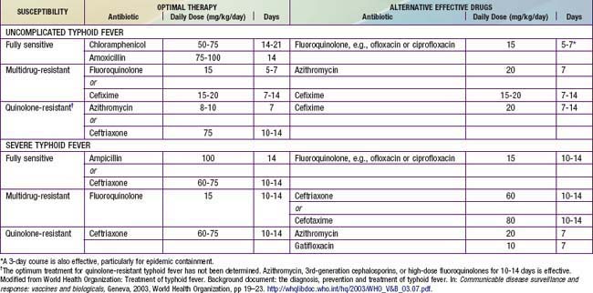
The propensity to become a carrier follows the epidemiology of gallbladder disease, increasing with patient age and the antibiotic resistance of the prevalent strains. Although limited data are available, rates of chronic carriage are generally lower in children than adults.

Diagnosis

The mainstay of the diagnosis of typhoid fever is a positive result of culture from the blood or another anatomic site. Results of blood cultures are positive in 40-60% of the patients seen early in the course of the disease, and stool and urine culture results become positive after the 1st wk. The stool culture result is also occasionally positive during the incubation period. However, the sensitivity of blood cultures in diagnosing typhoid fever in many parts of the developing world is limited because widespread liberal antibiotic use may render bacteriologic confirmation difficult. Although bone marrow cultures may increase the likelihood of bacteriologic confirmation of typhoid, collection of the specimens is difficult and relatively invasive.

Results of other laboratory investigations are nonspecific. Although blood leukocyte counts are frequently low in relation to the fever and toxicity, there is a wide range in counts; in younger children leukocytosis is common and may reach 20,000-25,000 cells/mm3. Thrombocytopenia may be a marker of severe illness and may accompany DIC. Liver function test results may be deranged, but significant hepatic dysfunction is rare.

The classic Widal test measures antibodies against O and H antigens of S. Typhi but lacks sensitivity and specificity in endemic areas. Because many false-positive and false-negative results occur, diagnosis of typhoid fever by Widal test alone is prone to error. Other relatively newer diagnostic tests using monoclonal antibodies have been developed that directly detect S. Typhi–specific antigens in the serum or S. Typhi Vi antigen in the urine. However, few have proved sufficiently robust in large-scale evaluations. A nested polymerase chain reaction analysis using H1-d primers has been used to amplify specific genes of S. Typhi in the blood of patients; it is a promising means of making a rapid diagnosis, especially given the low level of bacteremia in enteric fever. Despite these innovations, the mainstay of diagnosis of typhoid remains clinical in much of the developing world, and several diagnostic algorithms have been evaluated in endemic areas.

Treatment

An early diagnosis of typhoid fever and institution of appropriate treatment are essential. The vast majority of children with typhoid can be managed at home with oral antibiotics and close medical follow-up for complications or failure of response to therapy. Patients with persistent vomiting, severe diarrhea, and abdominal distention may require hospitalization and parenteral antibiotic therapy.

There are general principles of management of typhoid. Adequate rest, hydration, and attention are important to correct fluid and electrolyte imbalance. Antipyretic therapy (acetaminophen 10-15 mg/kg every 4-6 hr by mouth [PO]) should be provided as required. A soft, easily digestible diet should be continued unless the patient has abdominal distention or ileus. Antibiotic therapy is critical to minimize complications (Table 190-7). It has been suggested that traditional therapy with either chloramphenicol or amoxicillin is associated with relapse rates of 5-15% and 4-8%, respectively, whereas use of the quinolones and third-generation cephalosporins is associated with higher cure rates. The antibiotic treatment of typhoid fever in children is also influenced by the prevalence of antimicrobial resistance. Over the past 2 decades, emergence of multidrug-resistant strains of S. Typhi (i.e., isolates fully resistant to amoxicillin, trimethoprim-sulfamethoxazole, and chloramphenicol) has necessitated treatment with fluoroquinolones, which are the antimicrobial drug of choice for treatment of salmonellosis in adults, or cephalosporins. The emergence of resistance to quinolones has placed tremendous pressure on public health systems because alternative therapeutic options are limited.

Although some investigators have suggested that children with typhoid should be treated with fluoroquinolones like adults, others have questioned this approach on the basis of the potential development of further resistance to fluoroquinolones and the fact that quinolones are still not approved for widespread use in children. A Cochrane systematic review of the treatment of typhoid fever also indicates that there is little evidence to support the carte blanche administration of fluoroquinolones in all cases of typhoid.

In addition to antibiotics, the importance of supportive treatment and maintenance of appropriate fluid and electrolyte balance must be underscored. Although additional treatment with dexamethasone (3 mg/kg for the initial dose, followed by 1 mg/kg every 6 hr for 48 hr) has been recommended for severely ill patients with shock, obtundation, stupor, or coma, corticosteroids should be administered only under strict controlled conditions and supervision, because their use may mask signs of abdominal complications.

Prevention

Of the major risk factors for outbreaks of typhoid, contamination of water supplies with sewage is the most important. Other risk factors for development of typhoid fever are congestion, contact with another patient or a febrile individual, and lack of water and sanitation services. During outbreaks, central chlorination as well as domestic water purification is important. In endemic situations, consumption of street foods, especially ice cream and cut fruit, has been recognized as an important risk factor. The human-to-human spread by chronic carriers is also important, and attempts should therefore be made to target food handlers and high-risk groups for S. Typhi carriage screening. Once identified, chronic carriers must be counseled as to the risk for disease transmission and the importance of handwashing.

The classic heat-inactivated whole-cell vaccine for typhoid is associated with an unacceptably high rate of side effects and has been largely withdrawn from public health use. Globally, 2 vaccines are currently available for potential use in children. An oral, live-attenuated preparation of the Ty21a strain of S. Typhi has been shown to have good efficacy (67-82%) for up to 5 yr. Significant adverse effects are rare. The Vi capsular polysaccharide can be used in people ≥2 yr of age. It is given as a single intramuscular dose, with a booster every 2 yr, and has a protective efficacy of 70-80%. The vaccines are currently recommended for anyone traveling into endemic areas, but a few countries have introduced large-scale vaccination strategies. Previous studies in South America have demonstrated protection against typhoid among schoolchildren with the use of an oral attenuated Ty21 strain vaccine.

Several large-scale demonstration projects using the Vi polysaccharide vaccine in Asia have demonstrated protective efficacy against typhoid fever across all age groups, but the data on protection among young children (<5 yr) showed important differences between studies. The recent Vi-conjugate vaccine has been shown to have a protective efficacy exceeding 90% in younger children and may offer protection in parts of the world where a large proportion of preschool children are at risk for the disease enteric or typhoid fever.

Bibliography

Bhutta ZA. Current concepts in the diagnosis and treatment of typhoid fever. Br Med J. 2006;333:78-82.

Crump JA, Barrett TJ, Nelson JT, et al. Reevaluating fluoroquinolone breakpoints for Salmonella enterica serotype Typhi and for non-Typhi salmonellae. Clin Infect Dis. 2003;37:75-81.

Crump JA, Luby SP, Mintz ED. The global burden of typhoid fever. Bull WHO. 2004;82:346-353.

Crump JA, Mintz ED. Global trends in typhoid and paratyphoid Fever. Clin Infect Dis. 2010;50:241-246.

Dolecek C, Tran TP, Nguyen NR, et al. A multi-center randomised controlled trial of gatifloxacin versus azithromycin for the treatment of uncomplicated typhoid fever in children and adults in Vietnam. PLoS One. 2008;3:e2188.

Effa EE, Bukirwa H: Azithromycin for treating uncomplicated typhoid and paratyphoid fever (enteric fever) (review), Cochrane Data System Rev (4):CD006083, 2008.

Fangtham M, Wilde H. Emergence of Salmonella paratyphi A as a major cause of enteric fever: need for early detection, preventive measures, and effective vaccines. J Travel Med. 2008;15:344-350.

Gasem MH, Keuter M, Dolmans WM, et al. Persistence of salmonellae in blood and bone marrow: randomized controlled trial comparing ciprofloxacin and chloramphenicol treatments against enteric fever. Antimicrob Agents Chemother. 2003;47:1727-1731.

House D, Bishop A, Parry C, et al. Typhoid fever: pathogenesis and disease. Curr Opin Infect Dis. 2001;14:573-578.

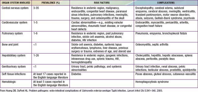
Huang DB, DuPont HL. Problem pathogens: extra-intestinal complications of Salmonella enterica serotype Typhi infection. Lancet Infect Dis. 2005;5:341-348.

Luby SP, Faizan MK, Fisher-Hoch SP, et al. Risk factors for typhoid fever in an endemic setting, Karachi, Pakistan. Epidemiol Infect. 1998;120:129-138.

Lynch MF, Blanton EM, Bulens S, et al. Typhoid fever in the United States, 1999–2006. JAMA. 2009;302:859-865.

Parry CM, Hien TT, Dougan G, et al. Typhoid fever. N Engl J Med. 2002;347:1770-1782.

Prakash P, Mishra OP, Singh AK, et al. Evaluation of nested PCR in diagnosis of typhoid fever. J Clin Microbiol. 2005;43:431-432.

Sinha A, Sazawal S, Kumar R, et al. Typhoid fever in children aged less than 5 years. Lancet. 1999;354:734-737.

Sur D, Ochiai RL, Bhattacharya SK, et al. A cluster-randomized effectiveness trial of Vi typhoid vaccine in India. N Engl J Med. 2009;361:335-344.

Thaver D, Zaidi AK, Critchley J, et al. A comparison of fluoroquinolones versus other antibiotics for treating enteric fever: meta-analysis. BMJ. 2009;338:b1865.

Wain J, House D, Parkhill J, et al. Unlocking the genome of the human typhoid bacillus. Lancet Infect Dis. 2002;2:163-170.

World Health Organization. Background document: The diagnosis, prevention and treatment of typhoid fever. Communicable disease surveillance and response: vaccines and biologicals. World Health Organization, Geneva, 2003;19-23. http://whqlibdoc.who.int/hq/2003/WHO_V&B_03.07.pdf.