Chapter 6 Reproductive system
Methods of imaging the female reproductive system
Akin O., Mironov S., Pandit-Taskar N., et al. Imaging of uterine cancer. Radiol. Clin. North Am.. 2007;45(1):167-182.
Barakat R.R., Hricak H. What do we expect from imaging. Radiol. Clin. North Am.. 2002;40(3):521-526. vii
Fütterer J.J., Heijmink S.W., Spermon J.R. Imaging the male reproductive tract: current trends and future directions. Radiol. Clin. North Am.. 2008;46(1):133-147. vii
Hysterosalpingography
Indications
Contraindications
Patient preparation
Technique
N.B. Opiates increase pain by stimulating smooth muscle contraction.
Aftercare
Complications
Due to the contrast medium
Allergic phenomena – especially if contrast medium is forced into the circulation.
Due to the technique
Ascher S.M., Reinhold C. Imaging of cancer of the endometrium. Radiol. Clin. North Am.. 2002;40(3):563-576.
Bates J. Practical Gynaecological Ultrasound, 2nd edn. Cambridge: Cambridge University Press, 2006.
Bhatt S., Ghazale H., Dogra V.S. Sonographic evaluation of ectopic pregnancy. Radiol. Clin. North Am.. 2007;45(3):549-560.
Funt S.A., Hann L.E. Detection and characterization of adnexal masses. Radiol. Clin. North Am.. 2002;40(3):591-608.
Ultrasound of the Scrotum
Indications
Equipment
7.5–15 MHz transducer. Linear array for optimum imaging. Stand-off may be helpful in some cases.
Technique
CT of the Reproductive System
MAGNETIC RESONANCE IMAGING OF THE REPRODUCTIVE SYSTEM
Indications
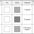
Pulse sequences
Perfusion imaging of the uterus can be used to assess the effectiveness of uterine fibroid therapy.
MRI is also used to measure the pelvic outlet in pelvimetry in order to avoid radiation dose.
Scrotal MR is generally performed with the scrotum supported as in US, using a surface coil. High-resolution axial, sagittal and coronal T2-weighted spin echo scans are obtained with a T1-weighted scan to identify haemorrhage. Large field-of-view (FOV) scans should be performed to assess the inguinal canal for the presence of a hernia. Gadolinium i.v. can be given if necessary to assess perfusion. Scans should include the pelvis and kidneys if an undescended testis is being investigated.
Brown M.A., Martin D.R., Semelka R. Future directions in MR imaging of the female pelvis. Magn. Reson. Imaging Clin. N. Am.. 2006;14(4):431-437.
Burn P.R., McCall J.M., Chinn R.J., et al. Uterine fibroleiomyoma: MR imaging appearances before and after embolization of uterine arteries. Radiology. 2000;214(3):729-734.
Coakley F.V. Staging ovarian cancer: role of imaging. Radiol. Clin. North Am.. 2002;40(3):609-636.
Hamm B., Forstner R., Kim E.S., et al. MRI and CT of the female pelvis. J. Nucl. Med.. 2008;49(5):862.
Humphries P.D., Simpson J.C., Creighton S.M., et al. MRI in the assessment of congenital vaginal anomalies. Clin. Radiol.. 2008;63(4):442-448.
Martin D.R., Salman K., Wilmot C.C. MR imaging evaluation of the pelvic floor for the assessment of vaginal prolapse and urinary incontinence. Magn. Reson. Imaging. Clin. N. Am.. 2006;14(4):523-535.
Scheidler J., Heuck A.F. Imaging of cancer of the cervix. Radiol. Clin. North Am.. 2002;40(3):577-590.