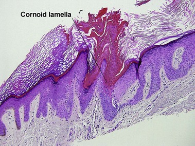
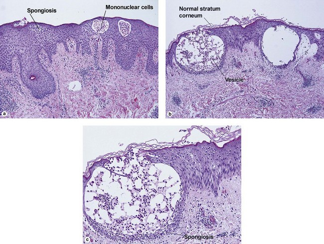
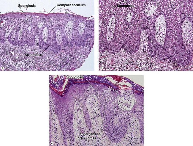
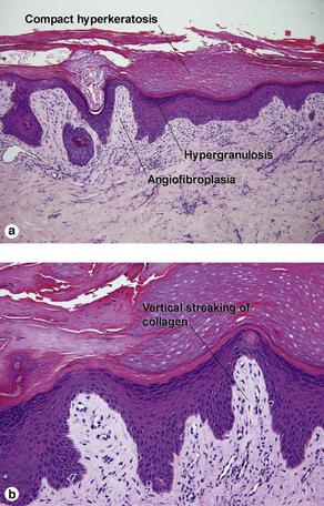
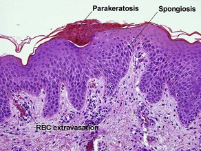
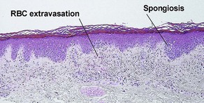
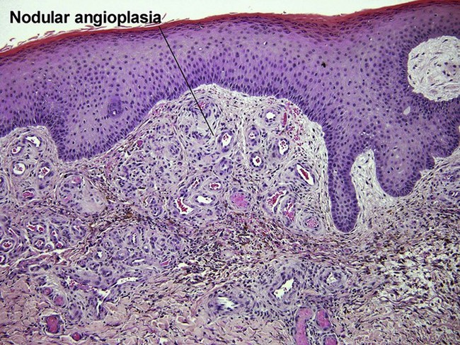
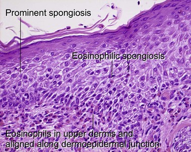
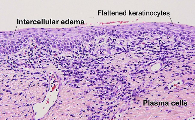
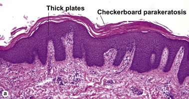
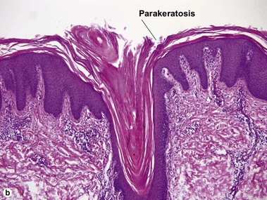
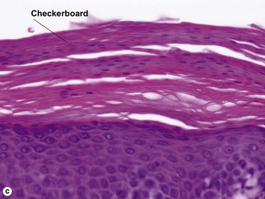
Psoriasiform and spongiotic dermatitis
Behrhof, W, Springer, E, Bräuninger, W, et al. PCR testing for Treponema pallidum in paraffin-embedded skin biopsy specimens: test design and impact on the diagnosis of syphilis. J Clin Pathol. 2008; 61(3):390–395.
Chen, CY, Chi, KH, George, RW, et al. Diagnosis of gastric syphilis by direct immunofluorescence staining and real-time PCR testing. J Clin Microbiol. 2006; 44(9):3452–3456.
Hugel, H. Histological diagnosis of inflammatory skin diseases. Use of a simple algorithm and modern diagnostic methods. Pathologe. 2002; 23(1):20–37.
Meymandi, S, Silver, SG, Crawford, RI. Intraepidermal neutrophils – a clue to dermatophytosis? J Cutan Pathol. 2003; 30(4):253–255.
Pujol, RM, Wang, CY, el-Azhary, RA, et al. Necrolytic migratory erythema: clinicopathologic study of 13 cases. Int J Dermatol. 2004; 43(1):12–18.