Proximal, Distal, and Generalized Weakness

Published on 12/04/2015 by admin

Filed under Neurology

Last modified 22/04/2025

Print this page

rate 1 star rate 2 star rate 3 star rate 4 star rate 5 star
Your rating: none, Average: 0 (0 votes)

This article have been viewed 10152 times

Chapter 25 Proximal, Distal, and Generalized Weakness

Muscle weakness may be due to disorders of the central nervous system (CNS) or peripheral nervous system (PNS). The PNS includes the primary sensory neurons in the dorsal root ganglia, nerve roots, peripheral nerves, neuromuscular junctions, and muscles. Although not strictly peripheral, the primary motor neurons (anterior horn cells) in the brainstem and spinal cord are also conventionally included as part of the PNS. The neurological examination allows separation of the causes of weakness arising at these different locations. If the pattern of weakness is characteristic of upper motor neuron (UMN) dysfunction (i.e., weakness of upper-limb extensors and lower-limb flexors) together with hyperreflexia and extensor plantar responses, the weakness clearly is of CNS origin. Weakness with sensory loss may occur in both CNS disorders and disorders of the nerve roots and peripheral nerves. Weakness without sensory loss may also occur from CNS disorders, but in the PNS, this pattern of weakness occurs in disorders of the anterior horn cell, neuromuscular junction, or muscle. Rarely in the PNS, peripheral motor fibers will be the site of pathology (e.g., as occurs in multifocal motor neuropathy with conduction block). Although fatigue often accompanies most disorders of weakness, marked fatigue, especially when involving the extraocular, bulbar, and proximal upper limb muscles, often indicates a disorder of the neuromuscular junction.

The motor unit is the primary building block of the PNS and includes the anterior horn cell, its motor nerve, terminal nerve fibers, and all their accompanying neuromuscular junctions and muscle fibers. This chapter concentrates on disorders of the motor unit and disorders that may also involve the peripheral sensory nerves. The pattern of weakness often localizes the pathological process to the primary neurons, nerve roots, peripheral nerves, neuromuscular junctions, or muscles. Muscle weakness changes functional abilities that are more or less specific to the muscle groups affected. Recognizable patterns of symptoms and signs often allow a reasonable estimation of the anatomical involvement. Identifying these patterns is the first step in the differential diagnosis of weakness, because certain disorders affect specific muscle groups. This chapter begins with a review of the symptoms and signs of muscular weakness with respect to the muscle groups affected. A discussion follows of the bedside examinations, functional examinations, and laboratory tests often used in evaluating patients with muscle weakness. The chapter concludes with an approach to the differential diagnosis of muscle weakness based on which muscle groups are weak, whether the muscle weakness is constant or fluctuating, and whether the disorder is genetic or acquired.

Clinical Presentation by Affected Region

General Considerations

As muscles begin to weaken, the associated clinical features depend more on which muscles are involved than on the cause of involvement. A complicating factor in evaluating weakness is the patient’s interpretation of the term weak. Although physicians use this term to denote a loss of muscle power, patients tend to apply it more loosely in describing their symptoms. Even more confusing, many people use the words numb and weak interchangeably, so the clinician should not accept a complaint of weakness at face value; the patient should be questioned further until it is clear that weakness means loss of muscle strength.

If the patient has no objective weakness when examined, the clinician must rely on the history. In patients with weak muscles, a fairly stereotypical set of symptoms emerges according to which muscle groups are weak (discussed later in this section). The patient whose weakness is caused by depression or malingering has vague symptoms, avoids answering leading questions, and the stereotypical symptoms of weakness are seldom volunteered. Instead, these patients make such statements as “I have no strength to do the housework,” “I just can’t do (the task),” or “I can’t climb the stairs because I get so tired and have to rest.” When pressed regarding these symptoms, it soon becomes apparent that specific details are lacking. Patients who cannot get out of a low chair because of real weakness explain exactly how they have to maneuver themselves into an upright position (e.g., pushing on the chair arms, leaning forward in the seat, and bracing their hands against the furniture). The examiner should avoid providing patients with clinical details they appear to be searching for. Asking whether pushing on the arms of the chair is required to stand up provides the patient with key information that may later be used in response to the questions of baffled successive examiners. In addition, it often is difficult to differentiate true muscle weakness from apparent weakness that accompanies tendon or joint contractures or is secondary to pain. For example, patients with primary orthopedic conditions often complain of weakness. In these patients, however, pain with passive or active motion often is a prominent part of the symptoms.

In evaluating weakness, the first key task is to discern which muscle groups are affected. In this regard, it is helpful to consider the clinical presentation with involvement of specific body regions: ocular; facial and bulbar; neck, diaphragm, and axial; proximal upper extremity; distal upper extremity; proximal lower extremity; and distal lower extremity.

Facial and Bulbar Muscles

Patients experience facial weakness as a feeling of stiffness or sometimes as a twisting or altered perception in the face (note that patients often use the word numbness in describing facial weakness). Drinking through a straw, whistling, and blowing up balloons all are particularly difficult tasks for these patients and may be sensitive tests for facial weakness, particularly when such weakness dates from childhood. Acquaintances may notice that the patient’s expression is somehow changed. A pleasant smile may turn into a snarl because of weakness of the levator anguli oris muscles. In lower facial weakness, patients may have difficulty with drooling and retaining their saliva, often requiring them to carry a tissue in the hand—the so-called napkin sign—which often accompanies bulbar involvement in amyotrophic lateral sclerosis (ALS). A common observation in mild long-standing facial weakness, as with facioscapulohumeral (FSH) muscular dystrophy, is a tendency for the patient to sleep with the eyes open from weakness of the orbicularis oculi. Weakness of masticatory muscles may result in difficulty chewing, sometimes with a sensation of fatigue and discomfort, as may occur with myasthenia gravis (MG). Pharyngeal, palatal, and tongue weakness disturbs speech and swallowing. A flaccid palate is associated with nasal regurgitation, choking spells, and aspiration of liquids. Speech may become slurred or acquire a nasal or hoarse quality. In contrast with central lesions, no problem with fluency or language function is observed.

Neck, Diaphragm, and Axial Muscles

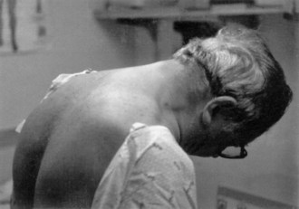
Neck muscle weakness becomes apparent when the patient must stabilize the head. Riding as a passenger in a car that brakes or accelerates, particularly in emergencies, may be disconcerting for the patient with neck weakness, because the head rocks forward or backward. Similarly, when the patient is stooping or bending forward, weakness of the posterior neck muscles may cause the chin to fall on the chest. A patient with neck-flexion weakness often notices difficulty lifting the head off the pillow in the morning. As neck weakness progresses, patients may develop the dropped head syndrome, in which they no longer can extend the neck, and the chin rests against the chest (Fig. 25.1). This posture leads to several secondary difficulties, especially with vision and swallowing.

Shortness of breath often develops when diaphragm muscles weaken, especially when individuals lie flat or must exert themselves. These symptoms can be mistakenly attributed to lung or heart disease. Severe diaphragmatic weakness leads to hypoventilation and carbon dioxide retention. This may first be manifested as morning headaches or vivid nightmares. Later, hypercapnia results in sedation and a depressed mental state. Rarely, axial and trunk muscles can be involved early in the course of a neuromuscular disorder. Weakness of the abdominal muscles may make sit-ups impossible. Focal weakness of the lower abdominal muscles results in an obvious protuberance that superficially mimics an abdominal hernia. Patients with weakness of the paraspinal muscles are unable to maintain a straight posture when sitting or standing, although they can do so when lying on the bed (so-called bent spine syndrome).

Bedside Examination of the Weak Patient

The neurological examination of patients with muscle weakness is the same as that used for patients with other neurological problems. Special attention to the observational and functional components of the evaluation, however, is particularly rewarding in the patient with weakness.

Observation

It is useful to spend a few moments observing the patient and noting natural posture and motion. When patients, particularly children, are aware of the examination, they often concentrate on performing as normally as possible. When unaware of scrutiny, their posture and movements may be more natural. At one time or another, we have heard the parent’s exasperated cry, “He never does it that way at home.” For example, ptosis may be obvious on inspection of the head and neck. The more severe the ptosis, the greater the patient’s tendency to throw the head backward. The eyebrows are elevated and the forehead wrinkled in an attempt to raise the upper lids. This sometimes is so successful that ptosis is apparent only when the examiner smoothes out the wrinkled forehead and allows the eyebrows to assume a more normal position. Psychogenic ptosis is easy to detect: the lower lid elevates with contraction of both parts of the orbicularis oculi muscles (i.e., blepharospasm) to accompany the lowered upper lid.

Weakness of the facial muscles present since childhood may give a smooth, unlined appearance to the adult face. In addition, facial expression diminishes or changes. A smile may become a grimace or a snarl, with eversion of the upper lip. The normal blink may slow, or eyelid closure may be incomplete so that the sclera is always visible. The normal preservation of the arch of the upper lip may be lost, and the mouth may assume either a tented or a straight-line configuration. Actual wasting of the facial muscles is difficult to see, but temporalis and masseter atrophy produce a characteristic scalloped appearance above and below the cheekbone. Because rearranging the hair style may cover the wasting, the examiner should make a conscious effort to check the upper portion of the patient’s face. The tongue is inspected for atrophy and fasciculations. Inspecting the tongue at rest with the mouth open, looking for the random irregular twitching movements of fasciculations, is the best method. When the tongue is fully protruded, many patients have some normal quivering movements that can easily be mistaken for fasciculations. It is wise to diagnose fasciculations of the tongue only when there is associated atrophy.

Facial weakness causes the normal labial sounds (that of p and b) to be softened. The examiner with a practiced ear can detect other alterations of speech. Lower motor neuron (LMN) involvement of the palate and tongue gives the speech a hollow, nasal, echoing timbre, whereas UMN dysfunction causes the speech to be monotonous, forced, and strained. Laryngeal weakness also may be noticed in speech when the voice becomes harsh or brassy, often associated with loss of the glottal stop (the small sound made by the larynx closing, as at the start of a cough).

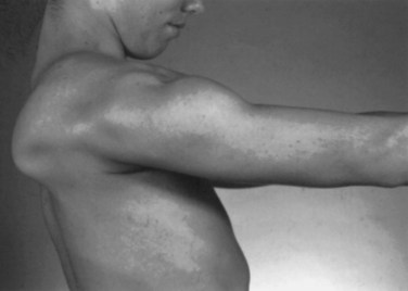
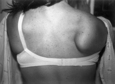
Weakness of the shoulder muscles causes a characteristic change in posture. Normally the shoulders brace back by means of the tone of the muscles, so the hands are positioned with the thumbs forward when the arms are by the side. As the shoulder muscles lose their tone, the point of the shoulder rotates forward. This forward rotation of the shoulder is associated with a rotation of the arm, so that the backs of the hands now are forward facing. Additionally, the loss of tone causes a rather loose swinging movement of the arms in normal walking. When shoulder weakness is severe, the patient may fling the arms by using a movement of the trunk, rather than lifting the arms in the normal fashion. In the most extreme example, the only way the patient can get the hand above the head on a wall is to use a truncal movement to throw the whole arm upward and forward so the hand rests on the wall, and then to creep the hand up the wall using finger movements. Atrophy of the pectoral muscles leads to the development of a horizontal or upward sloping of the anterior axillary fold. This is especially the case in facioscapulohumeral (FSH) muscular dystrophy. The examiner may observe winging of the scapula, a characteristic finding in weakness of muscles that normally fix the scapula to the thorax (i.e., the serratus anterior, rhomboid, or trapezius). As these muscles become weak, any attempted movement of the arm causes the scapula to rise off the back of the rib cage and protrude like a small wing. The arm and shoulder act as a crane—the boom of the crane is the arm, and the base is the scapula. Obviously, if the base is not fixed, any attempt to use the crane results in the whole structure’s falling over. This is the operative mechanism with attempts to elevate the arm; the scapula simply pops off the back of the chest wall in a characteristic fashion. In the most common type of winging, the entire medial border of the scapula protrudes backward. In some diseases, particularly FSH muscular dystrophy, the inferomedial angle juts out first, and the entire scapula rotates and rides up over the back. This often is associated with a trapezius hump, in which the middle part of the trapezius muscle in the web of the neck mounds over the upper border of the scapula (Fig. 25.2). Note that when examining a slender person or a child, in whom prominent shoulder blades are common, the shoulder configuration returns to normal with forcible use of the arm, as in a push-up.

Muscle Bulk and Deformities

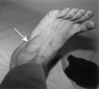
Assessment of muscle bulk looking for atrophy and hypertrophy is an important part of the neuromuscular examination. Prominent muscle wasting usually accompanies neurogenic disorders associated with axonal loss. However, severe wasting also occurs in chronic myopathic conditions. Wasting is best appreciated in the distal hand and foot muscles and around bony prominences. In the arm, wasting of the intrinsic hand muscles produces a characteristic hand posture in which the thumb rotates outward so that it lies in the same plane as that of the fingers (the simian hand), and the interphalangeal joints flex slightly with slight extension of metacarpophalangeal joints (the claw hand). Wasting of the small muscles leaves the bones easily visible through the skin, resulting in the characteristic guttered appearance of the back of the hand. In the foot, one of the easier muscles to inspect is the extensor digitorum brevis, a small muscle on the lateral dorsum of the foot that helps dorsiflex the toes (Fig. 25.3). It often wastes early in neuropathies and anterior horn cell disorders. In myopathic conditions in which proximal muscles are affected more than distal muscles, the extensor digitorum brevis may actually hypertrophy to try to compensate for weakness of the long toe dorsiflexors above it.

Muscle mass of the leg is so variable among individuals that it is sometimes difficult to decide whether wasting of the muscles has occurred. Any marked asymmetry indicates an abnormality, but distinguishing a slender thigh from quadriceps muscle atrophy often is difficult. One way to try to distinguish these conditions is to ask the patient to tighten the knee as firmly as possible. The firm medial and lateral bellies of the normal quadriceps that bunch up in the distal part of the thigh just above the knee fail to appear in the wasted muscle. The same technique can be used to evaluate anterior tibial wasting. In a severely wasted muscle, a groove on the lateral side of the tibia (which normally is filled by the anterior tibial muscles) is apparent. A moderate degree of wasting is difficult to distinguish from thinness of the leg, but if the patient dorsiflexes the foot, the wasted muscle fails to develop the prominent belly seen in a normal muscle.

Abnormal muscle hypertrophy is uncommon but may be a key finding when present. Beyond the expected increase in muscle bulk that accompanies exercise, generalized muscle hypertrophy is a feature of myotonia congenita and paramyotonia congenita, giving the appearance of the extreme development typically seen in weight lifters. Hypertrophy is a common finding in the rare syndrome of acquired neuromyotonia, in which the continuous discharge of motor axons results in the muscle effectively exercising itself. Exceptionally, hypertrophy occurs in some chronic denervating disorders, especially in the posterior calf muscle in S1 radiculopathies. Electromyography (EMG) in affected patients often reveals spontaneous discharges in these muscles (usually complex repetitive discharges) consequent to chronic denervation. By contrast, conditions exist in which muscle hypertrophy is not from true muscle enlargement but from infiltration of fat, connective tissue, and other material (i.e., pseudohypertrophy). Pseudohypertrophy occurs in calf muscles of patients with Duchenne and Becker muscular dystrophy, as well as in patients with limb-girdle muscular dystrophy, spinal muscular atrophy (SMA), and some glycogen storage disorders. Similarly, pseudohypertrophy occurs rarely in sarcoidosis, cysticercosis, amyloidosis, hypothyroid myopathy, and focal myositis. Palpable masses in muscles occur with muscle tumors, ruptured tendons, or muscle hernias.

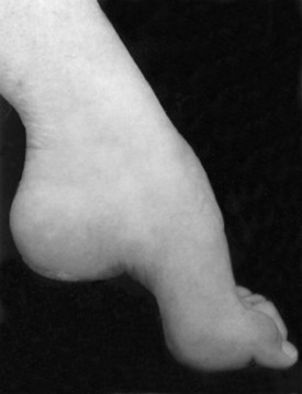
Several bony deformities often provide important clues to the presence of neuromuscular conditions. Proximal and axial muscle weakness often leads to scoliosis. Intrinsic foot muscle weakness present from childhood often leads to the characteristic foot deformity of pes cavus, in which the foot is foreshortened with high arches and hammer toes (Fig. 25.4). Pes cavus is a sign that weakness has been present at least since early childhood and implies a genetic disorder in most patients. Likewise, a high-arched palate often develops from chronic neuromuscular weakness present from childhood.

Muscle Palpation, Percussion, and Range of Motion

Palpation and percussion of muscle provide additional information. Fibrotic muscle may feel rubbery and hard, whereas denervated muscle may separate into discrete strands that roll under the fingers. Muscle in inflammatory myopathies or rheumatological conditions may be tender to palpation, but severe muscle pain on palpation is unusual. An exception to this rule is in the patient experiencing an acute phase of viral myositis or rhabdomyolysis, whose muscles may be very sensitive to either movement or touch. Percussion of muscle may produce the phenomenon of myotonia, in which a localized contraction of the muscle persists for several seconds after percussion. Percussing the thenar eminence and watching for a delayed relaxation of the thumb abductors will best show this phenomenon. This defining characteristic of myotonic dystrophy and myotonia congenita is distinguishable from myoedema, which occasionally occurs in patients with thyroid disorders and other metabolic problems. In myoedema, the development of a dimple in the muscle, which then mounds to form a small hillock, follows the percussion.

In addition to its diagnostic value, the presence of muscle contracture across a joint may cause disability, even in the absence of weakness. Thus, an evaluation of range of motion at major joints is an important part of the clinical examination. A standard examination includes evaluation for contractures at the fingers, elbows, wrists, hips, knees, and ankles. At the hips, both flexion and iliotibial band contractures should be looked for.

Muscle Tone

The physiological origin of muscle tone is complex and outside the scope of this chapter. In examining the weak patient, however, muscle tone offers valuable information regarding the origins of the weakness. Variations from a normal muscle tone result in increased tone (hypertonicity) or decreased tone (hypotonicity). Increased tone results from the loss of CNS influences on the tonic contraction of muscle. Decreased tone usually implicates a problem with the proprioceptive or peripheral motor innervation of a muscle but also may result from an acute spinal cord or cerebral lesions. Patients usually do not complain directly of increased or decreased tone; for example, the spastic patient may complain of heaviness, stiffness, or slowness of movement.

Several methods are used to examine tone. First is the spontaneous posture of the extremities. With spasticity, the upper limbs often are in a fixed flexed posture, and affected muscles are firm to palpation. The examiner should attempt to relax the patient to allow free passive movement; helpful instructions may include statements such as “Don’t try to help me do the work.” Normally, resistance is the same throughout the range of motion and does not change with changes in the velocity of the movement. In a patient with spasticity, rapid passive displacement of the extremity results in increased resistance followed by relaxation (clasp-knife phenomenon). Resistance varies with the speed and direction of passive motion. Examination of tone in the legs should include supine examination, because with the patient in this position, the examiner easily accomplishes hip and knee flexion. In spasticity, the heel elevates off the examination table, while normally the heel remains in contact with the table. Hypotonia is the loss of normal tone and is felt as increased ease of passive movements during these maneuvers, or floppiness. In patients with severe hypotonia, the joints may be passively hyperextended.

Strength

Evaluation of individual muscle strength is an important part of the clinical examination. Many methods are available. Fixed myometry has become popular within the research community. This method uses a strain gauge attached to a rigid supporting structure, often integrated into the examining couch on which the patient lies. The patient then uses maximum voluntary contraction, quantitated in newtons (N). The merits of this method are debatable, and for the average clinician, the equipment expense is prohibitive.

In an office situation and in many clinical drug trials, manual muscle testing gives perfectly adequate results and is preferable to fixed myometry in young children. The basis is the Medical Research Council grading system, with some modification (Table 25.1). This method is adequate for use in an office situation, particularly if supplemented by the functional evaluation. A scale of 0 to 5 is used, in which 5 indicates normal strength. A grade of 5 indicates that the examiner is certain a muscle is normal and never used to compensate for slightly weak muscles. Muscles that can move the joint against resistance may vary quite widely in strength; grades of 4+, 4, and 4− often are used to indicate differences, particularly between one side of the body and the other. Grade 4 represents a wide range of strength, from slight weakness to moderate weakness, which is a disadvantage. For this reason, the scale has been more useful in following the average strength of many muscles during the course of a disease, rather than the course of a single muscle. Averaging many muscle scores smoothes out the stepwise progression noted in a single muscle. This may demonstrate a steadily progressive decline. A grade of 3+ is assigned when the muscle can move the joint against gravity and can exert a tiny amount of resistance but then collapses under the pressure of the examiner’s hand. It does not denote the phenomenon of sudden give-way, which occurs in conversion disorders and in patients limited by pain. Grade 3 indicates that the muscle can move the joint throughout its full range against gravity, but not against any added resistance. Sometimes, particularly in muscles acting across large joints such as the knee, the muscle is capable of moving the limb partially against gravity but not through the full range of movement. A muscle that cannot extend the knee horizontally when the patient is in a sitting position but can extend the knee to within 30 to 40 degrees of horizontal is graded 3−. Grades 2, 1, and 0 are as defined in Table 25.1.

Table 25.1 The Medical Research Council Scale for Grading Muscle Strength

Grade Description
0 No contraction
1 Flicker or trace of contraction
2 Active movement with gravity eliminated
3 Active movement against gravity
4 Active movement against gravity and resistance
5 Normal power

Although it is commendable and sometimes essential to examine each muscle separately, most clinicians test muscle groups rather than individual muscles. In our clinic, we test neck flexion, neck extension, shoulder abduction, internal rotation, external rotation, elbow flexion and extension, wrist flexion and extension, finger abduction and adduction, thumb abduction, hip flexion and extension, knee flexion and extension, ankle dorsiflexion and plantar flexion, and dorsiflexion of the great toe.

Fasciculations, Cramps, and Other Abnormal Muscle Movements

All limbs are examined to determine the presence or absence of fasciculations. A fasciculation is a brief twitch caused by the spontaneous firing of one motor unit. Fasciculations may be difficult or impossible to see in infants or obese patients. They can be present in normal people, so their presence in the absence of wasting or weakness is of no significance (benign fasciculations). Fasciculations that are widespread and seen on every examination may indicate denervating disease, particularly anterior horn cell disease. Mental or physical fatigue, caffeine, cigarette smoking, or drugs such as amphetamines exacerbate fasciculations.

In some patients who have been careful to avoid exposure to exacerbating factors, disease-related fasciculations may be absent or appear benign. This should be kept in mind during the evaluation. Abundant fasciculations may be difficult to differentiate from myokymia, which is a more writhing, bag of worms–like motion of muscle. Myokymia results from repetitive bursting of a motor unit (i.e., grouped fasciculations) and characteristically is associated with certain neuromuscular conditions (e.g., radiation injury, Guillain-Barré syndrome).

Similar to fasciculations, cramps may be benign or accompany several neuropathic conditions. A cramp is a painful involuntary muscle contraction. Cramps occur when a muscle is contracting in a shortened position. During a cramp, the muscle becomes hard and well defined. Stretching the muscle relieves the cramp. Superficially, a muscle contracture that occurs in a metabolic myopathy may resemble a cramp, although these two entities are completely different on electrophysiological testing. During a contracture, electrical silence is characteristic, whereas numerous motor units fire at high frequencies during a cramp.

Functional Evaluation of the Weak Patient

Walking

Alteration of gait may occur with weakness of the muscles of the hip and back, leg, and shoulder. In normal walking, when the heel hits the ground, the action of the hip abductors, which stabilize the pelvis, serves to counteract the shock. Thus in a sense, the hip abductors act as shock absorbers. Weakness of these muscles disturbs the normal fluid movement of the pelvis during walking, so when the heel hits the ground, the pelvis dips to the other side; bilateral weakness produces a waddle. Additionally, weakness of the hip extensors and back extensors makes it difficult for the patient to maintain a normal posture. Ordinarily the body is carried so that the center of gravity is slightly forward of the hip joint. To maintain an erect posture, the hip and back extensors are in continual activity. If these muscles become weak, the patient often throws the shoulders back so that the weight of the body falls behind the hip joints. This postural adjustment accentuates the lumbar lordosis. Alternatively, with pronounced weakness of the quadriceps muscles, the patient stabilizes the knee by throwing it backward. When the knee is hyperextended, it locks, deriving its stability from the anatomy of the joint rather than from muscular support. Finally, weakness of the muscles of the lower leg may result in a steppage gait, in which a short throw at the ankle midswing affects dorsiflexion of the foot. The foot then rapidly comes to the ground before the toes fall back into plantar flexion. Shoulder weakness is noted as the patient walks; the arms hang loosely by the sides and tend to swing in a pendular fashion rather than with a normal controlled swing.

Arising from the Floor

The normal method for arising from the floor depends on the age of the patient. The young child can spring rapidly to the feet without the average observer being able to dissect the movements. The elderly patient may turn to one side, place a hand on the floor, and rise to a standing position with a deliberate slowness. Despite such variability, abnormalities caused by muscle weakness are easily detectable. The patient with hip muscle weakness will turn to one side or the other to put the hand on the floor for support. The degree of turning is proportional to the severity of the weakness. Some patients must turn all the way around until they are in a prone position before they draw their feet under them to begin the standing process. Most people arise to a standing position from a squatting position, but the patient with hip extensor and quadriceps muscle weakness finds it easier to keep the hands on the floor and raise the hips high in the air. This has been termed the butt-first maneuver; the patient forms a triangle with the hips at the apex and the base of support provided by both hands and feet on the floor, and then laboriously rises from this position, usually by pushing on the thighs with both hands to brace the body upward. The progress of recovery or progression of weakness can be documented by noting whether the initial turn is greater than 90 degrees, whether unilateral or bilateral hand support is used on the floor and thighs, whether this support is sustained or transitory, and whether a butt-first maneuver is used. The entire process is known as the Gower maneuver, but it is useful to break it up into its component parts (Fig. 25.5).

Stepping Onto a Stool

For a patient with hip and leg weakness, stepping onto an 8-inch-high footstool is equivalent in difficulty to a normal person’s stepping up onto a coffee table. This analogy is apt because the required maneuvers are similar in both cases. Whereas the patient with normal strength readily approaches a footstool and easily steps onto it, the patient with weakness often hesitates in front of the stool while contemplating the task. A curious little maneuver occurs, known colloquially as the fast-foot maneuver. Normal persons can easily take the weight of the body on one leg, straightening out the knee as they stand on the footstool. Patients with weakness feel unsafe. They like to get both feet under them before straightening the knees and rising to their full height. To accomplish this, they place one foot on the footstool. While the knee of this leg is still bent, they quickly transfer the other foot from the floor to the footstool and then straighten the knees. This gives the impression of a hurried transfer of the trailing foot from floor to footstool, hence the term fast foot. As the weakness increases, the pelvis may dip toward the floor as the leading leg takes up the strain and the patient’s weight transfers from the foot on the floor to the foot on the stool, the so-called hip dip. Finally, if the weakness is severe, patients may either use hand support on the thighs or appear to gather themselves in and throw the body onto the footstool. Analysis of the various components—the hesitation, fast foot, hip dip, and throw—together with the presence or absence of hand support may provide a sensitive measure of changes in disease.

Psychogenic Weakness

An experienced examiner should be able to differentiate real weakness from psychogenic weakness. The primary characteristic of psychogenic weakness is that it is unpredictable and fluctuating. Muscle strength may suddenly give out when a limb is being evaluated. The patient has difficulty knowing the exact muscle strength expected and cannot adequately counter the examiner’s resistance. This gives rise to a wavering, collapsing force. Tricks are useful to bring out the discrepancy in muscle performance. For example, if the weak thigh cannot lift off the chair in a seated position, then the legs should not swing up onto the mattress when being seated on the examining table. When the examiner suspects that weakness of shoulder abduction is feigned, the patient’s arm is placed in abduction. With the examiner’s hand on the elbows, the examiner can instruct the patient to push toward the ceiling. At first, the downward pressure is very light, and the patient is unable to move the examining hand toward the ceiling. However, the arm does not fall down either, and as the downward pressure is gradually increased, continued exhortation to push the examiner’s hand upward results in increasing resistance to the downward pressure. The examiner ends up putting maximum weight on the outstretched arm, which remains in abduction. The logical conclusion is that the strength is normal. Patients do not realize this because they believe that because they did not move the examiner’s hand upward, they must be weak.

Clinical Investigations in Muscular Weakness

In the investigation of diseases of the motor unit, the most helpful tests are measurement of the serum concentration of creatine kinase (CK), electrodiagnosis, and muscle biopsy. These are available to all physicians. Genetic testing increasingly provides definitive diagnosis. In addition, if facilities are available, exercise testing can provide useful information.

Serum Creatine Kinase

The usefulness of measuring the serum CK concentration in the diagnosis of neuromuscular diseases is in differentiating between neurogenic disease, in which normal or mild to moderate elevations of CK may be seen, and myopathies, in which the CK concentration often is markedly increased. Notable exceptions exist. CK concentrations may be elevated as high as 10 times normal in patients with spinal muscular atrophy and occasionally in those with ALS (see Chapter 74). Measurements of serial CK concentrations follow the progress of the disease. Problems have been recognized with both of these uses. Foremost is the determination of the normal level. A survey of 250 hospitals in Ontario, Canada, showed a surprising ignorance of the basic mechanisms involved in the test and the way to derive normal values. Some hospital laboratories were unaware that race, gender, age, and activity level are important in determining normal values. Blood samples obtained from truly normal controls and not from inactive hospital patients not showing overt muscle disease show a higher normal serum CK concentration than would be anticipated. Furthermore, all studies on CK concentration show that gender and race affect values. A log transformation does much to convert this to a normal distribution curve, but even then, the results are not perfect.

In a survey of 1500 hospital employees, using carefully standardized methods, it was possible to detect three populations, each with characteristic CK values. The upper limits of normal (97.5th percentile) were as follows:

The nonblack population included Hispanics, Asians, and Caucasians. Because expression of the upper limit is as a percentile of the mean, by definition, levels in 2.5% of the normal population will be above that. Although this does not seem like a large proportion, in a town of 100,000, 2500 people would have abnormal levels. The point is that the upper limit of normal CK concentration is not rigid and requires intelligent interpretation. Although the serum CK concentration can be useful in determining the course of an illness, judgment is required because changes in CK values do not always mirror the clinical condition. In treating inflammatory myopathies with immunosuppressive drugs or corticosteroids, a steadily declining CK concentration is a reassurance, whereas concentrations that are creeping back up again when the patient is presumably in remission may be concerning.

Serum CK concentrations are also useful to determine whether an illness is monophasic. A bout of myoglobinuria may be associated with very high concentrations of CK. The concentration then declines steadily by approximately 50% every 2 days. This pattern indicates that a single episode of muscle damage has occurred. Patients with CK concentrations that do not decline in this fashion or that vary from high to low on random days have an ongoing illness. Finally, exercise may cause a marked elevation in CK, which usually peaks 12 to 18 hours after the activity but may occur days later. CK concentrations are more likely to increase in people who are sedentary and then undertake unaccustomed exercise than in a trained individual.

Muscle Biopsy

The use of muscle biopsy is important for establishing the diagnosis in most disorders of the motor unit. Histochemical evaluation is available at most hospitals and is particularly useful, and electron microscopy may provide a specific diagnosis. An important newer aspect of the muscle biopsy study is the analysis of the muscle proteins. Individual muscle proteins, including dystrophin, sarcoglycans, and other structural proteins may be missing in specific illnesses, and the diagnosis is often definitive with these analyses.

Chapter 83 reviews the details of muscle biopsy, but a word about the selection of the muscle to be biopsied is appropriate here. All biopsy procedures carry a risk of sampling error. Not all muscles are equally involved in any given disease, and it is important to select a muscle that is likely to give the most useful information. The gastrocnemius muscle, often chosen for muscle biopsy, is not ideal because it demonstrates a predominance of type 1 fibers in the normal person and often shows denervation changes caused by minor lumbosacral radiculopathy. In addition, it has more than its fair share of random pathological changes, such as fiber necrosis and small inflammatory infiltrates, even when no clinical suspicion of a muscle disease exists. For this reason, it is preferable to select either the quadriceps femoris or the biceps brachii if either of these muscles is weak. A biopsy should never be performed on a muscle that is the site of a recent EMG or intramuscular injection, because these procedures produce focal muscle damage. If such a muscle has to be biopsied, at least 2 to 3 months should elapse after the procedure before the biopsy is performed. In the patient with a relatively acute (duration of weeks) disease, it is wise to select a muscle that is obviously clinically weak. In patients with long-standing disease, it may be better to select a muscle that is almost normal to avoid an “end-stage” muscle. Sometimes an apparently normal muscle is biopsied. For example, in a patient who is suspected of having motor neuron disease and has wasting and weakness of the arms, with EMG changes of denervation in the arms but no apparent denervation of the legs, biopsy of the biceps muscle would show the expected denervation and would add no useful information. Biopsy evidence of denervation in a quadriceps muscle, however, would be consistent with widespread involvement, supporting the diagnosis of motor neuron disease. On the other hand, if biopsy of the quadriceps muscle yielded normal results, this would make the diagnosis of motor neuron disease less likely, because even strong muscles in patients with motor neuron disease usually show some denervation. Motor neuron disease is not usually an indication for biopsy unless the diagnosis is in question.

Genetic Testing

Chapter 40 covers the details of genetic testing and counseling. Genetic analysis has become a routine part of the clinical investigation of neuromuscular disease and in many situations has supplanted muscle biopsy and other diagnostic tests. This is a distinct advantage to the patient if a blood test can substitute for a muscle biopsy. The use of genetic testing for diagnosis in a specific patient implies that the genetic cause of a specific disease is established, and that intragenic probes are available that allow the determination of whether the gene in question is abnormal. Examples of such abnormalities are deletions in the dystrophin gene, seen in many cases of Duchenne muscular dystrophy, and the expansion of the triplet repeat in the myotonic dystrophy gene.

Linkage studies are useful when the gene has a known location, but tests for mutations of the gene itself are not available. The success of such studies depends on having probes that are close to the gene. With use of these closely situated probes, it often can be demonstrated that a person does or does not carry the part of the chromosome on which an involved gene must have occurred in another affected family member. For linkage studies to be successful, a sufficient number of family members both with and without the illness must be available for testing to allow an identification of the segment of the chromosome at fault. Hampering this type of study is the tendency of parts of the chromosome to become detached during meiosis and exchange with parts of another chromosome, a phenomenon known as recombination. The closer the probe is to the actual gene, the less likely recombination is to separate them. Genetic counseling based on linkage studies is less likely to be successful when only one or two patients with the illness and few family members are available. It is difficult to keep up with the mushrooming list of genes known to be associated with neuromuscular diseases, yet maintaining current knowledge is imperative if patients are to be provided with suitable advice. Useful references can be found in the journal, Neuromuscular Disorders, which carries a list of all known neuromuscular genetic abnormalities each month, and on the websites Online Mendelian Inheritance in Man (www.ncbi.nlm.nih.gov/omim/) and GeneTests-GeneClinics (http://www.ncbi.nlm.nih.gov/sites/GeneTests/).

Exercise Testing

Exercise testing may be an important part of the investigation of muscle disease, particularly in metabolic disorders. The two types of exercise tests used are forearm exercise testing and bicycle exercise ergometry. Forearm (grip) exercise protocols are designed to provide a test of glycolytic pathways, particularly those involved in power exercise. Incremental bicycle ergometry gives additional information regarding the relative use of carbohydrates, fats, and oxygen.

Forearm exercise testing is used in accordance with any of several schedules. The traditional method has been to have the patient grip a dynamometer repetitively, with a blood pressure cuff on the upper arm raised above systolic pressure. The necessity of the blood pressure cuff is now questionable. If the work performed by the patient is sufficiently strenuous, the cuff is unnecessary because the muscle is working at a level that surpasses the ability of bloodborne substances to sustain it. In addition, ischemic exercise may result in rhabdomyolysis in patients with defects in the glycolytic enzyme pathway.

After an adequate level of forceful exercise is maintained for 1 minute, samples of venous blood can be obtained at intervals after exercise to monitor changes in metabolites. In normal persons, the energy for such short-duration work derives from intramuscular glycogen. Thus, lactate forms when exercise is relatively anaerobic, as with strenuous activity. Additionally, serum concentrations of hypoxanthine and ammonia, as well as lactate, are elevated with short-duration strenuous activity. Patients with defects in the glycolytic pathways produce normal to excessive amounts of ammonia and hypoxanthine, but no lactate. Patients with adenylate deaminase deficiency show the reverse: neither ammonia nor hypoxanthine appears, but lactate production is normal. Patients who cannot cooperate with the testing and show poor effort produce neither high lactate nor ammonia concentrations.

In mitochondrial disorders and other instances of metabolic stress, the production of both lactate and hypoxanthine is excessive. More recently, a modified ischemic forearm test has been used as a sensitive and specific screen for mitochondrial disorders. During exercise in normal persons, mitochondrial oxidative phosphorylation increases 100-fold from that measured during rest. In mitochondrial disorders, the disturbed oxidative phosphorylation results in an impaired systemic oxygen extraction. In one study comparing 12 patients with mitochondrial myopathy, 10 patients with muscular dystrophy and 12 healthy subjects, measurement was made of cubital venous oxygen saturation after 3 minutes at 40% of maximal voluntary contraction of the exercised arm. Oxygen desaturation in venous blood from exercising muscle was markedly lower in patients with mitochondrial myopathy than in patients with other muscle diseases and healthy subjects. Measurement of serum lactate was not reliable at differentiating patients with mitochondrial myopathy from normal subjects.

Incremental bicycle ergometry allows the measurement of oxygen consumption and carbon dioxide production associated with varying workloads. The patient pedals a bicycle at a steady rate. The workload is increased every minute or two. Excessive oxygen consumption for a given work level suggests an abnormality in the energy pathway in muscle. In addition, the respiratory exchange ratio (RER)—the ratio of carbon dioxide produced to oxygen consumed—is characteristic for various fuel sources. Carbohydrate metabolism results in an RER of 1.0. Fat, on the other hand, has an RER of 0.7. The resting RER in normal persons is approximately 0.8. For complex reasons, at the end of an incremental exercise test in normal volunteers, the RER can be as high as 1.2. Patients with disorders of lipid metabolism often have an unusually high RER because they preferentially metabolize carbohydrates, whereas patients with disorders of carbohydrate metabolism may never increase RER to more than 1.0 because they preferentially metabolize lipids.

Differential Diagnosis by Affected Region and Other Manifestations of Weakness

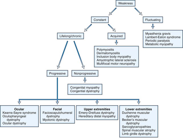
Once the presence of weakness has been established by means of either the history or physical examination, the clinical features may be so characteristic that the diagnosis is obvious. At other times, the cause of the weakness may be less certain. Fig. 25.6 displays an outline of diagnostic considerations based on the characteristics of the weakness, such as whether it is fluctuating or constant. This approach can be used in the differential diagnosis of weakness affecting specific body regions and with other manifestations of weakness, as described next.

Disorders with Prominent Ocular Weakness

In oculopharyngeal muscular dystrophy, slowly progressive weakness of the eye muscles, causing ptosis and external ophthalmoplegia, is associated with difficulty in swallowing. This disorder is inherited as an autosomal dominant condition, with symptom onset usually after the age of 50 years. Many patients also have facial weakness and hip and shoulder weakness. Swallowing difficulty may become severe enough to necessitate cricopharyngeal myotomy or gastrostomy tube placement; however, lifespan in this condition appears to be normal.

Kearns-Sayre syndrome is a distinctive collection of features including ptosis, external ophthalmoplegia, cardiac conduction defects, pigmentary degeneration of the retina, cerebellar ataxia, pyramidal tract signs, short stature, and mental retardation, with symptom onset before age 20 years. These findings accompany an abnormality of the mitochondria in muscle and other tissues. Kearns-Sayre syndrome usually is sporadic. It may be slowly progressive or nonprogressive. Other mitochondrial disorders also may include external ophthalmoplegia as a feature.

In addition, several other disorders may display prominent extraocular muscle involvement. Among these is centronuclear myopathy, one of the congenital myopathies. This condition is not restricted to the eye muscles and has prominent involvement of the limbs as well. External ophthalmoplegia of subacute progressive onset, with or without other bulbar and limb muscle involvement, may occur in variant forms of Guillain-Barré syndrome (i.e., Miller Fisher syndrome) and in botulism. Finally, isolated ptosis or extraocular muscle weakness often is a presenting feature of MG and occasionally of Lambert-Eaton myasthenic syndrome.

Disorders with Distinctive Facial or Bulbar Weakness

The diagnosis of FSH muscular dystrophy may be delayed until early adult life. Weakness of the face may lead to difficulty with whistling or blowing up balloons and may be severe enough to give the face a smooth, unlined appearance with an abnormal pout to the lips (Fig. 25.7, A). Weakness of the muscles around the shoulders is constant, although the deltoid muscle is surprisingly well preserved and even pseudohypertrophic in its lower portion. When the patient attempts to hold the arms extended in front, winging of the scapula occurs that is quite characteristic. The whole scapula may slide upward on the back of the thorax. The inferomedial border always juts backward, producing the appearance of a triangle at right angles to the back, with the base of the triangle still attached to the thorax. In addition, a discrepancy in power often occurs between the wrist flexors, which are strong, and the wrist extensors, which are weak. Similarly, the plantar flexors may be strong, whereas the dorsiflexors of the ankles are weak. It is common for the weakness to be asymmetrical, with one side much less involved than the other (Fig. 25.8). Inheritance of the disorder is as an autosomal dominant trait, although mild forms of the illness often are asymptomatic.

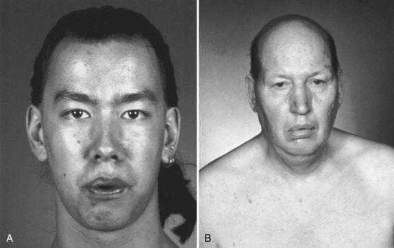
Myotonic dystrophy type I is a common illness with distinctive features including distal predominance of weakness. Inheritance is as an autosomal dominant trait, but often the family history is negative because patients may be unaware that other family members have the illness. This is due to the phenomenon of anticipation, whereby more severe syndromes appear in successive generations because of the expansion of the trinucleotide repeat. This diagnosis is suggested in any patient with muscular dystrophy and predominantly distal weakness. The neck flexors and temporalis and masseter muscles often are wasted. More characteristic than the distribution of the weakness is the long, thin face with hollowed temples, ptosis, and frontal balding (see Fig. 25.7, B). Percussion myotonia and grip myotonia occur in most patients after the age of 13 years. An EMG study can be diagnostic. Muscle biopsy usually is not necessary but may show characteristic changes. Genetic testing is now preferred to muscle biopsy for diagnosis of the disorder and is almost 100% accurate. The genetic defect is an amplified trinucleotide repeat in the 3′ untranslated region of the myotonin–protein kinase gene on chromosome 19.

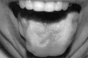
A subset of patients with ALS present with isolated bulbar weakness of LMN type (i.e., progressive bulbar palsy) or UMN type (i.e., progressive pseudobulbar palsy). Frequently the condition shows a combination of UMN and LMN involvement. In these patients, dysarthria, dysphagia, and difficulty with secretions are the prominent symptoms. On examination, the tongue often is atrophic and fasciculating (Fig. 25.9), and the jaw and facial reflexes are exaggerated. The voice often is harsh and strained as well as slurred, reflecting the coexistent UMN and LMN dysfunction. In patients with X-linked spinobulbar muscular atrophy (Kennedy disease), bulbofacial muscles also are prominently affected. Patients often have a characteristic finding of chin fasciculations.

image

Fig. 25.9 Tongue atrophy in a patient with amyotrophic lateral sclerosis.

(Reprinted with permission from Katirji, B., Kaminski, H.J., Preston, D.C., et al., (Eds.), 2002. Neuromuscular Disorders in Clinical Practice. Butterworth Heinemann, Boston.)

Disorders with Prominent Respiratory Weakness

Disorders with prominent respiratory muscle weakness include inherited and acquired myopathies, disorders of the neuromuscular junction or peripheral nerves, motor neuronopathies, and CNS processes involving the brainstem or high cervical spinal cord. Adult-onset acid maltase deficiency (i.e., adult-onset Pompe disease), a glycogen storage disorder, frequently manifests with respiratory system–related symptoms of dyspnea or excessive daytime sleepiness, although proximal muscle weakness is present in most patients. Chronic progressive respiratory weakness occurs in Duchenne muscular dystrophy late in the course. In the intensive care unit (ICU) setting, critical illness myopathy may result in difficulty weaning from a ventilator, although limb muscles also are weak in this condition. Myasthenia gravis occasionally manifests with respiratory failure, although usually myasthenic crisis occurs in patients already known to have myasthenia. Botulism results in respiratory compromise when severe, but the onset usually is stereotypical, with oculobulbar weakness followed by descending weakness, aiding in diagnosis. Guillain-Barré syndrome is a frequent cause of neuromuscular respiratory failure, with subacute onset of ascending weakness and numbness as the most common presentation. ALS leads to respiratory muscle weakness, usually late in the course of the disease. The occasional patient with weakness in the ICU setting, however, is found to have ALS after evaluation for failure to wean from a ventilator. In a patient with limb and respiratory muscle weakness but normal bulbar muscle strength, the possibility of a high cervical cord lesion should be considered.

Disorders with Distinctive Shoulder-Girdle or Arm Weakness

In Emery-Dreifuss muscular dystrophy, clinical features include prominent early contractures of the elbows, posterior neck, and Achilles tendons, with atrophy and weakness of muscles around the shoulders, upper arms, and lower part of the legs. Cardiac conduction abnormalities are common, and acute heart block is a frequent cause of death.

Distal muscular weakness and atrophy are most common in neurogenic disorders. Among these is Charcot-Marie-Tooth disease, which usually manifests with distal weakness and wasting that starts in the distal lower limbs before involving the hands. ALS often begins as weakness and wasting in one distal limb. More important to identify, because it is treatable, is multifocal motor neuropathy with conduction block, a rare demyelinating polyneuropathy that may be confused clinically with ALS with LMN dysfunction. The initial features often are weakness, hyporeflexia, and fasciculations, especially of the hands. Clues to the diagnosis are a slow indolent course, weakness out of proportion to the amount of atrophy, and asymmetrical involvement of muscles of the same myotome but with a different peripheral nerve supply (e.g., weakness of ulnar nerve–innervated C8 muscles out of proportion to weakness of median nerve–innervated C8 muscles). Benign focal amyotrophy, also known as Sobue disease or monomelic amyotrophy, manifests with the insidious onset of weakness and atrophy of the hand and forearm muscles, predominantly in men between the ages of 18 and 22 years.

Distal muscular dystrophies that may manifest with upper-extremity complaints include myotonic dystrophy, discussed earlier, and Welander myopathy, a hereditary distal myopathy. Welander myopathy, transmitted as an autosomal dominant trait, has a predilection for the finger and wrist extensor muscles. Other hereditary distal myopathies typically present first in the lower extremities.

Insidious onset of weakness of the finger flexors with relative preservation of finger extensor strength is common in inclusion-body myositis, a condition that generally manifests after the age of 50 years. In patients with this disorder, however, weakness is also prominent in the lower extremities, especially the quadriceps.

Disorders with Prominent Hip-Girdle or Leg Weakness

Although patients with these disorders often have diffuse weakness including arm and shoulder-girdle weakness, hip and leg weakness brings them to medical attention.

The SMAs are hereditary neuronopathies manifesting with prominent proximal weakness. The atrophy results from the death of anterior horn cells in the spinal cord. This condition spares extraocular muscles, and reflexes are absent. The classification of the SMAs is by age at onset and severity; most forms share a defect in the survival motor neuron gene on chromosome 5q and are of autosomal recessive inheritance. Acute infantile SMA (Werdnig-Hoffmann disease) is a severe and usually fatal illness characterized by marked weakness of the limbs and respiratory muscles. Children with the intermediate form of SMA (chronic Werdnig-Hoffmann disease or spinal muscular atrophy type 2) also have severe weakness, rarely maintaining the ability to walk for more than a few years. The progression of the illness is not steady. The condition may plateau for some years, with periods of more rapid deterioration. Scoliosis is common. A fine tremor of the outstretched hands is characteristic. The chronic juvenile form of SMA (Kugelberg-Welander syndrome) begins sometime during the first decade of life, and patients walk well into the second decade or even into early adult life. Scoliosis is less common than in the infantile form. This condition is consistent with a normal lifespan. Finally, adult-onset SMA leads to slowly progressive proximal muscle weakness after the age of 20 years.

The inherited muscular dystrophies cause progressive, nonfluctuating weakness. Aside from the inherited distal muscular dystrophies discussed earlier in the chapter, other muscular dystrophies manifest with proximal muscle weakness. Duchenne muscular dystrophy, inherited as an X-linked recessive trait, is associated with an absence of dystrophin. Clinically, the combination of proximal weakness in a male child with hypertrophic calf muscles and contractures of the Achilles tendons gives the clue to the diagnosis. The serum CK concentration is markedly elevated. Although muscle biopsy is diagnostic, genetic testing is now preferred to confirm the diagnosis (see Chapter 40). The clinical features of Becker muscular dystrophy are identical except for later onset and slower progression. Cardiomyopathy also is a feature. Female carriers of the gene usually are free of symptoms but may present with limb-girdle distribution weakness or cardiomyopathy.

The limb-girdle dystrophies constitute a well-accepted diagnostic classification despite their clinical and genetic heterogeneity. Weakness begins in the hips, shoulders, or both and spreads gradually to involve the rest of the limbs and the trunk. Knowledge concerning the genetics of these disorders recently expanded (see Chapter 40), and genetic testing is now available for some limb-girdle dystrophies.

Severe early-onset limb-girdle dystrophy similar in phenotype to Duchenne muscular dystrophy, including calf hypertrophy, occurs in the sarcoglycanopathies. The cause is a deficiency in one of the dystrophin-associated glycoproteins (sarcoglycans α, β, γ, and δ). The inheritance pattern in these disorders is autosomal recessive, not X-linked, and the sarcoglycanopathies affect both genders equally. Cardiac involvement is rare, and mental retardation is not part of the phenotype. Another cause of a severe Duchenne-like phenotype is mutation of the FKRP gene, also inherited in an autosomal recessive manner.

With less severe limb-girdle phenotypes, several genetic causes have been recognized, and inheritance is both autosomal recessive and autosomal dominant. In general, the phenotype in the autosomal recessive group is clinically more severe, with earlier onset of weakness and more rapid progression.

Diagnostic evaluation of limb-girdle muscular dystrophies is rapidly evolving and covered in greater depth in Chapter 79. Genetic testing for dystrophin, sarcoglycans, and other genes may be appropriate before performance of muscle biopsy. If the appropriate genetic tests are uninformative, then muscle biopsy is indicated. The biopsy specimen will show dystrophic changes, separating limb-girdle dystrophy from other (inflammatory) myopathies and from denervating diseases such as SMA. Immunohistochemical analysis of dystrophic muscle may provide a specific diagnosis, but not in all cases. Unfortunately, many patients with limb-girdle muscular dystrophies do not receive a specific diagnosis.

With the exception of Welander myopathy, predominantly lower-extremity weakness is the usual presentation of hereditary distal myopathies. Among these disorders are the Markesbery-Griggs-Udd, Nonaka, and Laing myopathies, which affect anterior compartment muscles in the leg, and Miyoshi myopathy, which affects predominantly the posterior calf muscles.

In patients with inclusion-body myositis, the quadriceps and forearm finger flexor muscles often are preferentially involved. In some patients, this involvement may be asymmetrical at the onset. The other inflammatory myopathies—polymyositis and dermatomyositis—affect proximal, predominantly hip-girdle muscles in a symmetrical fashion. Although rare, the Lambert-Eaton myasthenic syndrome manifests with proximal lower-extremity weakness in more than half of patients, similar to a myopathy. Hyporeflexia and autonomic and sensory symptoms may suggest the diagnosis. EMG often is diagnostic.

Ascending weakness of subacute onset with hyporeflexia, usually with numbness, is the hallmark of Guillain-Barré syndrome. The examiner should take care to look for a spinal sensory level and UMN signs, because a spinal cord lesion can mimic this presentation. When present, bulbar weakness is helpful in the diagnosis. Respiratory weakness may result. As discussed earlier, multiple neuromuscular causes of weakness of subacute onset with respiratory failure are recognized.

Distal muscle weakness and atrophy are the hallmarks of neurogenic disorders. In both the demyelinating and axonal forms of Charcot-Marie-Tooth disease, the problem in the legs antedates that in the hands. In ALS, the weakness often is asymmetrical and may combine with UMN signs.

Disorders with Fluctuating Weakness

An important consideration in the differential diagnosis is whether the weakness is constant or fluctuating. Even constant weakness may vary somewhat in degree, depending on how the patient feels. It is well recognized that an individual’s physical performance is better on days when they feel energetic and cheerful and is less optimal on days when they feel depressed or are sick. Such factors also can be expected to affect the patient with neuromuscular weakness. The examiner should make specific inquiries to determine how much variability exists. Does the strength fluctuation relate to exercise or time of day? Symptoms and signs provoked by exercise imply a disorder in the physiological or biochemical mechanisms governing muscle contraction. Pain, contractures, and weakness after exercise often are characteristic of abnormalities in the biochemistry of muscle contraction. Pathological fatigue is the hallmark of neuromuscular junction abnormalities.

Factors other than exercise may result in worsening or improvement of the disease. Some patients notice that fasting, carbohydrate loading, or other dietary manipulations make a difference in their symptoms. Such details may provide a clue to underlying metabolic problems. Patients with a defect in lipid-based energy metabolism are weaker in the fasting state and may carry a candy bar or sugar with them. The patient with hypokalemic periodic paralysis may notice that inactivity after a high-carbohydrate meal precipitates an attack.

The usual cause of weakness that fluctuates markedly on a day-to-day basis or within a space of several hours is a defect in neuromuscular transmission or a metabolic abnormality (e.g., periodic paralysis), rather than one of the muscular dystrophies. Most neurologists recognize that the cardinal features of MG are ptosis, ophthalmoparesis, dysarthria, dysphagia, and proximal weakness (see Chapter 78). On clinical examination, the hallmark of MG is pathological muscle fatigue. Normal muscles fatigue if exercised sufficiently, but in MG, fatigue occurs with little effort. Failure of neuromuscular transmission may prevent holding the arms in an outstretched position for more than a few seconds or maintenance of sustained upgaze. Frequently the patient is relatively normal in the office, making the diagnosis of myasthenia more difficult; the history and ancillary studies (assay for acetylcholine receptor antibodies, anti-MuSK antibodies, and EMG with repetitive stimulation or single-fiber EMG) must be relied on to establish the diagnosis.

In the Lambert-Eaton myasthenic syndrome, fluctuating weakness also may occur, but the fluctuating character is less marked than in MG. Weakness of the shoulder and especially the hip girdle predominates, with the bulbar, ocular, and respiratory muscles relatively spared. Exceptions to this latter rule have been recognized, and some presentations of Lambert-Eaton myasthenic syndrome mimic MG. Typically, reflexes are reduced or absent at rest. After a brief period of exercise, weakness and reflexes often are improved (facilitation), which is the opposite of the situation in MG. The electrophysiological correlate of this phenomenon is the demonstration of a marked incremental response to rapid, repetitive nerve stimulation or brief exercise. The underlying pathophysiology of Lambert-Eaton myasthenic syndrome is an autoimmune or paraneoplastic process mediated by anti–voltage-gated calcium channel antibodies; commercial testing for these antibodies is available.

Patients with periodic paralysis note attacks of weakness, typically provoked by rest after exercise (see Chapter 78). Inheritance of the primary periodic paralyses is as an autosomal dominant trait secondary to a sodium or calcium channel defect (see Chapter 64). In the hyperkalemic (sodium channel) form, patients experience weakness that may last from minutes to days; beginning in infancy to early childhood, the provocation is by rest after exercise or potassium ingestion. Potassium levels generally are high during an attack. In the hypokalemic (calcium channel) form, weakness may last hours to days, is quite severe beginning in the early teens, manifests more in males than in females, and the provocation is by rest after exercise or carbohydrate ingestion. Potassium levels generally are low during an attack.

Secondary hypokalemic periodic paralysis occurs in a subset of patients with thyrotoxicosis. The syndrome is clinically identical to primary hypokalemic periodic paralysis, except for the age at presentation, which usually is in adulthood. In both types of primary periodic paralysis, paralysis may be total, but with sparing of bulbofacial muscles. Respiratory muscle paralysis is rare in hypokalemic periodic paralysis. Patients with paramyotonia congenita also may experience attacks of weakness, especially in the cold. EMG with special protocols for exercise and cooling may be diagnostic; genetic testing also is available for these disorders.

Disorders Exacerbated by Exercise

Fatigue and muscle pain provoked by exercise, the most common complaints in patients presenting to the muscle clinic, often are unexplained. Diagnoses such as fibromyalgia (see Chapter 26) may confound the examination. Biochemical defects are being detected in an increasing number of patients with exercise-induced fatigue and myalgia. The metabolic abnormalities that impede exercise are disorders of carbohydrate metabolism, lipid metabolism, and mitochondrial function (see Chapter 79). The patient’s history may give some clue to the type of defect.

Fatty acids provide the main source of energy for resting muscle. Initiation of vigorous exercise requires the use of intracellular stores of energy because bloodborne metabolites initially are inadequate. It takes time for the cardiac output to increase, for capillaries to dilate, and for the blood supply to muscle to be increased, and an even longer time to mobilize fat stores in the body in order to increase the level of fatty acids in the blood. Because muscle must use its glycogen stores for energy in this initial phase of heavy exercise, defects of glycogen metabolism cause fatigue and muscle pain in the first few minutes of exercise. As exercise continues, the blood supply increases, resulting in an increased supply of oxygen, glucose, and fatty acids. After 10 to 15 minutes, the muscle begins to use a mixture of fat and carbohydrate. The use of carbohydrate is not tolerated for long periods, however, because it would deplete the body’s glycogen stores, potentially resulting in hypoglycemia. After 30 to 40 minutes of continued endurance exercise, the muscle is using chiefly fatty acids as an energy source. Patients with defective fatty acid metabolism easily tolerate the initial phase of exercise. With endurance exercise lasting 30 to 60 minutes, however, they may become incapacitated. Similarly, in the fasting state, the body is more dependent on fatty acids, which it uses to conserve glucose. Thus, the patient with a disorder of fatty acid metabolism may complain of increased symptoms when exercising in the fasting state. Ingestion of a candy bar may give some relief because this quickly boosts the blood glucose level. Patients with fatty acid metabolism defects often have well-developed muscles, because they prefer relatively intense, brief, power exercise such as weight lifting.

Disorders of mitochondrial metabolism vary in presentation. In some types, recurrent encephalopathic episodes occur, often noted in early childhood and resembling Reye disease (see Chapter 63). In other types, particular weakness of the extraocular and skeletal muscles is a presenting feature. In still other types, usually affecting young adults, the symptoms are predominantly of exercise intolerance. Defects occur in the electron transport system or cytochrome chain that uncouples oxygen consumption from the useful production of adenosine triphosphate (ATP). The resulting limit on available ATP causes metabolic pathways to operate at their maximum with even a light exercise load. Resting tachycardia, high lactic acid levels in the blood, excessive sweating, and other indications of hypermetabolism may be noted. This clinical picture may lead to an erroneous diagnosis of hyperthyroidism. It is essential always to measure the serum lactic acid concentration when a mitochondrial myopathy is suspected, even though the level is normal in some patients. In addition to lactate, ammonia and hypoxanthine concentrations also may be elevated.

Patients with suspected metabolic defects should undergo forearm exercise testing. A blood pressure cuff should not be used for the ischemic portion of the test, because this may be hazardous in patients with defects in the glycolytic pathway.

Acquired Disorders Causing Weakness

The usual acquired disorders that produce weakness are motor neuron diseases; inflammatory, toxic, or endocrine disorders of muscle; neuromuscular transmission disorders; and peripheral neuropathies with predominantly motor involvement. The first task is to determine whether the weakness is neuropathic, myopathic, or secondary to a neuromuscular transmission defect. In some instances this is straightforward, and in others it is very difficult. For instance, some cases of motor neuron disease with predominantly LMN dysfunction may mimic inclusion-body myositis, and Lambert-Eaton myasthenic syndrome may mimic polymyositis. If fasciculations are present, the disorder must be neuropathic. If reflexes are absent and muscle bulk is preserved, suspect a demyelinating neuropathy, although presynaptic neuromuscular junction disorders (e.g., Lambert-Eaton myasthenic syndrome) also show hyporeflexia with normal muscle bulk. The presence of sensory signs or symptoms, even if mild, may indicate a peripheral neuropathy or involvement of the CNS. Often, separating these conditions requires serum CK testing, EMG, and muscle biopsy.

ALS is the most common acquired motor neuron disease. Although peak age at onset is from 65 to 70 years, the disorder can occur at any adult age. It often follows a relatively rapid course preceded by cramps and fasciculations. Examination shows muscle atrophy and often widely distributed fasciculations. If the bulbar muscles are involved, difficulties with swallowing and speaking also are present. The diagnosis is relatively simple if unequivocal evidence of UMN dysfunction accompanies muscle atrophy and fasciculations. UMN signs include slowness of movement, hyperreflexia, Babinski sign, and spasticity. A weak, atrophic muscle associated with an abnormally brisk reflex is almost pathognomonic for ALS. The finding of widespread denervation on needle electrode examination in the absence of any sensory abnormalities or demyelinating features on nerve conduction testing supports the diagnosis. In all patients without bulbar involvement, it is important to rule out spinal pathology, because the combination of cervical and lumbar stenosis occasionally may mimic ALS with respect to clinical and electrophysiological findings.

In patients with only LMN dysfunction, it is essential to exclude the rare diagnosis of multifocal motor neuropathy with conduction block, a condition usually treatable with intravenous gamma globulin. Patients with multifocal motor neuropathy with conduction block usually have no bulbar features or UMN signs, and a characteristic finding includes demyelination (i.e., conduction block) on motor nerve conduction testing. Because the underlying pathophysiological process is conduction block, weakness usually is more severe than expected for the observed degree of atrophy. However, atrophy occurs, especially when the condition is of long duration.

Although most adults with motor neuron disease have ALS or one of its variants, sporadic forms of adult-onset SMA and especially X-linked spinobulbar muscular atrophy (Kennedy disease) can occur as well. In these cases, the progression of weakness is much slower, and UMN involvement is absent. Of importance, these latter cases, especially Kennedy disease, often have elevated CK levels in the range of 500 to 1500 U/liter.

If the patient has a myopathy, acquired and inherited causes should be considered. A discussion of the presentation of inherited myopathic disorders appears earlier in the chapter. Causes of acquired myopathies include inflammatory conditions and a large number of toxic, drug-induced, and endocrine disorders. Inflammatory myopathies include polymyositis, dermatomyositis, and inclusion-body myositis and often run a steadily progressive course, although some fluctuations occur, particularly in children. Onset of weakness in polymyositis and dermatomyositis is subacute, weakness is proximal, and serum CK levels usually are increased. If an associated rash is present, little doubt exists about the diagnosis of dermatomyositis. In its absence, polymyositis may be difficult to differentiate from any of the other causes of proximal weakness. Sometimes the illness occurs as part of an overlap syndrome in which fragments of other autoimmune diseases (e.g., scleroderma, lupus, rheumatoid arthritis) are involved. Polymyositis sometimes is difficult to differentiate from a muscular dystrophy, even after muscle biopsy; some inflammatory changes occur in muscular dystrophies, most notably in FSH muscular dystrophy. Other signs of systemic involvement such as malaise, transitory aching pains, mood changes, and loss of appetite are more common in polymyositis than in limb-girdle dystrophy.

Inclusion-body myopathy typically has a chronic, insidious onset. It occasionally mimics polymyositis but more often mimics ALS associated with LMN dysfunction. Clues to the diagnosis are male gender, onset after the age of 50 years in most patients, slower progression, and characteristic involvement of the quadriceps and long finger flexors. Some patients may have proximal muscle weakness, as in polymyositis, whereas others may have predominantly distal weakness mimicking that of ALS and other neuropathic conditions. Serum CK generally is elevated but occasionally may be normal. As with other chronic inflammatory myopathies, interpreting the EMG study may be difficult and requires an experienced examiner, because inclusion-body myopathy often shows a combination of myopathic and neuropathic features. Inclusion-body myopathy, unlike polymyositis, often is unresponsive to immunosuppressive therapy. Pathological features include rimmed vacuoles and intracytoplasmic and intranuclear filamentous inclusions.

Toxic, drug-induced, and endocrine disorders are always considerations in the differential diagnosis for acquired myopathies. Among toxins, alcohol is still one of the most common and may produce both an acute and a chronic myopathic syndrome. Several prescription medicines are associated with myopathies. Most prominent are corticosteroids, cholesterol-lowering agents (i.e., statins), and colchicine.

Although neuromuscular transmission disorders are always diagnostic considerations in patients with fluctuating symptoms, the Lambert-Eaton myasthenic syndrome may be an exception. It often manifests with progressive proximal lower-extremity weakness without fluctuations. Clues to the diagnosis include a history of cancer, especially small-cell lung cancer (although in many patients the myasthenic syndrome may predate the discovery of the cancer), hyporeflexia, facilitation of strength and reflexes after brief exercise, and coexistent autonomic symptoms, especially urinary and sexual dysfunction in men.

Sensory features separate peripheral neuropathies from disorders of the motor unit. The notable exception is multifocal motor neuropathy with conduction block, discussed earlier. Other neuropathies also may manifest with predominantly motor symptoms. Among these are toxic neuropathies (from dapsone, vincristine, or lead, or an acute alcohol-related neuropathy) and some variants of Guillain-Barré syndrome (especially the acute motor axonal neuropathy syndrome).

Lifelong Disorders

Most patients presenting to the neuromuscular clinic will have lifelong or at least very chronic, presumably inherited, disorders. These include inherited disorders of muscle (e.g., dystrophies, congenital myopathies), anterior horn cell (e.g., spinal muscular atrophies), peripheral nerves (e.g., Charcot-Marie-Tooth polyneuropathy), or very rarely, neuromuscular transmission (e.g., congenital myasthenic syndromes). In some of these disorders, the responsible genetic abnormality has been identified. An important point in the differential diagnosis is to determine whether the weakness is truly progressive. The examiner should ask questions until the progressive or nonprogressive nature of the disease is ascertained. The severity of the disease is not proof of progression. It is difficult to imagine that a 16-year-old girl confined to her wheelchair with spinal muscular atrophy and scoliosis and having difficulty breathing has a relatively nonprogressive disorder, but careful questioning may reveal no loss of function for several years. Furthermore, it is not sufficient to ask the patient in vague and general terms whether the illness is progressive. Questioning should be specific; for example, “Are there tasks you cannot perform now that you could perform last week (month, year)?” The examiner also must be alert for denial, which is common in young patients with increasing weakness. The 18-year-old boy with limb-girdle dystrophy may claim to be the same now as in years gone by, but questioning may reveal that he was able to climb stairs well when he was in high school, whereas he now needs assistance in college.

Lifelong Nonprogressive Disorders

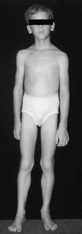
Some patients complain of lifelong weakness that has been relatively unchanged over many years. Almost by definition, such disorders have to start in early childhood. Nonprogression of weakness does not preclude severe weakness. Later-life progression of such weakness may occur as the normal aging process further weakens muscles that have little functional reserve. One major group of such illnesses is the congenital nonprogressive myopathies, including central core disease, nemaline myopathy, and congenital fiber-type disproportion. The typical clinical picture in these diseases is that of a slender dysmorphic patient with diffuse weakness (Fig. 25.10). Other features may include skeletal abnormalities such as high-arched palate, pes cavus, and scoliosis, which are supportive of the presence of weakness in early life. Deep tendon reflexes are depressed or absent. Though unusual, severe respiratory involvement may occur in all these diseases. The less severe (non-X-linked) form of myotubular (centronuclear) myopathy is suggested by findings of ptosis, extraocular muscle weakness, and facial diplegia. Muscle biopsy usually provides a specific morphological diagnosis in the congenital myopathies; specific genetic testing is now available for some congenital myopathies.

Several varieties of congenital muscular dystrophy (CMD) are recognized. The weakness in CMD manifests in the newborn period, with the affected child presenting as a floppy baby. Arthrogryposis may be present. The current classification is based on the presence or absence of associated structural CNS abnormalities. Such disorders without CNS defects constitute classic CMD. These disorders are subdivided into merosin-positive and merosin-negative disorders. Patients with classic CMD who are merosin-positive have the mildest phenotype. The disorders with CNS structural abnormalities are very severe; for example, characteristics of Fukuyama CMD include microcephaly, mental retardation, and seizures with severe disability. The serum CK concentration may be markedly elevated in CMDs. The muscle biopsy specimen shows dystrophic changes, and immunohistochemistry often provides a specific diagnosis.