Proliferative Diabetic Retinopathy
Clinical Features:
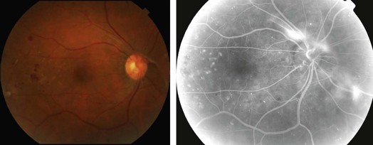
Retinal neovascularization is a hallmark of PDR, which can be seen at the slit lamp as fine networks of blood vessels extending from the retina into the vitreous cavity (Fig. 13.3.1). These vessels can cause visual loss secondary to vitreous hemorrhage, and can induce preretinal fibrosis leading to tractional retinal detachment, retinoschisis, macular edema and combined traction/rhegmatogenous RD
OCT Features:
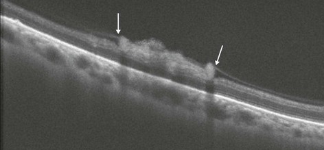
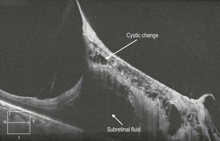
The typical findings of NPDR are seen in PDR as well. In addition, NVD and NVE may manifest as loops of hyper-reflective blood vessels projecting from the retina into the vitreous, either at the disc or elsewhere (Fig. 13.3.2). In contrast, areas of IRMA are seen as disorganization of the inner retinal vascular architecture with occasional projection beyond the internal limiting membrane, but with no disruption of the hyaloid face (Fig. 13.3.3). The hyaloid may be thickened in these cases. In some cases with NVD or NVE, traction of the retina with or without retinal detachment may be seen (Fig. 13.3.4).
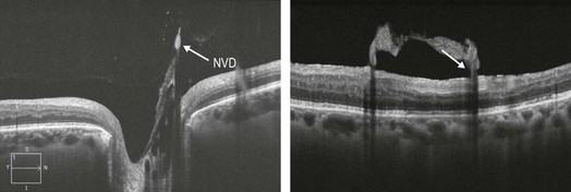
Figure 13.3.2 OCT section through the area of the neovascularization of the optic disc reveals hyper-reflective neovascularization into the vitreous cavity (arrow). The adjacent picture shows a high resolution OCT scan through an area of NVE.