Principles of Toxicology

Published on 08/02/2015 by admin

Filed under Basic Science

Last modified 22/04/2025

Print this page

rate 1 star rate 2 star rate 3 star rate 4 star rate 5 star
Your rating: none, Average: 0 (0 votes)

This article have been viewed 2121 times

Chapter 8 Principles of Toxicology

Abbreviations
ATP Adenosine triphosphate
CNS Central nervous system
CO Carbon monoxide
DNA Deoxyribonucleic acid
EDTA Ethylenediamine tetraacetic acid
GI Gastrointestinal

Therapeutic Overview

Industrial chemical and pharmaceutical development and environmental contamination present significant health hazards to the general population. It is estimated that each year in the United States approximately 8 million people suffer acute poisoning, accounting for as much as 10% to 20% of hospital admissions. In addition to acute incidences, chronic toxicity resulting from long-term exposure to very low doses of environmental or occupational chemicals presents a larger problem, because it is often difficult to identify detrimental effects, which may take years to develop.

Any substance injurious to humans may be classified as a poison. Sodium chloride, oxygen, and many other substances generally considered nontoxic can be dangerous under certain conditions. The most important axiom of toxicology is that “the dose makes the poison,” because any chemical can be toxic if the dose or exposure is high enough. Similarly, the degree of injury increases as the dose increases.

The terms poison, toxic substance, toxic chemical, and toxicant are synonymous. Toxicity refers to the adverse effects manifested by an organism in response to a substance. In the assessment of toxicity in humans, it is important to understand how the dose of a chemical alters biochemical and physiological processes, and at which dose toxicity is manifest.

The time between ingestion of or exposure to a chemical and the onset of its deleterious effects can vary considerably. Generally, if a toxic response results from a single dose or exposure, it is considered acute toxicity. Subacute or subchronic toxicity usually refers to that occurring after several days to weeks of exposure, and chronic toxicity refers to that occurring after months to years. Most acutely toxic agents have an immediate effect on critical cellular processes. For example, cyanide causes immediate injury by inhibiting cellular respiration. In contrast, peripheral neuropathy that may be manifest after exposure to certain organophosphate insecticides takes months or longer to develop.

Mechanisms of Action

A chemical causing direct toxicity injures a cell after coming in direct contact with it. Chemicals causing indirect toxicity do so by injuring one group of cells, which precipitates injury in others. Interference with normal physiological processes may injure cells dependent on that process, while other processes are able to repair tissues damaged by toxicants.

Some substances, such as strong acids or bases, act locally. Most other toxic substances produce systemic effects or a combination of local and systemic effects. For example, hydrofluoric acid, a common industrial chemical, produces an extremely painful local penetrating injury at the site of skin exposure. In larger exposures, enough fluoride ions enter the blood to bind Ca++ and produce hypocalcemia.

Many toxic substances are known primarily to affect a specific target organ, such as the liver, nervous system, lungs, or kidneys, whereas others target the immune system or may affect normal fetal and cell growth and development. It is important to note, however, that although certain anatomical or functional characteristics may predispose organs to injury, designation of toxicants as “organ specific” (e.g., neurotoxic, cardiotoxic) is somewhat misleading, for several reasons. First, toxic substances may have adverse effects throughout the body, and although a chemical may primarily affect one organ, other organs may also incur less notable injury. Second, a toxic response in one tissue will undoubtedly have serious consequences for other tissues depending on the characteristics of the chemical (physical state, site of exposure, dose, and duration of exposure) and of the patient (general health, nutritional state, age, sex, enzyme induction, immune response, antioxidant concentrations). Thus a minor insult to a compromised organ may cause unexpected injury to that organ and may also affect other organs.

Hepatic Toxicity

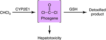
Because of a dual blood supply from the hepatic artery and the portal vein, the liver may be exposed to toxicants entering from the systemic circulation (e.g., via inhalation) or from the splanchnic circulation (absorbed from the gastrointestinal [GI] tract). The leaky capillary system of the hepatic sinusoids promotes the extraction of toxicants from the blood into the liver. The liver contains enzymes that metabolize many endogenous and exogenous chemicals (see Chapter 2). In some cases chemical modification produced by biotransformation results in bioactivation (production of toxic-reactive metabolites). This is exemplified by the bioactivation of chloroform to phosgene (Fig. 8-1). Compounds that enhance the activity of CYP2E1 (enzyme-inducing agents) or reduce the hepatic concentration of glutathione can exacerbate chloroform-induced liver injury. Other compounds that can cause liver injury after metabolism include several solvents (carbon tetrachloride, halogenated benzenes) and carcinogens (aflatoxin B, aromatic amines).

Pulmonary Toxicity

Injury to the lung results primarily from respiratory exposure to toxicants, although blood-borne compounds may also contribute. The pulmonary system is composed of the nasopharyngeal and tracheobronchial airways and the pulmonary parenchyma (alveoli). Airways are directly exposed to many gaseous or particulate toxicants. Gases may react with airway mucosal cells or penetrate to the alveoli. For example, chlorine gas reacts with upper airway fluids and produces hydrochloric acid, causing mucosal injury. Cyanide and chloroform penetrate to the alveoli, are well absorbed, and cause systemic, not pulmonary, toxicity. Particulates can also become impacted in the airway or, if small enough (less than 5 μm), can reach the alveoli. If trapped in the airway, they may be cleared by the mucociliary apparatus. Substances reaching the alveoli can be eliminated only by absorption into the blood, by macrophage phagocytosis, or by biotransformation. Macrophages phagocytose toxicants in particulate form and then enzymatically degrade them or simply transport them out of the alveolus. This function may actually underlie asbestos toxicity, because macrophages engulf but cannot degrade asbestos particles. When the macrophages ultimately die, they release degradative enzymes into the interstitium, which damage adjacent cells. Repetition of this process eventually leads to progressive fibrosis and restrictive respiratory dysfunction. The pulmonary biotransformation of chemicals may also lead to toxicity. The Clara cells, located in terminal bronchioles, and the alveolar type II cells possess cytochrome P450, which can produce toxic metabolites of certain chemicals. Inhalation of benzo[a]pyrene and other polycyclic aromatic hydrocarbons causes lung cancer by this mechanism.

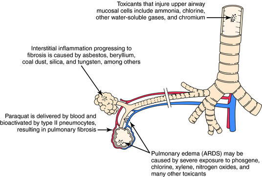
The blood can serve as the route of exposure of the lung in the case of paraquat, a herbicide taken up by type II pneumocytes. Biotransformation in the lung yields a reactive intermediate that undergoes redox cycling, which produces reactive oxygen species that injure the cell. Although exposure to paraquat usually occurs after ingestion or skin contamination, death results from pulmonary injury. Similarly, certain pyrrolidine alkaloids are metabolized in the liver to compounds that circulate in the blood and produce toxicity in the lung. The actions of toxicants on pulmonary tissue are shown in Figure 8-2.

Renal Toxicity

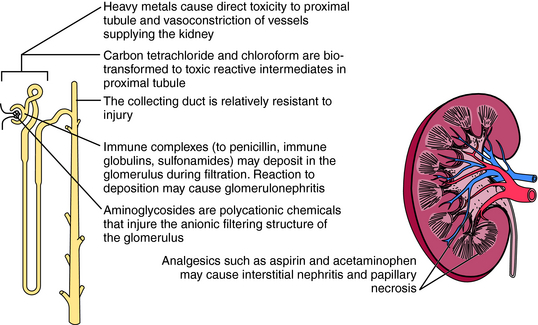
The kidney is also susceptible to toxicants. The mechanisms of renal injury are similar to those in other organs but also include unique mechanisms. Delivery of blood-borne toxicants to the kidney is high, because the kidney receives 25% of cardiac output, and its functions include filtering, concentrating, and eliminating toxicants. As water is reabsorbed, the concentration of chemicals in the tubule can increase to toxic levels. In some cases the concentration may exceed the solubility of a chemical and lead to precipitation and obstruction of the affected area (Fig. 8-3).

The kidney also biotransforms chemicals, although to a lesser extent than the liver. Cytochrome P450 is located in the proximal tubule, which may explain the susceptibility of this region to chemical injury. For example, carbon tetrachloride and chloroform, two halogenated hydrocarbons that injure the proximal tubule, must be biotransformed by the cytochrome P450 system to be toxic. In addition, many heavy metals damage the proximal tubules. Because they are not metabolized by cytochrome P450, other mechanisms must be involved. Heavy metals may concentrate in renal tubular cells and may also injure blood vessels supplying the proximal tubule cells, a combination of direct and indirect toxicity.

The kidney can compensate for excessive chemical exposure because, like many organs, it has a reserve mass. Thus tissue injury equal to one entire kidney must occur before loss of function is clinically apparent. The kidney also replaces lost functional capacity by hypertrophy. In addition, the kidney has developed binding proteins to remove heavy metals. For example, metallothioneins avidly bind cadmium, protecting the kidney and other organs from toxicity. However, renal toxicity will develop if exposure exceeds the binding capacity or if a large amount of the cadmium-metallothionein complex accumulates.

Immunotoxicity

The immune system can be affected by a wide variety of toxicants. These agents often cause immunosuppression by interfering with cell growth or proliferation. Other compounds may directly destroy immune system components. Some chemicals distort normal signaling mechanisms that ultimately reduce the immune response. For example, benzene causes lymphocytopenia but also affects other bone marrow elements. The functional result is a deficiency in cell-mediated immunity. Workers exposed to benzene show decreased humoral immunity, as evidenced by depressed complement and immunoglobulin concentrations.

In humans, immunosuppression may lead to an increased incidence of bacterial, viral, and parasitic infections. Theoretically, immunosuppression may also interfere with the immune system’s surveillance function, resulting in an increased incidence of cancer, although this is still being investigated.

It is becoming increasingly apparent that activation and recruitment of phagocytic cells to sites of chemical-induced injury play a major role in the progression of tissue injury. Therapeutic interventions that could minimize the effects of these activated phagocytic cells include preventing the adhesion of phagocytic cells at the site of injury, reducing the release of cytotoxic factors, or inactivating these cytotoxic factors.

In addition to being a target for chemical-mediated injury, the immune system may mediate injury by producing hypersensitivity reactions—adverse events caused by an immune response to foreign antigens (see Chapter 6). Indeed, cell-mediated hypersensitivity initiated by T lymphocytes may lead to contact dermatitis as a consequence of exposure to nickel and several industrial chemicals.

Cell Toxicity

Cancer cells are cells that escape from the control mechanisms that govern growth, development, and division of normal cells (see Chapter 53). Some human cancers are of environmental origin, caused by radiation, viral infection, or chemical exposure.

Because cancer often develops in rats and mice exposed to extremely large doses of chemicals for long periods, many people believe that synthetic chemicals such as drugs, pesticides, or industrial agents are a primary cause of cancer. In fact, however, most chemicals that produce cancer in laboratory animals after large doses do not produce cancer in humans. Exceptions are vinyl chloride, benzene, and naphthylamine, which can cause cancer in humans after prolonged occupational exposure.

Lifestyle choices also result in significant exposure to chemical carcinogens. For example, cigarette smoke contains many potent cancer-causing chemicals and is believed to be a causative factor of lung cancer. Charcoal broiling contaminates food with polycyclic aromatic hydrocarbons, which are the same as those in coal tars and soot. It is possible that many human cancers could be prevented or delayed by “lifestyle” changes.

Duration, dose, and frequency of exposure are important variables in chemical-induced cancers. Because cancer may take 20 or more years to develop, cause-and-effect relationships are difficult to establish. However, some chemicals interact directly and covalently with deoxyribonucleic acid (DNA), whereas others must be metabolically transformed before they can do so. If cell division occurs before enzymatic repair of the damaged DNA, a permanent mutation is encoded in the genome. Because cellular damage undoubtedly kills some cells in the target tissue, the stimulus for division of adjacent cells is high. The result is a new cell type with altered genotypic and phenotypic properties. Additional mutational events can then convert transformed cells to malignant cells.

Chemicals can also increase the incidence of cancers or decrease the latency for tumor development without interacting with DNA or producing mutations. These agents are referred to as promoters and manifest effects only when administered repeatedly after an initial insult to a cell. Certain carcinogens that have direct effects may also act as promotors, creating an environment conducive to the proliferation of insulted cells and supporting autonomous cell division and tumor growth.

Pharmacokinetics

Generally the means by which a toxicant reaches its target organ is governed by the same pharmacokinetic principles that govern the actions of therapeutic drugs (see Chapter 2). These principles also provide the basis for methods to reduce, reverse, or prevent toxicity of many substances.

Chemicals may be absorbed by dermal, GI, or pulmonary routes, and measures to prevent absorption may reduce the concentration of a toxicant at its site of action. Washing the skin, stomach lavage, and oral administration of charcoal are examples of ways to reduce dermal or GI absorption of toxicants. After absorption, toxic substances are distributed to tissues through the blood, and in some cases it is possible to intercept the toxicant before it reaches its target. This can be accomplished by the following mechanisms:

It is generally too late to prevent cell or organ damage after the toxicant reaches its target, although the effects of many toxicants may still be minimized at this stage. Compounds metabolized to reactive intermediates are good examples. An antidote that prevents biotransformation can minimize injury produced by a reactive intermediate. The competitive antagonism of methanol or ethylene glycol by ethanol is based on this principle (see Chapter 32). Similarly, drugs that inhibit certain cytochrome P450 isozymes can reduce biotransformation of some toxicants, either reducing or enhancing their effects.

Toxic metabolites produced by biotransformation may injure the cell in which they are produced, or they may diffuse in the blood and affect other areas. For a few substances this offers a final opportunity to eliminate toxic metabolites by using hemodialysis to clear the blood.

Another strategy for reducing cellular injury is to prevent the reactive intermediate from interacting with important cell constituents (e.g., enzymes, DNA). Other interventions will be developed as the sequence of events involved in the progression of chemical-induced tissue injury becomes better understood. For example, agents that can consume reactive oxygen species or down regulate an inflammatory response may be effective if administered up to several hours after a drug overdose or chemical exposure.

Relationship of Mechanisms of Action to Chemical Toxicity

Clinical diagnosis and treatment of patients suffering from chemical toxicity is beyond the scope of this book. However, the toxicity of gases and heavy metals, which are most common, are discussed in the following text. In addition, because individuals are often exposed to multiple toxic compounds, particularly in the environment, resulting in synergistic, additive, or antagonistic effects, the concepts of interactive and environmental toxicology are presented.

TOXIC GASES

Among the most toxic gases are carbon monoxide (CO) and volatile cyanides. Both are toxic because they deprive cells of energy. Other gases such as ozone, oxides of nitrogen, and phosgene are toxic because of their chemical reactivity. They are irritating to mucous membranes and may trigger asthma-like symptoms in susceptible people.

Carbon Monoxide

CO is the leading cause of death by poisoning. It also inflicts sublethal injuries, including myocardial infarction and cerebral atrophy. CO acts primarily by displacing oxygen from hemoglobin and impairing oxygen release from hemoglobin. CO displaces oxygen from hemoglobin because it has more than 200 times higher affinity for hemoglobin than oxygen. Even at a concentration in air of only 0.5%, CO displaces oxygen to produce 50% carboxyhemoglobin. However, CO produces more injury than that predicted on the basis of the simple replacement of oxygen. This is explained by the fact that normal hemoglobin binding of oxygen shows cooperativity. Binding of one oxygen molecule promotes binding of subsequent molecules, and hemoglobin also shows cooperativity in releasing oxygen. However, carboxyhemoglobin does not release oxygen normally. CO therefore shifts the oxyhemoglobin dissociation curve to the left and reduces oxygen release, causing enhanced anaerobic metabolism and cell death if not reversed.

The symptoms of CO poisoning reflect the effects of oxygen deprivation. Early symptoms of nervous system dysfunction resemble those of the flu and include nausea, headache, malaise, light-headedness, and dizziness. Later signs and symptoms are more ominous, including depressed sensorium, loss of consciousness, seizures, and death.

The heart is particularly susceptible to CO poisoning. It has high oxygen requirements, and because it normally extracts more oxygen from the blood than other organs, it compensates poorly for decreased delivery. When CO decreases oxygen delivery, severe myocardial ischemia may develop.

As CO dissociates from the hemoglobin, it is expired. However, this is a slow process because of its high affinity for hemoglobin. The t1/2 of carboxyhemoglobin without treatment is 3 to 4 hours, depending on the patient’s ventilation. Administration of 100% oxygen by face mask shortens the time to 90 minutes. Hyperbaric oxygen reduces the t1/2 to 20 minutes. The patient’s outcome depends on the duration and severity of the hypoxic episode.

Cyanide

Exposure to cyanide commonly occurs through the inhalation of smoke produced by the burning of plastics. Certain paints may also contribute. Other potential sources are fruit seeds (e.g., apricots and cherries), which contain toxic amounts of soluble cyanide that must be metabolized by intestinal bacteria to release it. Salts of cyanide have been used in suicide or homicide poisoning.

Cyanide produces toxicity by binding avidly to ferric iron (Fe+++), which prevents its reduction to the ferrous form (Fe++) involved in the cytochrome oxidase electron-transport system. Transfer of electrons from cytochromes to molecular oxygen is prevented, which in turn inhibits adenosine triphosphate (ATP) production and forces the cell to produce energy by anaerobic metabolism. As in all cases of hypoxia, anaerobic glycolysis produces only small amounts of ATP and large quantities of lactic acid.

Thus victims of cyanide poisoning show symptoms of hypoxia. Similar to CO toxicity, cyanide toxicity first affects organs that have a large oxygen requirement. However, onset is often faster but depends on route of exposure. Inhalation may produce a rapid demise, whereas symptoms may be delayed for 30 minutes or more if the cyanide is ingested orally. CNS dysfunction causes loss of consciousness and respiratory arrest.

Treatment is focused on preventing cyanide from reaching its target, cytochrome oxidase. Because cyanide has a high affinity for ferric iron, ferric iron in the form of oxidized hemoglobin can be provided by administration of sodium nitrite. This converts hemoglobin to methemoglobin, the ferric form, which effectively competes with cyanide for cytochrome a3, forming cyanmethemoglobin. Cyanide is removed from the body after being converted to thiocyanate by the enzyme rhodanese and is ultimately excreted in the urine. Sodium thiosulfate is administered to facilitate thiocyanate formation. An alternative is to administer hydroxocobalamin, which reacts with cyanide to produce cyanocobalamin. Because hydroxocobalamin and cyanocobalamin have little toxicity, they hold promise for the treatment of cyanide poisoning.

HEAVY METALS

The widespread occurrence of metals in the environment and their numerous industrial and medical uses make them important potential toxicants. The rate at which heavy metals are absorbed depends on their physical state. Metals may exist in their elemental state or may be bound to inorganic or organic ligands. The elemental and inorganic forms of metals may be well absorbed because of their physical similarity to nutritionally essential metals. For example, lead is absorbed by the normal transport protein for iron located in the GI tract mucosa. Organs containing these transport systems are prone to injury from these metals. Commonly injured organs include liver, kidney, and GI tract mucosa. Organic forms of metals are more lipid soluble than inorganic forms and may be well absorbed without specific transport systems.

The state of the metals also affects their distribution, and lipid-soluble forms reach higher concentrations in areas such as the brain. Inorganic and elemental forms of mercury primarily injure the kidney, whereas organic forms such as methylmercury injure the brain.

The body has developed specific defense mechanisms against certain metals. For example, as noted earlier, the kidney has a binding protein for cadmium called metallothionein, which strongly binds cadmium, concentrates it in the kidney, and reduces its excretion. The binding prevents toxicity until the protein is saturated, at which time additional cadmium accumulation causes injury.

Mechanisms involved in heavy metal toxicity are poorly understood. Many metals function as essential enzyme cofactors. Substitution by a similar but toxic metal may produce enzymatic dysfunction. Metals are generally very reactive and may bind key sulfhydryl groups in active centers of enzymes. Besides causing direct metal toxicity, metals can also produce hypersensitivity reactions. Nickel, chromium, gold, and others cause cell-mediated (type IV) hypersensitivity reactions (Table 8-1).

TABLE 8–1 Mechanisms of Heavy Metal Toxicity

Metal Mechanism Target Organs
Arsenic Reacts with sulfhydryl groups Peripheral neurons
  GI tract
Interferes with oxidative phosphorylation Liver
    Cardiovascular system
Lead Reacts with sulfhydryl groups Hematopoietic system
  Central and peripheral nervous systems
Interferes with heme synthesis Kidney
Direct toxic effect Central nervous system
Mercury Reacts with sulfhydryl groups Central and peripheral nervous systems
Some forms have direct cytotoxic effects Kidney
  GI tract
  Respiratory tract

Treatment for metal toxicity focuses on increasing excretion of the metal from the body. Chelator drugs bind the metal between two or more functional groups to form a complex that is excreted in urine. Specific chelators work best in the removal of certain metals. The uses of chelators are summarized in Table 8-2.

TABLE 8–2 Metals Chelated by Therapeutic Agents

Chelator Metal
Succimer Lead, arsenic, mercury
Deferoxamine Iron
EDTA Lead
Penicillamine Copper, lead
British anti-Lewisite Lead, arsenic, mercury

EDTA, Ethylenediamine tetraacetic acid.

Lead

Lead is one of the oldest known poisons but continues to pose a significant health problem. Industrialization, mining, and leaded gasoline have dramatically increased the amount of lead in the environment and consequently in humans. However, the switch to unleaded gasoline has resulted in decreases in environmental lead contamination. Lead is also present in some ceramic glazes and paints. In older homes, paint containing lead flakes off or is present in dust and may be ingested or inhaled by children, producing toxicity. Adults are exposed to toxic concentrations in certain work environments.

Lead binds to sulfhydryl and other active sites in many enzymes, leading to inactivation. Although its effects are diffuse, certain manifestations predominate. Two enzymes in the heme biosynthetic pathway are inhibited by lead, and inhibition of heme synthesis can result in anemia.

Lead is particularly toxic to the nervous system, especially in children. Early signs and symptoms include anorexia, colicky abdominal pain, lethargy, and vomiting. If lead exposure continues, children are more likely than adults to develop encephalopathy, manifested by irritability progressing to seizures and coma. Approximately 30% will develop permanent neurological sequelae.

Low-concentration lead exposure may pose special risks for children. Subtle neurological injuries including depressed IQ scores and learning disorders have been reported. This may be due to enhanced accumulation in the immature nervous system. Lead may cross the blood-brain barrier more easily in children, and their CNS may also be less capable of removing it. Children with blood lead concentrations exceeding 10 µg/dL are considered to be at risk.

Vague complaints of headache and lightheadedness develop in adults exposed to lead. With increased exposure, a peripheral neuropathy develops.

The whole-blood lead concentration is the best indicator of exposure in the patient with lead-poisoning signs and symptoms. As the concentrations increase, the danger of encephalopathy increases, although overt signs and symptoms do not usually occur until lead concentrations approach 50 µg/dL.

Lead is slowly excreted from the body. The primary treatment of lead poisoning is removal from the source. Excretion may be hastened by the use of chelators such as British anti-Lewisite.

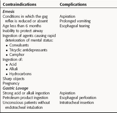
A summary of some common antidotes for several types of chemical toxicants is provided in Table 8-3. In addition, because emesis and gastric lavage are often used for the treatment of toxicity, the main contraindications and problems associated with these procedures are presented in Table 8-4.

TABLE 8–3 Common Antidotes

Toxicant Antidote Mechanism
Cyanide Cyanide kit (sodium nitrite, sodium thiosulfate) Induction of methemoglobinemia; cyanide binds preferentially to methemoglobin
Metals Chelators Binding of metal with subsequent urinary excretion
Methanol, ethylene glycol Ethanol Competition for alcohol dehydrogenase
CO O2 Displaces CO molecules from carboxyhemoglobin

TABLE 8–4 Contraindications and Complications of Emesis and Gastric Lavage

image

ENVIRONMENTAL TOXICOLOGY

Chemical contaminants in the environment (soil, water, food, indoor/outdoor air) are a hazard to human health. Petroleum mixtures contain benzene, a known human carcinogen commonly found in air and water. Agricultural chemicals, industrial waste, and incineration by-products also contribute pollutants such as pesticides, polychlorinated biphenyls, dioxins, and polyaromatic hydrocarbons. Naturally occurring chemicals such as arsenic (found in drinking water), methylmercury (found in fish), and aflatoxin B (produced by a fungus that grows on corn and peanuts) are examples. Chronic exposure of humans to high doses over a lifetime is likely to produce adverse health effects.

Chemical Exposure and Health Outcomes

Exposure to environmental toxicants may occur in several ways and under different circumstances. Catastrophic accidents producing immediate morbidity and mortality are infrequent. In 1984 an explosion in a pesticide factory produced more than 2000 deaths and 10,000 injuries in Bhopal, India. High concentrations of methylisocyanate, a chemically reactive gas, were released in an urban setting and caused immediate lung damage and death in both workers and residents of the city.

Continual exposure over a long period of time can also result in accumulation of chemicals to toxic levels. Accumulation may result from slow elimination of the harmful chemical, a lack of cellular repair mechanisms, or both. This can contribute to chronic diseases such as cancer, heart disease, and neurodegenerative disorders. Well-documented examples include the multiple health threats from tobacco use and exposure to second-hand smoke, the development of rare hepatic angiosarcomas in factory workers exposed to vinyl chloride, mesothelioma as a result of asbestos exposure, and male infertility caused by occupational exposure to the fumigant 1,2-dibromo-3-chloropropane. The current incidence of these cases is relatively low due to environmental advocacy, governmental regulation, and improved manufacturing and waste-handling processes.

Associating chemical exposure with a particular disease or set of symptoms is problematic. The low-dose environmental exposure situation often creates a dilemma, because a causal relationship often cannot be made with adequate certainty, confounding diagnosis, treatment, and subsequent remedial action to inhibit further exposure.

Estimation of Health Risks

A lower incidence or even an absence of harmful effects from hazardous chemicals is expected if exposure is low enough. Strategies for prevention of toxic effects require knowledge of the threshold dose above which chemical toxicity will likely occur. Unfortunately, severe difficulties are involved in determining the toxicological threshold exposure for a particular chemical in humans. Data from laboratory animal studies are usually available, but species differences make extrapolation of results to humans uncertain. Differences among individuals (age, health status, genetics) also make selection of a single threshold value questionable. Epidemiology studies in exposed and control human populations can yield useful information, but such studies are rarely available.

Government agencies, particularly the United States Environmental Protection Agency, are mandated to develop regulatory controls to limit exposure of human and wildlife populations to harmful chemicals. This is accomplished by selecting a threshold value of daily exposure above which the risk of toxicity from a particular chemical is unacceptable. Because these are rarely clearly established, safety factors are applied to lower the threshold exposure value to an acceptable or tolerable daily intake. This value, smaller by 10-fold to 1000-fold (depending on the adequacy of the data), is adopted to protect the health of individuals with extreme sensitivity to the chemical (e.g. infants, aged, diseased). The adjusted value is used to calculate how much of a chemical can be released into the environment or to select an allowed concentration in drinking water, air, or food. The public and health professionals must realize that these regulatory procedures, known as “risk assessment,” do not provide values that can be equated with actual risk to a single individual. Instead they are used to ensure that public health, as it relates to exposure to toxic chemicals in the environment, will be maintained with a high margin of safety.

Share this: