Principles of surgery and management of intraoperative complications

Published on 09/03/2015 by admin

Filed under Obstetrics & Gynecology

Last modified 22/04/2025

Print this page

rate 1 star rate 2 star rate 3 star rate 4 star rate 5 star
Your rating: none, Average: 0 (0 votes)

This article have been viewed 4449 times

CHAPTER 8 Principles of surgery and management of intraoperative complications

Introduction

In major surgery, occasional damage to vital structures is unavoidable, but with good training, appropriate experience and careful application, such damage should be rare. Timely recognition of potential problems has a major impact on the long-term outcome for patients. The request for appropriate assistance from a more experienced colleague, either within the specialty or from another specialty, and early repair, preferably during the original operation, can make the difference between complete and rapid recovery and long-term morbidity and further surgery.

To minimize complications, a surgical technique should be developed that involves careful and accurate identification of tissue planes, preferably with sharp dissection, with the aim of causing the minimum of damage to tissues and structures that are to be preserved. A gentle approach should be used and tissues handled in such a way as to maintain optimum viability.

One should operate under direct vision at all times. The temptation to push an instrument deep into a plane of dissection should be avoided, since inadvertent damage may be caused and bleeding down a deep hole is more difficult to control. Sufficient time should be allowed for each operation, and surgery should not be hurried. Particularly when in training, greater speed will develop by a methodical attention to detail of technique rather than by hurrying individual cases, and outcome is always more important than operative time.

Every case undertaken should be seen as an opportunity to improve technique. Straightforward cases are particularly useful for the practice of sharp dissection to the correct tissue planes. When tissue planes are more difficult to identify, this practice will be invaluable.

One should aim to keep the operation under control at all times; it is much better to spend a little time controlling bleeding, so that the operation can proceed unhurriedly and with good visibility, than to rush on hoping to stop the bleeding as the operation progresses.

This chapter will discuss the common intraoperative complications, and the aspects of technique that reduce the likelihood of these complications occurring. The chapter will also highlight the range of methods used to repair damage, although for the most part, detailed descriptions are not appropriate in a textbook of this type.

Opening and Closing the Abdomen

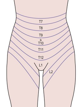
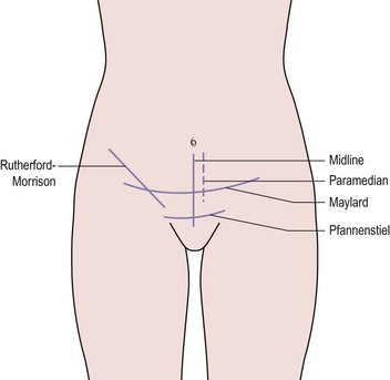
The layers of the anterior abdominal wall include skin, subcutaneous fat, rectus sheath and oblique abdominal muscles, transversalis fascia and peritoneum. Within these structures lie segmental arteries, veins and nerves that supply the dermatomes and myotomes, and also the epigastric vessels (Figure 8.1). Abdominal incisions have been developed that preserve function as well as possible and which heal rapidly with good strength. Commonly used incisions are shown in Figure 8.2.

It should not be forgotten that inappropriate wound closure technique can increase the risk of pulmonary complications and death (Niggebrugge et al 1999). While some surgeons prefer to use electrocautery rather than a cold scalpel to open the wound, there seems to be no difference in the early or late complications in midline incisions (Franchi et al 2001).

Incisions

The vertical midline incision avoids all major nerves, vessels and muscles by dividing the rectus sheath. It gives good access to the whole of the abdomen except the subdiaphragmatic areas, and is a very fast incision to make. Its principal drawback is that it heals slowly and suffers from a high incidence of wound dehiscence and incisional hernia. The development of improved sutures and technique has dramatically reduced these complications. A midline incision should always be closed using the ‘mass closure’ technique. This involves 1-cm-deep bites of the rectus muscle and peritoneum in a continuous closure, with sutures placed 1 cm apart. The suture material used does not affect the rates of dehiscence or infection, but incisional hernia is more common if braided absorbable material is used (Rucinski et al 2001). The same meta-analysis showed that incision pain and suture sinus formation are more common after non-absorbable material is inserted. Absorbable monofilament used in a continuous mass closure gave the best results.

The paramedian incision is only included for historical interest. The layered closure that was used improved strength when catgut sutures were employed, and reduced the incidence of dehiscence. It is inferior to the mass closure technique described above, and does not provide improved access to either side of the abdomen.

The majority of gynaecological surgery is performed through a transverse incision, usually a Pfannenstiel incision. By dividing each layer in a different direction, the function is well preserved. Even the muscle-cutting variants of the transverse incision heal more rapidly than vertical incisions. The cosmetic result is superior and, if correctly placed, access to the pelvis is good, although access to the pelvic brim is limited. This incision is difficult to extend if improved abdominal access is required.

The Maylard incision involves dividing all layers in the line of the skin incision and also divides the inferior epigastric vessels. The rectus sheath is not separated from the muscle and closure is in layers, leaving the muscle to be drawn together by the sheath. This incision provides improved access to the pelvic side wall when compared with the Pfannenstiel incision, and is useful for oncological surgery. Many centres now use this incision or a Pfannenstiel incision for radical hysterectomy (Orr et al 1995, Scribner et al 2001).

The Cherney incision is similar to the Pfannenstiel incision but the rectus muscles are divided 1 cm from their insertion into the symphysis pubis. The incision is closed in layers, but the muscle is repaired using a continuous suture in the membranous distal portion of the muscle. This incision can also be useful in oncological surgery or, if placed lower, is useful for complex urogynaecological procedures.

The Rutherford–Morrison incision involves dividing all layers in the line of the incision and is particularly useful for approaching ovarian masses in the second half of pregnancy. The incision is closed in layers and heals well.

Closure of peritoneum

This practice has been questioned recently. Research in animal models has suggested that closure of the peritoneum increases rather than decreases peritoneal adhesions. Human studies support this finding, showing that closure of the pelvic peritoneum does not reduce the incidence of postoperative adhesions or obstruction (Table 8.1; Tulandi et al 1988). The peritoneum spreads rapidly across any raw areas left after surgery, although devascularized tissue, such as pedicles, are a focus for adhesion formation. Practice still varies with regard to peritoneal closure, although it probably results in increased adhesion formation (Royal College of Obstetricians and Gynaecologists 1998). However, if a suction drain is used in the wound, the peritoneum must be closed to avoid drawing the bowel into the wound.

Table 8.1 The effect of peritoneal closure on wound infection and adhesion formation.

  Closed (%) Not Closed (%)
Wound infection 3.6 2.4
Adhesions 22.2 15.8
Obstruction 1.6 0

Source: From Tulandi T, Hum HS, Gelfand MM 1988 Closure of laparotomy incisions with or without peritoneal suturing and second look laparoscopy. American Journal of Obstetrics and Gynecology 158:536, with permission of Elsevier.

Ureteric Damage

Ureters are the organs most respected by gynaecologists. They lie close to the genital tract and repair of a damaged ureter is technically demanding, with results that are not always satisfactory. By recognizing when and where the ureters are at greatest risk and by adopting a safe technique, the risk of damage to the ureter should be minimized.

Anatomical relations

The urinary and genital tracts are closely related in embryological development, and their anatomy and physiology, not surprisingly, are intertwined. The ureter develops from a bud on the posterolateral border of the mesonephric duct near the cloaca, which elongates and eventually fuses with the developing kidney. The ventral portion of the cloaca develops into the urethra, bladder and lower portion of the vagina.

The ureters enter the pelvis by crossing the common iliac arteries in the region of their bifurcation. They descend on the pelvic side wall, medial to the branches of the internal iliac arteries and lateral to the ovarian fossae. From there, they run on the anterior surface of the levator ani muscles lateral to the uterosacral ligaments to pass beneath the uterine arteries, 1–1.5 cm lateral to the cervix and vagina. The ureters then swing medially around the vagina, to enter the bladder 2–3 cm below the anterior vaginal fornix.

The ureter is accompanied by a plexus of freely anastomosing fine vessels running in the loose tissue surrrounding it. The blood supply in the upper portion is derived from the renal and ovarian vessels; in the middle third from branches of the aorta, common iliac and internal iliac arteries; and in the lower part from branches of the uterine, vaginal, middle haemorrhoidal and vesical arteries. The ureter may be mobilized extensively provided this plexus of vessels is preserved.

The ureter is at risk during gynaecological surgery in four regions: at the pelvic brim where it can be confused with the infundibulopelvic ligament; lateral to the ovarian fossa where it can be adherent to an ovarian mass; in the ureteric tunnel beneath the uterine artery; and anterior to the vagina where it runs into the bladder.

The ureter at the pelvic brim and ovarian fossa

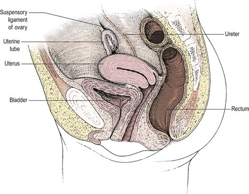
At the pelvic brim, the infundibulopelvic ligament with the ovarian vessels also crosses the iliac vascular bundle, usually 1–2 cm distal to the bifurcation of the common iliac artery and the ureter (Figure 8.3). At this point, the ureter and the ovarian vessels are running in parallel in a similar plane and may be confused if care is not taken. Occasionally, the ureter may be duplex. Where the ureter runs lateral to the ovary, it will almost inevitably be associated with any inflammatory or malignant mass. Happily, it will lie on the lateral aspect of the mass where it can be identified and dissected free, usually without difficulty, although in patients with endometriosis, the ureter may be fixed in dense fibrosis.

The key to the safe identification of the ureter on the pelvic side wall is to open the peritoneum and dissect in the retroperitoneal space. This is most easily done by dividing the round ligament between two clips, dividing the peritoneum in a cranial direction 1.5 cm lateral to the ovarian vessels and in a caudal direction down towards the uterovesical fold. The loose areolar tissue then encountered should be separated by blunt dissection with careful diathermy of any small vessels. The ureter will lie on the lateral aspect of the leaf of peritoneum reflected by this manoeuvre. At this stage, the infundibulopelvic ligament can be safely divided with the ureter under direct vision. If the anatomy of the pelvic side wall has been distorted, this technique will almost always allow the ureter to be identified and followed within the pelvis. If an ovarian mass is present, the ureter may be dissected free from the mass in this retroperitoneal plane.

The lower ureter

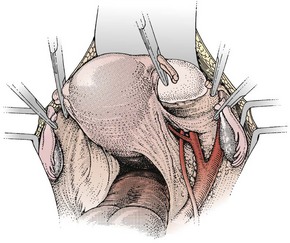
The third position where the ureter is at risk is in the ureteric tunnel, where the ureter crosses beneath the uterine artery but superior to the cardinal ligament and approximately 1–1.5 cm lateral to the angle of the vagina (Figure 8.4). It is possible to palpate the ureter in this position by gripping the paracervical tissue between one finger in the pouch of Douglas and the thumb placed laterally in front of the uterine pedicle. The ureter, which is felt as a firm cord running across the cardinal ligament, is surprisingly far lateral to the cervix if the bladder has been properly reflected and the anatomy is normal. However, adhesions, fibrosis or inadequate dissection may disrupt these relationships, causing the ureter to remain fixed near the lateral margin of the cervix.

Damage to the ureter in this site is prevented by carefully reflecting the bladder and by not taking a large pedicle that includes both the uterine artery and the paracervical tissue. The author prefers to take the uterine artery relatively high, at the level of the internal os, which allows the parametrium to fall laterally, taking the ureter with it. The ureter can then be palpated again and a second pedicle taken medial to the first, to include the cardinal ligament.

In most cases, damage to the ureter occurs at this site because the pedicle slips or the original ligature is not adequate. This can be avoided by preparing the pedicle carefully before clamping it. Modern clamps, such as Roget’s or Zeppelin’s parametrial clamps, rarely slip, and ties should be placed accurately. If bleeding does occur, the ureter should be palpated again after replacing the clamp to ensure that it has not been included.

The ureter is occasionally damaged in its course across the anterior surface of the vagina. This may occur when taking a cuff of vagina, during a vaginal hysterectomy or colposuspension. When dissecting the upper vagina, it is important to keep in the correct plane close to the vaginal wall. Stitches placed either vaginally or abdominally in this area must be in tissue that has been accurately identified.

Repairing ureteric damage

If a ureter is damaged, the presence of the contralateral kidney should be checked. If the ureter has not been cut across but merely crushed or ligated in error, and this is recognized on the table, it is acceptable to remove the ligature and insert a stent into the ureter in the hope that a stenosis will not result. If the ureter is divided, repair will be necessary. In the past, gynaecologists have resorted to simply tying off the divided ureter but this is rarely justifiable now.

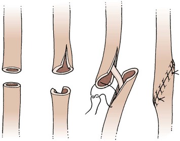
Repair of the ureter is technically demanding and may lead to long-term complications. It is not appropriate for a gynaecologist without urological training or extensive urological experience to undertake this. In principle, an end-to-end anastomosis can be performed for ureteric damage at the pelvic brim or above. The ureter is mobilized so that the ends can be brought together without tension. They are spatulated and the anastomosis performed using fine interrupted sutures over a Silastic stent (Figure 8.5). This is removed several weeks later. An alternative for damage at this level is to perform a uretero-ureteric anastomosis.

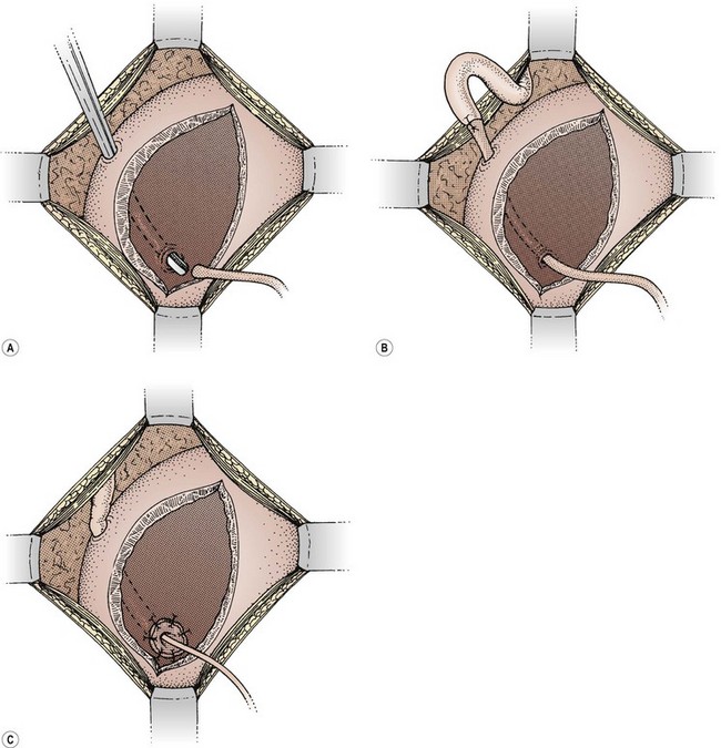
If the damage has occurred at the level of the ureteric tunnel or lower, the safest method of repair is to reimplant the ureter into the bladder. The bladder is opened between stay sutures. A submucosal tunnel is fashioned, taking care to avoid the other ureter. The distal end of the ureter is brought through the tunnel and, after spatulating the end, is sutured to the bladder mucosa. A couple of sutures into the serosal surface anchor the ureter to the outer layer of the bladder (Figure 8.6).

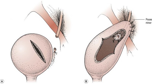
When damage has occurred higher on the pelvic side wall, the bladder can be elevated to the cut end of the ureter to allow reimplantation without tension, using either a psoas hitch or a Boari flap. In the former, an appropriate part of the bladder is elevated towards the end of the ureter. To facilitate this, the cystotomy is performed across the direction of elevation and is closed in the opposite direction to elongate the bladder. In addition, the contralateral superior vesical pedicle may be divided. The ureter is reimplanted as described previously and the bladder is fixed to the psoas muscle to relieve any tension. This technique will allow a ureter divided at any level in the pelvis up to the pelvic brim to be safely reimplanted (Figure 8.7).

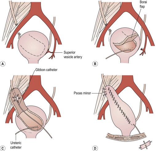
The Boari flap is an alternative to the psoas hitch. A flap of bladder is elevated, the ureter is reimplanted into this and the flap is closed as a tube (Figure 8.8). This allows good elevation of the bladder at the expense of reduced bladder capacity.

Alternatively, a uretero-ureteric anastomosis to the opposite ureter can be performed. This has the disadvantage that both ureters may be compromised but may be the only method if access to the bladder is difficult.

Bladder Damage

Avoiding damage

The bladder must always be emptied prior to pelvic surgery and the peritoneum should be opened superiorly, away from the bladder, when making a transverse incision. It can be palpated as a thickening in the peritoneum, and if there is any doubt about its position, careful dissection should allow the detrusor muscle to be recognized before the mucosa is opened. Opening the peritoneum by blunt dissection with fingers will not necessarily protect the bladder, as it is possible to dissect bluntly into it. Similarly, the technique of tearing the parietal peritoneum open by ‘stretching’ the wound once the abdominal cavity has been opened can lead to damage to the top of the bladder, particularly after previous surgery.

An essential part of any hysterectomy is separation of the bladder from the cervix and upper vagina. This plane can be found after division of the uterovesical fold of peritoneum merely by pushing caudally with a swab applied firmly to the anterior surface of the cervix cranial to the bladder. This is very quick but inevitably leaves a few small bleeding points on the back of the bladder that may be difficult to find. In addition, the bladder is occasionally damaged by this manoeuvre, particularly after previous surgery.

An alternative approach is to use a combination of sharp and blunt dissection. After dividing the uterovesical fold of peritoneum, the scissors are held slightly opened to begin separation in this plane by pushing the bladder off the cervix. Small blood vessels can be recognized and diathermied before cutting, to maintain haemostasis at all times. Once the correct plane has been identified in the midline, the dissection is extended laterally to mobilize the ureters and can be continued as far as necessary down the vagina. Although a little more time consuming, this technique is safe and can be used on simple cases to gain experience for more difficult situations.

When performing the operation vaginally, the same plane needs to be defined and dissected. If the plane does not develop easily, it is important to use sharp dissection, as merely pushing on the bladder may tear a hole. A sound in the bladder may help to define the correct plane. Alternatively, a finger over the fundus of the uterus or over the broad ligament to the side, once the pouch of Douglas is opened, can define the uterovesical fold of peritoneum and clarify the correct plane for dissection.

The bladder can also be damaged during closure of the vaginal vault, particularly if it has not been sufficiently mobilized. Sutures may be placed through the detrusor muscle or even into the bladder itself, although this rarely causes problems (Meeks et al 1997). However, if this is recognized at the time of surgery, such stitches should be removed and the bladder mobilized to allow closure of the vault without including the bladder. If damage to the bladder has occurred, this should be repaired and the bladder drained as discussed below.

Bowel Damage

Inadvertent damage to the bowel should be unusual, unless adhesions are present. The occasions when damage is most likely to occur are during opening of the peritoneal cavity or when dissecting dense intra-abdominal adhesions. In addition, the rectum lies close to the uterosacral ligaments and posterior wall of the vagina, and may be at risk during extensive pelvic dissection for cervical or vaginal carcinoma.

When reopening a previous incision, it is preferable to enter the peritoneal cavity away from the previous closure. If that is not possible, great caution should be used when approaching the peritoneum. Very occasionally, the peritoneal cavity is almost obliterated and it may be neccessary to dissect extraperitoneally around the adhesions to enter the peritoneal cavity.

When dense intraperitoneal adhesions are encountered, the adhesions can usually be divided safely with care and patience. Occasionally, the bowel is so matted together that resection of a small portion may be necessary. Particular care must be used in a patient who has been treated with radiotherapy, as the bowel is more friable and often densely adherent.

When dividing the uterosacral ligaments close to the pelvic side wall during a radical hysterectomy, the rectum is at risk of being included in the clamps. The rectovaginal space must be opened widely and the rectum dissected free. Care is necessary when entering this space to avoid direct damage to the rectum.

Repair of bowel

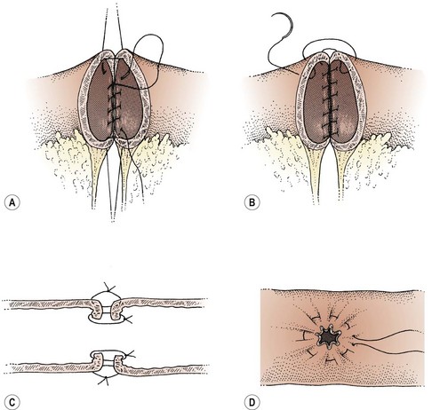
Repair of damaged bowel may involve primary closure of a small hole or may be facilitated by the excision of a portion of unhealthy bowel and reanastomosis (Figure 8.9). Consideration should be given to a defunctioning colostomy proximal to anastomosis of large bowel, particularly if the blood supply to the bowel wall is compromised in any way. Factors that have a bearing on this decision are previous radiotherapy, bowel obstruction, gross infection of the operative field or other medical factors such as diabetes, steroid therapy, malignancy and advanced age. If no adverse factors are present, a colostomy may not be necessary. Small bowel usually heals without the need for defunctioning. If there is any doubt about the safety of a repair, a surgeon who specializes in bowel should be involved to help with the repair.

Vascular Damage

Bleeding may occur at any stage of gynaecological surgery, especially if the anatomy is distorted or small tributaries are increased in size, providing collateral supply to fibroids or tumours. Careful and accurate surgical technique will avoid most, but not all, haemorrhagic complications.

Careful, gentle packing of the bowel out of the pelvis reduces the risk of haemorrhage by maximizing exposure of the operative field. While the bowel must be kept out of the pelvis as much as possible, the packs should not compress the inferior vena cava and obstruct the flow of blood out of the pelvis. Excessively tight packing results in distended pelvic veins and excessive venous bleeding. In the same way, a modest head-down tilt lowers the venous pressure in the pelvis and reduces blood loss.

Bleeding may occur from the main uterine or ovarian vessels while they are being identified, clamped and ligated. Using the technique of pelvic dissection described previously, the ovarian vessels can usually be identified and ligated without difficulty. However, the ovarian vein is particularly delicate and easily torn. A tie around the whole pedicle is preferable to transfixion, as the latter risks haematoma formation proximal to the tie. The proximity of the ureter to these vessels at the pelvic brim is an added problem when endometriosis, inflammatory disease, adhesions or tumours distort the anatomy. In contrast, the uterine artery is divided lateral to the uterus as a plexus of vessels in relatively tough tissue and sutures must be inserted. Care must be taken to include all branches in the ligature. The paracervical tissue and vaginal angle are also rich with arterial tributaries, and similar considerations apply. A figure-of-eight suture in the angle of the vagina is useful to ensure that the heel of the pedicle that contains these vessels does not slip out of the ligature. These pedicles should be adjacent to each other with no space between as haemorrhage can occur later, even if the area looks dry at the completion of the procedure.

Small bleeding points on peritoneum or bladder can be diathermied, but slight oozing from a pedicle signals inaccurate technique and should be corrected by oversewing the pedicle and the adjacent pedicle if necessary. The position of the ureter can be identified by palpation of the cardinal ligament lateral to the pedicle prior to the insertion of further sutures.

If bleeding from these vessels occurs, it will usually be controlled temporarily with pressure while appropriate instruments and ligatures are readied. The swab can then be removed gradually, exposing more of the field bit by bit until the bleeding point is revealed. Suction may be needed to keep the operative field clear. The location of the ureter must be determined by palpation or inspection before attending to the bleeding vessel. It is usually best to stop the bleeding by grasping the vessel gently with fine, long-handled artery forceps. A suture may then be inserted.

Damage to the large iliac vessels usually only occurs during surgery for cancer when lymph nodes or tumour are being removed from the surface of the vessels. However, deeply infiltrating endometriosis may become adherent to the internal iliac vessels, and any retroperitoneal tumour or postoperative adhesions may distort the anatomy so that the iliac vessels are exposed to trauma.

If the blood supply to the leg is compromised by damage to the external or common iliac vessels, serious morbidity can result and it is vital that repair is carried out to the highest standard. Usually, a vascular surgeon should be involved.

The internal iliac vessels may be safely ligated if damage occurs, but bleeding from the thin-walled veins on the pelvic side wall can be very difficult to control. Initially, bleeding should be controlled by direct pressure, either by a well-placed finger or using a swab. This allows time to assemble the necessary instruments, a fine sucker and fine sutures on small needles to repair the laceration or ligate the vessel, and also a chance to take a deep breath so that the subsequent suturing is not carried out in a panic. Great care should be taken, as rough handling of the vessels can make the situation worse. If in doubt, more experienced help should be obtained.

In most cases, tears in the common and external iliac veins can be repaired by suturing with fine Vicryl. Damage to the arteries is usually less of a problem because the muscular vessel wall constricts and contains the haemorrhage. Local pressure, sometimes supplemented by sutures, usually gives a very satisfactory result. The real danger comes from damage to the internal iliac veins and their branches, or ragged tears removing parts of the wall of the external or common iliac veins. The deeply placed veins retract into the muscle on the pelvic side wall where they become relatively inaccessible. Deeply placed mattress sutures are required to control the haemorrhage. Unfortunately, such sutures may involve one of the large pelvic nerves. This will only become apparent after the operation when the patient wakes up. It may prove impossible to control bleeding from these veins completely. The often-quoted technique of tying off the internal iliac artery in cases of troublesome bleeding is not without its own hazards. The origin of the artery lies over the bifurcation of the common iliac vein, and great care is needed to avoid damaging the internal iliac vein while dissecting behind the artery to pass the ligature. This technique is often less effective than might be hoped in controlling pelvic haemorrhage because such bleeding is often venous. Firm packing may prove to be the only alternative.

More common than either of the two dramatic scenarios above but equally dangerous is a steady ooze from many small venules and arterioles. This usually follows an extensive dissection such as may be required to remove an endometrioma or to mobilize an adherent bladder. This often happens at the end of a long operation during which there has been a slow but steady blood loss that, because it has not been particularly dramatic, has not been replaced fully. A consumptive coagulopathy results as the clotting factors are exhausted. The first step therefore is to hold a hot pack firmly on the bleeding area for 10 min. The second is to commence the prompt replacement of blood and clotting factors. When the pack is removed gently, all may be well or no more than two or three bleeders may persist. These may be controlled with judicious use of diathermy. If there is still significant generalized blood loss not obviously coming from a single significant vessel, a hot pack should be reinserted and kept firmly in place until the blood volume and clotting factors have been replaced adequately. This may take more than 1 h, during which it is often helpful for the surgical team to take a break. If necessary, the abdomen may be closed with the packs in place. Antibiotics are given and the packs removed gently the following day.

Uterine Perforation and False Passage

During dilatation of the cervix, pushing an instrument through the substance of the cervix rather than following the canal may create a false passage. Instruments may also be pushed through the fundus of the uterus into the peritoneal cavity.

When learning to dilate the cervix, one must develop a gentleness of touch that combines pressure with sensitivity or ‘feel’ for the tissues. It is important to avoid using force with instruments, but rather to feel for the path of least resistance. Due to the barrel-shaped contour of the endocervical canal, an exploring instrument can deviate from the axis of the cervix and not engage the internal os. The size of instrument chosen for this exploration is important, as a fine probe will create its own passage more easily and therefore less pressure should be used. It is said that right-handed people are more likely to perforate towards the right side of the cervical canal, although a false passage can be created in any direction. It then becomes very difficult to identify the true cervical canal and gain access to the uterine cavity.

If a false passage is suspected, one helpful manoeuvre is to perform a bimanual examination with the vulsellum attached and sound still within the passage. It is often possible to feel where the sound is in relation to the axis of the cervix and in which direction the true passage is likely to be found.

When a false passage has been created, provided damage to other organs is not suspected and the patient is not pregnant, the situation can usually be managed conservatively. In most cases, it is best to desist from further attempts at dilatation. In the pregnant patient, however, significant haemorrhage may occur, particularly following lateral perforation into the broad ligament and damage to the uterine vessels. If vaginal bleeding continues or the patient’s general condition deteriorates, laparotomy will be necessary. The broad ligament will be full of blood if the uterine vessels have been damaged, and hysterectomy may be necessary to control the blood loss, although ligation of the uterine artery and repair of the perforation may be an option. In some cases, bleeding into the retroperitoneal space may not become apparent for several hours. Careful postoperative observation is necessary to detect this at an early stage.

The consequences of fundal perforation depend on whether the patient is pregnant and what instrument has created the perforation. In a non-pregnant uterus, a dilator through the fundus is very unlikely to cause any problems. It is likely that most perforations go unrecognized by the surgeon. The patient should be observed for a period after recovery but is unlikely to need further intervention. In a pregnant patient, perforation is said to be more likely to cause haemorrhage but, in general, a simple perforation can be managed conservatively. The temptation to ‘confirm’ the perforation by repeatedly reintroducing instruments into the uterus should be avoided as this may make matters worse.

If there is significant risk of damage to intra-abdominal organs, a laparotomy is indicated because laparoscopy cannot be relied upon to exclude damage to bowel. For example, if a suction catheter has been inserted through a perforation with suction connected, laparotomy and careful examination of the bowel are mandatory.

Laparoscopy

The incidence of complications in laparoscopic surgery relates very closely to the experience of the surgeon and the training they have received. Gynaecologists have built up extensive experience in the specialty with simple procedures over the last generation. Most senior house officers and junior registrars are trained in diagnostic laparoscopy as one of their earliest procedures. Nevertheless, adoption of the more advanced operative techniques has been slow, and experience in these areas is still very limited. If a high incidence of complications is to be avoided, it is essential that individual surgeons are well trained in the procedures they undertake. General complications of laparoscopy will be discussed but consideration of complications encountered in advanced laparoscopic surgery is outside the scope of this chapter (see Chapter 4, Laparoscopy, for more information).

Damage to blood vessels

When inserting the Veress needle and the first trocar, it is vital that these instruments are kept away from the major vessels. The position of the sacral promontory can be palpated and the instruments angled in such a way that they pass below the promontory and into the free space in the midline of the pelvis. While damage to a major blood vessel may be all too obvious, the bleeding may be predominantly retroperitoneal and difficult to see laparoscopically. If the patient’s blood pressure has dropped and pulse rate risen sufficiently to suggest the possibility of vascular damage, a laparotomy should be performed immediately. If the abdomen has to be opened in a hurry, one technique is to lift the laparoscope up to the anterior abdominal wall and cut down on to it, allowing immediate access.

Blood vessels in the abdominal wall may also be damaged. To reduce this risk, the abdominal wall is usually transilluminated to visualize vessels prior to insertion of the secondary trocars. The location of the inferior epigastric vessels should be recognized, as they will bleed profusely and this may be difficult to control. Minor bleeding around a trocar may be controlled either by leaving the trocar in place to tamponade the vessel, or by inserting a Foley catheter through the trocar, inflating the balloon with 30–50 ml of saline and withdrawing the trocar and balloon to apply pressure to the bleeding point. If the vessel requires suturing, this is facilitated by a long J-shaped needle.

Diathermy damage

Problems with laparoscopic use of diathermy stem either from the heat generated at the site of tissue destruction or from current finding an alternative path to earth through an adjacent organ. Tissue coagulated by diathermy becomes very hot and may retain its heat for several minutes. This heat will spread to underlying structures and adjacent organs, and may cause damage. Diathermied tissue must be allowed to cool before coming into contact with adjacent structures. A further problem is the rising impedance in desiccated, diathermied tissue. This may result in the current arcing to a nearby organ that offers a low-resistance path to earth. In a similar way, current may arc to nearby organs if the current is activated before the electrode is in contact with the tissue to be treated.

References

Cruise PJE, Foord R. A 5 year prospective study of 23,649 surgical wounds. Archives of Surgery. 1973;107:206.

Franchi M, Ghezzi F, Benedetti-Panici PL, et al. A multicentre collaborative study on the use of cold scalpel and electrocautery for midline abdominal incision. Amerian Journal of Surgery. 2001;181:128-132.

Meeks GR, Sams JO4th, Field KW, Fulp KW, Margolis MT. Formation of vesicovaginal fistula: the role of suture placement into the bladder during closure of the vaginal cuff after transabdominal hysterectomy. American Journal of Obstetrics and Gynecology. 1997;177:1298-1303.

Niggebrugge AHP, Trimbos JB, Hermans J, Steup W-H, Van De Velde CJH. Influence of abdominal-wound closure technique on complications after surgery: a randomised study. The Lancet. 1999;353:1563-1567.

Orr JWJr, Garter JF, Kilgore LC, et al. Closed suction pelvic drainage following radical pelvic surgery. American Journal of Obstetrics and Gynecology. 1987;155:867.

Orr JWJr, Orr PJ, Bolen DD, Holimon JL. Radical hysterectomy: does the type of incision matter? American Journal of Obstetrics and Gynecology. 1995;173:399-405.

Royal College of Obstetrics and Gynaecologists. Peritoneal Closure. Guideline No. 15. RCOG, London, 1998.

Rucinski J, Margolis M, Panagopoulos G, Wise L. Closure of the abdominal midline fascia: meta-analysis delineates the optimal technique. American Surgeon. 2001;67:421-462.

Scribner DRJr, Kamelle SA, Gould N, et al. A retrospective analysis of radical hysterectomies done for cervical cancer: is there a role for the Pfannenstiel incision? Gynecological Oncology. 2001;81:481-484.

Tulandi T, Hum HS, Gelfand MM. Closure of laparotomy incisions with or without peritoneal suturing and second look laparoscopy. American Journal of Obstetrics and Gynecology. 1988;158:536.