[level-membership-for-pediatrics-category]
Chapter 695 Primary Chondrodystrophy (Metaphyseal Dysplasia)
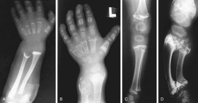
In primary chondrodystrophy, which is an autosomal dominant condition, bowing of the legs, short stature, and a waddling gait appear in the absence of abnormalities of serum levels of calcium and phosphate, alkaline phosphatase activity, or vitamin D metabolites. Metaphyseal chondrodysplasia (Jansen type) is typified by cupped and ragged metaphyses, which develop mottled calcification at the distal ends of bone over time (Fig. 695-1). Hypercalcemia, with serum values of 13-15 mg/dL, can occur. The spine can also be deformed by the irregular growth of vertebrae. Three different mutations have been identified in parathyroid hormone receptor type I as the molecular cause of this syndrome, as have some of the downstream target genes that can contribute to the pathogenesis of the disease. The Schmid type of metaphyseal chondrodysplasia is less severe, although the radiographic appearance of the knees and extreme bowing of the lower limbs resemble signs seen in patients with familial hypophosphatemia. It is associated with defects in collagen type X, and the hip abnormalities are more debilitating than in Jansen metaphyseal chondrodysplasia. Patients with both types of metaphyseal chondrodysplasia have lifelong short stature.
Metaphyseal dysostosis, or Pyle disease, results from defects in endochondral bone formation and metaphyseal modeling. The long ends of bones are splayed, resulting in an “Erlenmeyer flask” defect. Short stature is not necessarily characteristic, and serum chemical levels are normal. Leonine features often develop if the facial bones are involved. Metaphyseal dysostosis may be a clinical feature of Shwachman-Diamond syndrome, a rare autosomal recessive disorder characterized by neutropenia, pancreatic exocrine insufficiency, bone marrow dysfunction, and sometimes severe hematologic complications (Chapter 442). Allogeneic bone marrow transplantation has been used as a therapeutic approach, with mixed results.
Alanay Y, Rimoin DL. Chondrodysplasias. In: Rosen CJ, Compston JE, Lian JB, editors. Primer on the metabolic bone diseases and disorders of mineral metabolism. Washington, DC: American Society for Bone and Mineral Research; 2008:428-429.
Beier F, LuValle P. The cyclin D1 and cyclin A genes are targets of activated PTH/PTHrP receptor in Jansen’s metaphyseal chondrodysplasia. Mol Endocrinol. 2002;16:2163-2173.
[/level-membership-for-pediatrics-category][not-level-membership-for-pediatrics-category]
Chapter 695 Primary Chondrodystrophy (Metaphyseal Dysplasia)
In primary chondrodystrophy, which is an autosomal dominant condition, bowing of the legs, short stature, and a waddling gait appear in the absence of abnormalities of serum levels of calcium and phosphate, alkaline phosphatase activity, or vitamin D metabolites. Metaphyseal chondrodysplasia (Jansen type) is typified by cupped and ragged metaphyses, which develop mottled calcification at the distal ends of bone over time (Fig. 695-1). Hypercalcemia, with serum values of 13-15 mg/dL, can occur. The spine can also be deformed by the irregular growth of vertebrae. Three different mutations have been identified in parathyroid hormone receptor type I as the molecular cause of this syndrome, as have some of the downstream target genes that can contribute to the pathogenesis of the disease. The Schmid type
[/not-level-membership-for-pediatrics-category]
