Precocious puberty

Published on 21/03/2015 by admin

Filed under Pediatrics

Last modified 22/04/2025

Print this page

rate 1 star rate 2 star rate 3 star rate 4 star rate 5 star
Your rating: none, Average: 0 (0 votes)

This article have been viewed 1442 times

Chapter 35 PRECOCIOUS PUBERTY

Theodore X. O’Connell

General Discussion

Normal puberty begins between 8 and 14 years of age in girls and between 9 and 14 years of age in boys. Girls begin puberty with breast buds and skeletal growth, followed by the arrival of pubic hair, axillary hair, and menarche. Boys have testicular enlargement followed by the appearance of pubic hair, enlargement of the penis, and spermarche. The age at which pubertal milestones are attained varies among the population studied. In addition, it is influenced by activity level and nutritional status. Girls with low body fat may have a significant delay in menarche (up to a year or longer), whereas obese girls may have earlier onset of puberty.

Precocious puberty is defined as the development of secondary sexual characteristics before the age of 8 years in girls and 9 years in boys. It involves not only early physical changes of puberty, but also linear growth acceleration and acceleration of bone maturation, which leads to early epiphyseal fusion and short adult height.

There are two types of precocious puberty. Central precocious puberty (gonadotropin-releasing hormone [GnRH]-dependent precocious puberty) results from premature activation of the hypothalamic GnRH pulse generator-pituitary gonadotropin-gonadal axis. In peripheral precocious puberty, (GnRH-independent precocious puberty), sex steroid secretion is independent of the GnRH pulse generator. Pathologic causes of puberty are likely if sexual development occurs in very young children or if there is contrasexual development. Peripheral causes are always pathologic and tend to produce an atypical puberty with loss of synchronicity of pubertal milestones.

Central precocious puberty is more common by far in girls than in boys, and in girls it is idiopathic 95% of the time. Boys more commonly (i.e., more than 50%) have an identifiable pathologic peripheral cause for precocious puberty. Therefore, all boys with precocious puberty should undergo detailed investigation. In girls, additional investigation can be based on the clinical impression. Central nervous system (CNS) disorders account for a higher percentage of cases in boys but must also be excluded in girls.

Pubertal variants cause isolated development of one of the secondary sexual characteristics without accelerated skeletal maturation. Occasionally they can progress to precocious puberty.

Simple premature thelarche involves only breast development (unilateral or bilateral), without pubic hair growth, without accelerated bone maturation, and with a normal height outcome. No treatment is required. The disorder is usually self-limited and can resolve spontaneously or persist to normal puberty.

Simple premature adrenarche involves only pubic or axillary hair development, without the other manifestations of puberty. No treatment is required, although late-onset congenital adrenal hyperplasia should be ruled out with a measurement of the serum 17α-hydroxyprogesterone level. There is an increased incidence of simple premature adrenarche in patients with CNS abnormalities.

Unilateral or bilateral gynecomastia may occur in boys as puberty begins. In fact, this is a nearly universal finding among boys in middle to late puberty. It usually resolves within 2 years. Pathologic gynecomastia occurs in Klinefelter’s syndrome, prolactin-secreting adenomata, and as a result of drugs such as marijuana and phenothiazines.

Patients with precocious puberty and pubertal variants require an initial bone age as a baseline. In precocious puberty, the bone age is usually accelerated more than two standard deviations above the chronological age. In pubertal variants, the bone age should be within two standard deviations of the chronological age. The bone age should be obtained at periodic intervals to determine whether pubertal variants have progressed to precocious puberty. The bone age is also used to monitor the effectiveness of therapy. Additional evaluation of precocious puberty is outlined below.

Suggested Work-up

Radiograph of the left wrist To estimate physiologic age for comparison with the child’s chronologic age
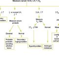
Serum follicle-stimulating hormone (FSH) and luteinizing hormone (LH), estradiol, testosterone, thyroid-stimulating hormone (TSH), thyroxine (T4), and human chorionic gonadotropin (hCG) To confirm the impression of idiopathic precocious puberty, to localize the abnormality of the pathologic cause of precocious puberty, or to guide the choice of imaging study
Serum 17-hydroxyprogesterone and dehydroepiandrosterone (DHEA) If a peripheral cause is suspected or if virilization is present in a female patient
Pelvic ultrasound Used to determine whether pubertal changes have occurred in the uterus and ovaries. Also indicated if a peripheral cause (ovarian tumor) is suspected.

Additional Work-up

Magnetic resonance imaging (MRI) of the brain and pituitary gland If a central pathologic cause is suspected on the basis of hormone measurements. Also indicated in boys with elevated hCG levels.
GnRH stimulation test 100 µg of GnRH is administered either intravenously or subcutaneously after an overnight fast. Serum levels of FSH and LH are measured at baseline just before the injection and at 15, 30, 45, and 60 minutes after the injection. Test interpretation is controversial, but a twofold to threefold rise in FSH and LH may be observed if the patient has central precocious puberty. A peak LH level of more than 15 international units per liter or a peak LH-to-peak FSH ratio of more than 0.66 are also criteria for defining a pubertal GnRH test.
Breast ultrasound Indicated in unilateral or asymmetric premature thelarche to exclude masses
Testicular ultrasound Indicated if asymmetric enlargement of the testes is present
Skeletal survey or radionuclidebone scan Indicated for patients with McCune-Albright syndrome to evaluate for lesions of fibrous dysplasia