CHAPTER 88 Postoperative Rehabilitation
INTRODUCTION
Patients with intractable lumbar radiculopathy, due to a lumbar disc herniation, require surgery. Lumbar discectomy is the standard of care. This invasive technique aims to release the pressure on the nerve caused by a prolapsed disc, while minimizing scar tissue formation, avoiding nerve damage and biomechanical destabilization.1 Rehabilitation following lumbar disc surgery has received far less attention than the operation itself. One could speculate that this is because after lumbar disc surgery the problems have been resolved and, therefore, rehabilitation is unnecessary. The reality is that in the long term too many patients still show considerable symptoms, reduced daily functional capacities, and disability.2,3 In other words, surgery to remove the lumbar disc protrusion does not solve all back or leg problems and a substantial number of patients continue to suffer residual complaints. That is, complaints remain or return after surgery.
In general, the success rate of lumbar disc surgery varies from 60% to 90%.4–6 It has been reported that, after lumbar disc surgery, 22–45% of patients experience residual leg pain and 30–70% have residual low back pain.7–9 The range of these statistics reflects the different inclusion criteria for lumbar disc surgery and the various definitions of a successful outcome. It is often not clear from these studies what kind of post surgical care, if any, was provided to subjects in a particular study.
A ‘FAILED’ SURGERY?
For the situation where complaints remain or return, the term ‘failed back surgery syndrome’ has been used.10,11 This is a controversial term.12,13 The authors agree with Verbeek13 that this term is not particularly helpful and even authors of articles who use these diagnostic terms admit that they are better avoided since they do not help to identify a cause or a treatment. After all, the only characteristics that these patients share are that they have been operated on for a lumbar disc surgery and that back-related complaints or leg pain remained or returned at some point after the operation. In contradistinction to Verbeek, the authors think that the term non-specific low back pain for this condition might also be somewhat misleading because that term is used for the general situation where the so-called ‘red flags’ have been excluded. The authors propose the term ‘residual complaints following lumbar disc surgery,’ because in the opinion of the authors that describes the situation most appropriately. First of all, it has not the negative connotation (both for surgeons and patients) that the surgery has ‘failed,’ but on the other hand it acknowledges the fact that there has been a surgery.
Before surgery
Before surgery, many patients have a long history of recurrent low back and leg pain.
Panjabi14 assumes that, as a result of injury, degenerative disc disease and muscle weakness, the control over the neutral zone of the spinal segment is decreased, which may lead to instability. The herniated disc as part of the degenerative cascade15 affects the stability of the spine: an increase in neutral zone to range of motion ratio in the different loading directions is reported.16 The stability around the spinal segment is conserved by the sensory-motor control system.17 There is extensive evidence of changes in both of these systems in patients with low back pain. After the first episode of low back pain, the lumbar multifidus, which is an important stabilizer, is inhibited and does not spontaneously recover, even when the patient is asymptomatic.18 Furthermore, as a result of a herniated disc, abnormal changes in the characteristics and the activity of the paraspinal muscles are observed on the involved side. In addition, the contraction of the tranversus abdominus muscle, the other important stabilizer, is altered and delayed in patients with low back pain when performing upper and lower limb movements, leading to inefficient muscular stabilization of the lumbar spine.19 Lumbar proprioception, postural control, and feed forward control of the paraspinal muscles seem to be impaired in patients with sciatica.20 In general, these patients often have a long medical history of pain and functional limitation, which can lead to long-term disability, antalgic posture, muscle imbalance, and reduced cardiovascular capacities.21 Other factors that play an important role, which are superimposed upon these neuromusculoskeletal abnormalities, include psychological influences such as anxiety or problems at work. Disability and patients’ expectation regarding the surgery have also been reported.10,22,23
During surgery
During surgery the functional spinal unit will be altered. The lumbodorsal fascia is incised, the paraspinal muscles are dissected and elevated off the spinous process, a portion or the entire lamina is removed, and the ligamentum flavum is incised. Depending on the size and location of the herniated fragment, a laminectomy or facet-otomy is necessary.24 The annulus fibrosis is also incised. It is obvious that injury to these tissues will necessarily interrupt the biomechanical integrity of the functional spinal unit. Another concern develops as a result of retraction of the paraspinal muscles. Even with careful dissection, damage to the medial branches of the posterior rami of the spinal nerves and denervation of the multifidus muscle can occur.25 In those instances in which the injury presumably results from ischemia, a risk factor for nerve root injury results and, moreover, the neural damage does not necessarily spontaneously recover.26 Of course, if the medial branch was surgically lysed, the probability of recovery is even lower.
After surgery
In the first period after surgery, local tissue response to the surgical intervention is detected on MRI and considered as normal.27 This postoperative edema gradually decreases at 3 weeks and may last up to 6 months. There is a plethora of evidence documenting the decreased stability of the lumbar motion segment as a result of surgical intervention. In the immediate postoperative period, disc height loss may be detected27 but generally no gross instability is reported. However, in the long term, even if an increase of motion does not occur shortly after surgery, reduced stiffness of the spine is observed, resulting in some degree of increased spinal motion.28 Repetitive loading of the spine and the presence of scar tissue appear to be two of the major factors for the development of spinal instability.29 Other factors that contribute to spinal instability include a higher load on the posterior elements,30 increased development of degenerative spondylosis,31 and instability of the level above.28 Evidence shows that the sensory-motor control system that, in normal conditions, can compensate for this acquired instability may fail to provide adequate control. A loss of muscle support and disturbed innervation is observed as a result of low back pain and sciatica in conjunction with the effects of the operative procedure.32 The deep back muscles not only stabilize the local segment, but also contribute to spinal stability as a whole. The instability is reinforced, leading to increased mechanical strain and further injury. With respect to the neurological deficits, immediate recovery is observed in a number of patients, as a result of the resolution of nerve root ischemia, but in a number of patients the paresis and the sensory disturbance do not spontaneously recover.33
For a portion of patients that experience residual complaints following lumbar disc surgery there is no real diagnosis and no specialist or operation which can simply stop the pain.10,11,34,35 For those patients, and certainly for any patient with primary postoperative axial pain, the main outcome or goal of the rehabilitation program should be aimed at restoring normal function during activities of daily living and/or work. But what is the evidence for these kinds of rehabilitation programs?
EVIDENCE REGARDING POSTOPERATIVE REHABILIATION PROGRAMS
In the international literature there is a surprisingly wide variation in the content of rehabilitation programs.36–38 Despite many plausible theories on the effectiveness of active, as well as passive, interventions following lumbar disc surgery, sound evidence is lacking. The choice of a specific method of rehabilitation is mainly based on the personal experience of care providers or on the results of studies of generally poor methodological quality. It has been suggested that passive treatment modalities should have no place in rehabilitation following first-time lumbar disc surgery and that active treatment is of paramount importance.39 Indeed, an active rehabilitation program is considered to enhance a patient’s independence from healthcare services in the long run. Here, the focus will be on the evidence regarding active rehabilitation programs.
Methods
In a systematic review that assessed the effectiveness of such programs used in the rehabilitation of patients following lumbar disc surgery,40 relevant randomized controlled trials (RCTs) and nonrandomized controlled clinical trials (CCTs) were included. This systematic review was conducted according to the method guidelines of the Cochrane Back Review Group.41 For this chapter, the literature was updated by searching PUBMED through December, 2003. RCTs or CCTs were included if they used at least one of the four primary outcome measures that were considered to be important, that is: (1) pain (e.g. VAS), (2) a global measure of improvement (overall improvement, proportion of patients recovered, subjective improvement of symptoms), (3) back pain-specific functional status (e.g. Roland Disability Questionnaire, Oswestry Scale), and (4) return to work (return to work status, days off work). Outcomes of physical examination (e.g. range of motion, spinal flexibility, degrees of straight leg raising or muscle strength), behavioral outcomes (e.g. anxiety, depression, pain behavior), and generic functional status (SF-36, Nottingham Health Profile, Sickness Impact Profile) were considered as secondary outcomes. Other outcomes such as medication use and side effects were also considered.
In assessing the quality of the included studies, the criteria list recommended in the method guidelines for systematic reviews by the Cochrane Back Review Group was applied.41 Only items reflecting the internal validity were used in assessing the quality of the included studies. One point was scored for each item in which a satisfactory response was obtained, and high quality was defined as fulfilling at least five of the validity criteria.
Results
Active rehabilitation programs that start immediately postsurgery
Two studies assessed neural mobilization. A high-quality RCT42 added an active and passive neural mobilization program to a standard 6-week physical therapy program that consisted of isometric and dynamic exercises. These exercises were increased in intensity from day 1 postsurgery as tolerated. The control group received the same standard physical therapy without the neural mobilization. This study revealed evidence that adding a neural mobilization program to a standard physical therapy program does not provide any additional benefits on any of the main outcome measures (level 3) on both short-term and long-term follow-up. This constitutes limited evidence (level 3) since there is only one high-quality RCT supporting this conclusion.
A low-quality RCT43 compared an intensive 7-day auto-assisted straight leg raise (SLR) regime for eight times a day with a 2-hour interval with a mild straight leg raise regimen that performed this same exercise only once a day for 7 days. This study revealed no significant differences between the two neural mobilization programs on pain, disability, or SLR at the 1-week and the 6-week measurement. No data were presented with regard to re-operation rates. Hence, there is limited evidence (level 3) that an intensive straight leg raise regimen is no more effective than a mild straight leg raise regimen.
A high-quality RCT44 evaluated an intensive exercise program that consisted of increasing daily activities, home training (mobilization, trunk strengthening) and later mainly intensive muscle strengthening exercises and cardiovascular exercises. The control group received no increase in daily activities, exercises only once a day, and no promotion of cardiovascular exercises. The results show that there are no statistically significant differences on any of the primary outcome measures, but some differences of the secondary outcomes. There was one re-operation (3.4%) in the intervention group and 2 re-operations (6.5%) in the control group. This is limited evidence (level 3) that there is no difference in effectiveness between an intensive exercise program and a less active program in the long term for global perceived effect, pain, and return to work.
Active rehabilitation programs that start 4–6 weeks postsurgery
Two high-quality RCTs39,45 and one low-quality RCT46 compared intensive exercise programs with mild exercise programs. The two high-quality RCTs reported no statistically significant differences between groups in overall improvement at 12-month follow-up. The low quality RCT did not include a long-term follow-up. All three studies showed a statistically significant difference on functional status in favor of the intensive exercise program on short-term follow-up and in one study45 on long-term follow-up. In the two high-quality studies return to work and daily activities were statistically significantly better in the intensive exercise programs in the short-term but not over the long term. The RCTs by Danielsen45 and Yilmaz46 revealed a statistically significant improvement in short-term pain relief in favor of the intensive program (but not long-term45). Furthermore, Danielsen45 observed 1-year re-operation rates that were negligible.
One high-quality RCT47 compared a behavioral graded activity program that was based on operant treatment principles and was time contingent (time dependent) with usual care as provided by physical therapists. On the post-treatment measurement, 67% of the patients treated with care as usual had recovered versus 48% of the patients treated with behavioral graded activity. This 19% difference was statistically significant. On long-term follow-up, 73% of patients receiving care as usual and 75% of patients receiving behavioral graded activity had recovered. This difference was no longer statistically significant. On all other main outcome measures there were no statistically significant or clinically relevant differences. This study provides limited evidence (level 3) that there are no long-term differences in effectiveness between a behavioral graded activity program and usual care as provide by physical therapists.
One low-quality RCT48 compared a supervised exercise program to home exercises. Both interventions incorporated the same exercises and at the same intensity. Re-operation rate was negligible in both groups. There is limited evidence (level 3) that supervised exercises and home exercises are equally effective on global perceived effect, disability, pain, and mobility.
One low-quality RCT49 added horseback riding (three times per week for 20 minutes per session) to an intensive 4-week rehabilitation program. There were no statistically significant differences for overall improvement in the short term. (No long-term follow-up was performed.) There was a statistically significant more rapid return to work in the intervention group. There were no statistically significant differences in pain and physical measures. This study provided limited evidence (level 3) that adding therapeutic horseback riding to a rehabilitation program is effective for return to work, but not for overall improvement.
One low-quality RCT50 compared a multidisciplinary rehabilitation program that consisted of sessions with a physical therapist, psychiatrist, occupational therapist, psychologist, social worker, and an intensive back training with usual care. At 1-year follow-up, there were no statistically significant differences between groups on global perceived effect, sick leave, or re-operation rate (3.7% in both groups). This study offered limited evidence (level 3) that multidisciplinary rehabilitation and usual care are equally effective.
One low-quality CCT51 added aerobic exercises to a treatment program. All outcome measures were performed only at 3 months. This is limited evidence (level 3) that adding aerobic exercises to a treatment program is not more effective on functional status, pain, and depression than exercises alone.
Rehabilitation in an occupational setting
Two studies specifically included patients in a work setting. One high-quality RCT52 compared a multidisciplinary rehabilitation-oriented approach in insurance medicine with usual care. This work provides limited evidence (level 3) that a rehabilitation-oriented approach in insurance medicine is more effective in affecting return to work at long-term follow-up. One low-quality CCT53 assessed a functional restoration program versus usual care. This study does not present comparisons between groups.
Comparisons among rehabilitation programs that start more than 12 months postsurgery
One low-quality RCT54 compared physical agents with joint manipulations, high-tech exercise, low-tech exercise, and no treatment. The low-tech exercise consisted of a McKenzie program under supervision in addition to spine stabilization. The high-tech exercise consisted of cardiovascular exercise (bicycle), isotonic trunk muscle training (DAPRE), and isokinetic exercises in flexion–extension and left–right rotation (Cybex TEF & Torso). There was limited evidence that low-tech and high-tech exercises, initiated more than 12 months after surgery, were more effective in improving low back functional status than physical agents, joint manipulation, or no treatment, but that there are no differences between the low-tech and high-tech exercise programs.
One high-quality RCT55 added hyperextension to an intensive exercise program. In the short term there was a statistically significant improvement in functional status that was equalized at long-term follow-up. There were no re-operations. There is moderate evidence (level 3) that adding hyperextension to an intensive exercise program is no more effective than intensive exercise alone on overall improvement or functional status.
PROPOSED TREATMENT PROTOCOL
As already mentioned, it is unclear what the exact content of postsurgery rehabilitation should be. The rationale for the currently described treatment protocol is based on knowledge of the consequences of the operative procedure, the phases of tissue healing, and the knowledge of the behavior of the functional spinal unit in non-specific low back pain and sciatica. Furthermore, in addition to the evidence as described in the previous part of this chapter, the evidence of the efficacy of physical therapy modalities used in chronic low back pain and sciatica have been taken into account.56,57,58 The emphasis is on biomechanical rehabilitation, which is supported by Ostelo40 who found no differences in the effectiveness of a behaviorally graded activity program and usual care in the long term. However, this doesn’t mean that a comprehensive bio-psycho-social approach and yellow flags should be neglected. Furthermore, red flags always have to be monitored. The complete treatment program is divided in different periods predicated on the classic phases of tissue healing. The main goals are to regain the maximal functional status without symptoms, and to prevent recurrences.
With this treatment program, a compliant patient, supported by a good social support system, should be able to return to work and recreational sport activities within 2 months. However, to prevent recurrence and to obtain automatic movement control, patients have to continue exercising until the autonomous stage of motor learning is reached. In the autonomous stage of motor learning, according to Fitts and Posner, the patient performs the skill automatically with a low degree of attention.59 Depending on the task, the performer, and the intensity of practice, this stage can take months to reach.
Finally, the described strategy forms the structure of the program which has to be adapted for each patient’s personal needs on a physical, psychological, and social level. The program should be individualized as much as possible: every patient has a different medical history, a slightly different operative procedure, a different ability to recover, and a different capacity for motor learning. Remember: ‘evidence-based practice is the integration of best research evidence with clinical expertise and patient values.’60
The protective phase of postoperative rehabilitation (hospital discharge to day 21)
In this period, phase 1, the surgical area is still vulnerable to injury and has to be protected. If possible, the rehabilitation program starts immediately after discharge with a home visit to provide back-care instructions and ergonomic advice in the home environment. At this stage, judgment of the social environment can be made and family support can be emphasized or moderated. The surgical procedure is once again explained to the patient. Donceel et al.52 demonstrated that an adequate explanation to the patient, together with a rehabilitation-oriented approach, increases the rate of return to work. They also stated that when poor outcome is predicted, i.e. a high score on the VAS scale for pain, the patient needs rehabilitation and has to be guided more intensively.
The surgical procedure is invasive for dorsal structures, which inherently need time to heal. In the first period, local tissue response is observed.27 This postoperative edema gradually decreases after 3 weeks and continues for up to 6 months. Consequently, for the first weeks, loaded flexion and rotation should be minimized to avoid tension on the healing tissues. To unload the disc, it is advised to avoid sitting and to load the spine as ergonomically correctly as possible in a lordotic posture.
General (muscle) activation, such as brisk walking, without provoking leg symptoms, is promoted. There is no evidence that advice to stay active is harmful for either acute low back pain, sciatica, or postsurgery patients. The benefit of increasing physical activity levels as early as feasible culminates in a significant reduction of sick leave and fear–avoidance behavior.40,61
The general rehabilitation phase of postoperative rehabilitation (day 21 to 6 weeks)
An active rehabilitation program (phase 2) is initiated from the fourth postoperative week onwards. As previously mentioned in the second part of this chapter (Evidence), there is strong evidence in favor of intensive exercise programs that are initiated at the 4–6-week postoperative interval. These will enhance short-term functional recovery and a faster return to work. Equally important to consider is that none of the proposed treatment strategies seems to carry a risk of re-herniation or re-operation. There are no studies that have investigated whether active rehabilitation programs should start immediately after surgery or 4–6 weeks later. Intuitively, when considering the time interval required to affect soft tissue healing, the 3-week benchmark appears to be reasonable. In this rehabilitation program, emphasis is first made on progressive local stabilization exercises to control the increased neutral zone. Although quality changes are found in the multifidus muscles, positive alterations are observed through physical therapy treatment after surgery.62 The tranversus abdominus muscle is an important stabilizer and often loses its anticipatory function in patients with low back pain,19 but to the authors’ knowledge, no information is available regarding its functioning after discectomy. Once the multifidus muscle regains the capability to contract, local stability can be (re)trained. As a result of the previously described denervation, retraining the multifidus muscle itself can be impossible, but local stability should be achieved by compensation strategies. Extensive literature regarding local motor control and stability retraining for patients with low back is available.56 In a short-term follow-up after lumbar discectomy, lumbar proprioception and feed forward control of the paraspinal muscles seems to recover, but the postural control remains impaired.20 Therefore, if on assessment postural control is reduced, retraining is integrated in the treatment program. Additional manipulative physical therapy for the surrounding restricted joints (e.g. hip, thoracic spine) can be useful and depends on the result of the individual assessment of the lower quadrant. Currently, no high-quality studies are available proving the efficacy of mobilization of restricted joint in postdiscectomy patients. But clinical experience and evidence in chronic non-specific low back pain63 support the use of mobilization for painful and restricted joints. When muscles are weak as the result of nerve compression, specific strengthening exercises are prescribed.
The two published studies on postoperative neural mobility exercises have not shown any improvement in the time it ultimately takes to recover.42,43 On the other hand, the authors’ clinical experience and that of Butler64 suggest the implementation of neural mobilization techniques when positive neural tension signs are present. In the literature, there seems to be no clear consensus regarding the fitness level of the patient with chronic low back pain. In contrast, patients with sciatica often dramatically decrease their normal exercise behavior resulting in reduced level of cardiovascular fitness.64a In view of the absence of agreement regarding the cardiovascular and muscular conditioning status of patients with low back and their predilection to limit physical endeavors, combined with the known diminished physical status of those with radicular complaints, it is the authors’ judgment that it is exceedingly appropriate to incorporate aerobic exercises.
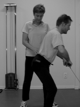
As preparation for return to work, individualized preventive back-care techniques should be taught. Individual back training appears to be most efficient in occupational settings and should be integrated when necessary (Fig. 88.1).
The specific rehabilitation phase of postoperative rehabilitation (6 weeks to 3 months)
In this stage (phase 3) of the rehabilitation program the patient is prepared for specific loading in activities of daily living, work, more intensive axial loading, and exposure to potentially provocative situations. Spinal stabilization is progressed to more complex weight-bearing positions and extended with exercises for the whole-body stability system. This functional stability depends upon integrated local and global muscle function and should provide active control over the available movement range.65 By now, the patient should reach the autonomous stage of task performance, in which a low degree of attention is required for the correct performance of motor tasks and dynamic stabilization of the spine, so it can be performed in an automatic manner during any movement pattern.66 The recovery of maximal endurance and explosive strength is incomplete 2 months after surgery.67,68 Given this fact, specific deficits in muscle strength, length, and aerobic conditioning must be identified. Focused exercises to address these inadequacies are weaved into the rehabilitation program. If the reaction of the patient to destabilizing forces is slow, specific exercises on an unstable surface are also incorporated.
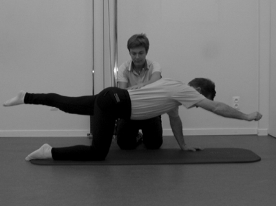
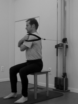
Although a basic notion, the need to evaluate the range of motion of the lumbar spine must be emphasized. Interestingly, there are no guidelines for mobility benchmarks due to the large variation in the population. Lumbar range does not give one the ability to discriminate between people with or without low back pain and with disability.69 Häkkinen et al.68 observed a decreased mobility of the spine 2 months postsurgery. If the mobility is judged to be restricted for that specific patient’s constitution, mobilizations of the spine are performed and self-mobilizations are taught to the patient. At this juncture, no evidence is available regarding the effect of spinal mobilization after surgery. Koes et al.63 stated that although no good randomized clinical trials are available, there certainly are indications that mobilizations are effective in some subgroups of patients with low back pain. Sport-specific drills are started with an eye towards eventual return to play. Only limited data are available regarding return to sports. Watkins and Williams70 followed professional and Olympic athletes after lumbar discectomy for 2 years. The athletes returned to their competitive level at an average of 5.2 months. The indicator of readiness to return to competition was superior trunk stability, excellent aerobic condition, good performance of sport-specific skills, and sport-specific strengthening and stretching exercises. Once the patient can perform the functional demands of daily living and professional and sport activities without symptoms, treatment sessions are ended and a singular control assessment is planned 3 weeks later. Typically, the patient can be discharged at that visit (Figs 88.2, 88.3).
1 Delamarter RB, Coyle J, Pakozdi D. Lumbar discectomy and rehabilitation. In: Maxey L, Magnusson J, editors. Rehabilitation for the postsurgical orthopedic patient: procedures and guidelines. St. Louis: Mosby; 2001:122-150.
2 Atlas JA, Keller RB, Chang Y, et al. Surgical and nonsurgical management of sciatica secondary to a lumbar disc herniation. Spine. 2001;26(10):1179-1187.
3 Gibson JNA, Grant ICG, Waddell G. The Cochrane review of surgery for lumbar disc prolapse and degenerative lumbar spondylosis. Spine. 1999;24(17):1820-1832.
4 Korres DS, Loupassis G, Stamos K. Results of lumbar discectomy: a study using 15 different evaluation methods. Eur Spine J. 1992;1:20-24.
5 Pappas CT, Harrington ET, Sonntag VKH. Outcome analysis in 654 surgically treated lumbar disc herniations. Neurosurgery. 1992;30(6):862-866.
6 Manniche C, Asmussen K, Vinterberg H, et al. Analysis of preoperative prognostic factors in first-time surgery for lumbar disc herniation, including Finneson’s and modified Spengler’s score systems. Dan Med Bull. 1994;41(1):110-115.
7 Weber H. Lumbar disc herniation. A controlled, prospective study with ten years of observation. Spine. 1983;8(2):131-140.
8 Dvorak J, Valach L, et al. The outcome of surgery for lumbar disc herniation. II. A 4–17 years’ follow-up with emphasis on psychosocial aspects. Spine. 1988;13(12):1423-1427.
9 Yorimitsu E, Chiba K, Toyama Y, et al. Long-term outcomes of standard discectomy for lumbar disc herniation: a follow-up study of more than 10 years. Spine. 2001;26(6):652-657.
10 Long DM. Failed back surgery syndrome. Neurosurg Clin N Am. 1991;2(4):899-919.
11 Fritsch EW, Heisel J, Rupp S. The failed back surgery syndrome: reasons, intraoperative findings, and long-term results: a report of 182 operative treatments. Spine. 1996;21(5):626-633.
12 Talbot L. Failed back surgery syndrome. Br Med J. 2003;327(7421):985-986.
13 Verbeek JH. Label is unhelpful. Br Med J. 2003;74(21):986-987.
14 Panjabi M. The stabilizing system of the spine: Part I: function, dysfunction, adaptation and enhancement. J Spinal Dis. 1992;5:383.. 289
15 Kirkaldy-Willis WH. The three phases of the spectrum of degenerative cascade. In: Kirkaldy-Willis WH, Burton CV, editors. Managing low back pain. 3rd edn. New York: Churchill Livingstone; 1992:105-119.
16 Mimaru M, Panjabi MM, Oxland TR, et al. Disc degeneration affects the multidirectional flexibility of the lumbar spine. Spine. 1994;19(12):1371-1380.
17 Panjabi M. The stabilizing system of the spine: Part II: neutral zone and instability hypothesis. J Spinal Dis. 1992;5:390-397.
18 Hides JA, Richardson CA, Jull GA. Multifidus muscle recovery is not automatic after resolution of acute, first-episode low back pain. Spine. 1996;21(23):2763-2769.
19 Hodges PW, Richardson CA. Altered trunk muscle recruitment in people with low back pain with upper limb movements at different speeds. Arch Phys Med Rehabil. 1999;80:1005-1012.
20 Leinonen V. Lumbar paraspinal muscle function, perception of lumbar position and postural control in disc herniation-related back pain. Spine. 2003;28(8):842-848.
21 Waddell G, Main CJ. A new clinical model of low back pain and disability. In: Waddell G, editor. The back pain revolution. London: Churchill Livingstone; 1998:223-240.
22 Mayer T, Gatchel R, Betancur J, et al. Trunk muscle endurance measurement. Isometric contrasted to isokinetic testing in normal subjects. Spine. 1995;20(8):920-926.
23 Lutz GK, Butzlaff ME, Atlas SJ, et al. The relation between expectations and outcomes in surgery for sciatica. J Gen Intern Med. 1999;14(12):740-744.
24 Goffin A. Microdiscectomy for lumbar disc herniation. Clincal Neurol Neurosurg. 1994;96:130-134.
25 Taylor H, McGregor AH, Medhis-Zadhen S, et al. The impact of self-retaining retractors on the paraspinal muscles during posterior spinal surgery. Spine. 2002;27(24):2758-2762.
26 Matsui H, Kitagawa H, Kawaguchi Y, et al. Physiologic changes of nerve root during posterior lumbar discectomy. Spine. 1995;20(6):654-659.
27 Babar S, Saifuddin A. MRI of the post-discectomy lumbar spine: review. Clinical Radiology. 2002;7:969-981.
28 Goel VK, Goyal S, Clark C, et al. Kinematics of the whole lumbar spine: effect of discectomy. Spine. 1985;10(6):543-554.
29 Kuroki H, Goek V, Holekamp S, et al. Contributions of flexion–extension cyclic loads to the lumbar spinal segment stability following different discectomy procedures. Spine. 2004;29(3):E39-E46.
30 Brinckmann P, Grootenboer H. Disc height, radial bulge, and intradiscal pressure from discectomy: an in vitro investigation on human lumbar discs. Spine. 1991;16(6):641-646.
31 Kambin P, Cohen LF, Brooks M, et al. Development of degenerative spondylosis of the lumbar spine after partial discectomy. Comparison of laminectomy, discectomy, and posterolateral discectomy. Spine. 1995;20(5):599-607.
32 Sihvonen T, Herno A, Paljarvi L, et al. Local denervation atrophy of paraspinal muscles in postoperative failed back syndrome. Spine. 1993;18(5):575-581.
33 Kobayashi S, Yoshizawa H, Yamada S. Pathology of lumbar nerve root compression. Part 2: Morphological and immunohistochemical changes of dorsal ganglion. J Orthop Res. 2004;22(1):180-188.
34 Schofferman J, Reynolds J, Herzog R, et al. Failed back surgery: etiology and diagnostic evaluation. Spine J. 2003;3:400-403.
35 Slipman CW, Shin CH, Patel RK, et al. Etiologies of failed back surgery syndrome. Pain Med. 2002;3(3):200-214.
36 Hansen JW. Postoperative management in lumbar disc protrusions. Acta Orto Scan. 1964;71(Suppl):1-47.
37 Zylbergold RS, Piper MC. Lumbar disc disease: comparative analysis of physical therapy treatments. Arch Phys Med Rehab. 1981;62(4):176-179.
38 Hurme M, Alaranta H. Factors predicting the result of surgery for lumbar intervertebral disc herniation. Spine. 1987;12(9):933-938.
39 Manniche C, Skall HF, Braendholt L, et al. Clinical trial of postoperative dynamic back exercises after first lumbar discectomy. Spine. 1993;18(1):92-97.
40 Ostelo RWJG, de Vet HCW, Waddell G, et al. Rehabilitation after lumbar disc surgery. Spine. 2003;28(3):209-218.
41 van Tulder MW, Assendelft WJJ, Koes BW, et al. Method guidelines for systematic reviews in the Cochrane Collaboration Back Review Group for Spinal Disorders. Spine. 1997;22(20):2323-2330.
42 Scrimshaw SV, Maher CG. Randomized controlled trial of neural mobilization after spinal surgery. Spine. 2001;26(24):2647-2652.
43 Kitteringham C. The effect of straight leg raise exercises after lumbar decompression surgery – a pilot study. Physiotherapy. 1996;82(2):115-123.
44 Kjellby Wendt G, Styf J. Early active training after lumbar discectomy. A prospective, randomized, and controlled study. Spine. 1998;23(21):2345-2351.
45 Danielsen JM, Johnsen R, Kibsgaard SK. Early aggressive exercise for postoperative rehabilitation after discectomy. Spine. 2000;25(8):1201-1206.
46 Yilmaz F, Yilmaz A, Medrol F, et al. Efficacy of dynamic lumbar stabilization exercise in lumbar microdiscectomy. J Rehabil Med. 2003;35(4):163-167.
47 Ostelo RWJG, de Vet HCW, Vlaeyen JWS, et al. Behavioral graded activity following first-time lumbar disc surgery: 1-year results of a randomized clinical trial. Spine. 2003;28(16):1757-1765.
48 Johannsen F, Remvig L, Kryger P, et al. Supervised endurance exercise training compared to home training after first lumbar diskectomy: a clinical trial. Clin Exp Rheumatol. 1994;12(6):609-614.
49 Rothaupt D, Laser T, Ziegelr H, et al. Die Orthopädische Hippotherapie in der postoperativen Rehabilitation von lumbalen Bandschiebenpatienten. Eine prospektive, randomisierte Therapiestudie. Sportverl. Sportschad. 1997;11:63-69.
50 Alaranta H, Hurme M, Einola S, et al. Rehabilitation after surgery for lumbar disc herniation: results of a randomized clinical trial. Inter J Rehab Res. 1986;9(3):247-257.
51 Brennan GP, Shultz BB, Hood RS. The effects of aerobic exercise after lumbar microdiscectomy. Spine. 1994;19(7):735-739.
52 Donceel P, Du Bois M, Laheye D. Return to work after surgery for lumbar disc herniation. A rehabilitation-oriented approach in insurance medicine. Spine. 1999;24(9):872-876.
53 Burke SA, Constas CK, Aden PS. Return to work/work retention outcomes of a functional restoration program. A multi-center, prospective study with a comparison group. Spine. 1994;19(17):1880-1885.
54 Timm KE. A randomized-control study of active and passive treatments for chronic low back pain following L5 laminectomy. JOSPT. 1994;20(6):276-286.
55 Manniche C, Asmussen K, Lauritsen B, et al. Intensive dynamic back exercises with or without hyperextension in chronic back pain after surgery for lumbar disc protrusion. A clinical trial. Spine. 1993;18(5):560-567.
56 Richardson C, Jull G, Hodges P, et al. Therapeutic exercise for spinal segmental stabilization in low back pain. Scientific basis and clinical approach. Edinburgh: Churchill Livingstone, 1999;191.
57 van Tulder MW, Ostelo RWJG, Vlaeyen JWS, et al. Behavioural treatment for chronic low back pain. Cochrane Database Syst Rev. 2000.
58 Hides JA, Jull GA, Richardson CA. Long-term effects of specific stabilizing exercises for first-episode low back pain. Spine. 2001;26(11):E243-E248.
59 Shumway-Cook A, Woollacott M, editors. Motor control: theory and practical applications. Baltimore: Williams & Wilkins, 1995.
60 Sacket DL, Rosenberg WMC, Muir Gray JM, et al. Evidence-based medicine: what it is and what it isn’t. Br Med J. 1996;312:71-72.
61 Carragee EJ, Helms E, O’Sullivan GS. Are postoperative activity restrictions necessary after posterior lumbar discectomy? A prospective study of outcomes in 50 consecutive cases. Spine. 1997;21(16):1893-1897.
62 Rantanen J, Hurme M, Fälck B, et al. The lumbar multifidus muscle five years after surgery for a lumbar intervertebral disc herniation. Spine. 1993;18(5):568-574.
63 Koes BW, Assendelft WJ, van der Heijden G, et al. Spinal manipulation for low back pain. An updated systematic review of randomized clinical trials. Spine. 1996;21(24):2860-2871.
64 Butler D. Adverse neural tension disorders centered in the spinal canal. In: Butler D, editor. Mobilisation of the nervous system. Melbourne: Churchill Livingstone; 1991:231-246.
64a Brennan GP, Ruhling Ro, Hood RS, et al. Physical characteristics of patients with herniated intervertebral lumbar discs. Spine. 1987;12(7):699-702.
65 Comerford M, Mottram L. Functional stability re-training: principles and strategies for managing mechanical dysfunction. Manual Ther. 2001;6(1):3-14.
66 Taylor JR, O’Sullivan P. Lumbar segmental instability: pathology, diagnosis, and conservative management. In: Twomey L, Taylor J, editors. Physical therapy of the low back. 3rd edn. New York: Churchill Livingstone; 2000:201-248.
67 Häkkinen A, Ylinene J, Tarvainne U, et al. Pain, trunk muscle strength, spine mobility and disability following lumbar disc surgery. J Rehabil Med. 2003;35(5):236-240.
68 Häkkinen A, Kuukkanen T, Tarvainen U, et al. Trunk muscle strength in flexion, extension, and axial rotation in patients managed with lumbar disc herniation surgery and in healthy control subjects. Spine. 2003;28(10):1068-1073.
69 Parks KA, Crichton KS, Goldford RJ, et al. A comparison of lumbar range of motion and functional ability scores in patients with back pain: assessment for range of motion. Spine. 2003;28(4):380-384.
70 Watkins G, Williams L. Microscopic lumbar discectomy results for 60 cases in professional and Olympic athletes. Spine J. 2003;3:100-105.