html xmlns=”http://www.w3.org/1999/xhtml”>
Chapter 15 Postmortem skeletal imaging
Introduction
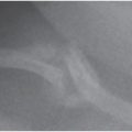
Fractures are common in fatally abused infants and young children and imaging assessments of skeletal injuries have been an important focus of postmortem studies (1–21). Computed tomography (CT) and magnetic resonance imaging (MRI) have also been used to assess for skeletal, visceral, central nervous system, and soft tissue injuries (22–28). Although a thorough autopsy provides information regarding the presence of fractures of the skull, bony thorax, and other major skeletal injuries, a thorough gross autopsy may offer little evidence of other associated skeletal injuries described in depth in this text (see Chapters 2–6). Radiologic evaluation is most critical in the assessment of high-specificity indicators of abuse, such as the classic metaphyseal lesion (CML), and other subtle injuries involving the extremities, spine, and shoulder girdle. The diagnosis of abuse in infant fatalities, as in living infants, frequently rests on the existence of prior injuries, and the recognition of early signs of healing at postmortem assessment is critical. In the author’s study of 31 abused infant fatalities, 93% of infants with extracranial fractures showed evidence of healing injuries on postmortem skeletal studies (6).
Even when the gross autopsy provides initial information regarding the presence of fracture involving the cranial vault, radiologic evaluation of the skull is an important complement to direct inspection. It is also true that gross inspection gives information regarding acute rib fractures, but a thorough radiologic evaluation of the rib cage, with specimen radiography, may reveal additional fractures, particularly those near the costovertebral articulations (7, 29). Recent healing fractures are usually detected upon careful gross inspection, but mature fractures may be missed. Furthermore, the radiologic criteria for assessment of the age of healing rib fractures are more precise than what can be determined by direct inspection. The radiologic evaluation should be followed by histologic analysis, and the ultimate assessment of the injury should be based on a combination of the gross autopsy, radiologic study, and histologic examination. Conventional CT has value in the postmortem assessment, particularly with respect to rib injuries, but at this time it should be seen as a complement and not a substitute for radiography. It may also miss fractures noted at gross autopsy (28). High-resolution CT is, however, useful for the detailed assessment of individual fractures (see below) (27).
It must be emphasized that once a child has died, and the custody of the deceased is assumed by the medical examiner/coroner, all further diagnostic imaging examinations performed by a radiology department must be ordered by the legally responsible agency. The satisfactory completion of the appropriate radiologic examinations and pathologic studies will best be achieved with a collegial and informed collaboration between these two entities.
Skeletal survey
Indications
Child abuse fatalities occur at any age but are most frequent in infants and toddlers (30–34). Although it is prudent to radiograph the entire skeleton in all cases of suspected abuse, radiologic examination is most critical in infants and young children. The indications for a skeletal survey (SS) are much the same as those for the forensic autopsy. The purpose is to assist in the determination of cause and manner of death. In the current context, the goal is to evaluate the possibility of skeletal injury, but it should be noted that other skeletal abnormalities may be encountered that support a naturally occurring illness. A search for skeletal injuries is an important component of the assessment of cases of sudden unexpected infant death (SUID), and the absence of skeletal injuries may support a determination of sudden infant death syndrome (SIDS) (see below) (http://www.cdc.gov/sids/).
The application of high-detail postmortem skeletal radiography has paralleled the evolution of the pediatric postmortem examination in cases of unexplained death in early childhood (3, 35, 36). In the past, it was common practice in some regions to assign a cause and manner of death to unexplained fatalities in children without performing an autopsy (37). The performance of postmortem skeletal radiography has been inconsistent, and these uneven practices continue despite education and legislation that has made autopsy mandatory (38). The “babygram,” a radiographic study that entails one or two images of the entire skeleton, is an inadequate examination to assess for skeletal injuries, particularly when they are inflicted. This view has been repeatedly stressed in the preceding chapters. The babygram should be strenuously rejected, as it may not only fail to identify critical forensic data, but the apparent absence of fractures may give unjustified reassurance that no trauma has occurred in cases of inflicted injury. The term “total body x-rays” is an ambiguous one, connoting a study ranging from a single “babygram” to a multiple view examination.
The literature supports the view that a complete analysis of the extent of inflicted skeletal injury in cases of suspected abuse is facilitated by high-quality skeletal imaging studies, and such exams should be interpreted by radiologists skilled in these assessments. The American College of Radiology (ACR) has developed standards for the performance of SSs in cases of suspected abuse (39). This approach has been detailed in Chapters 14 and 30. Although a similar approach entailing a thorough and meticulous postmortem SS has been advocated by the Society for Pediatric Radiology, the American Academy of Pediatrics (AAP), and the National Association of Medical Examiners (9, 17), compliance with these recommendations has been limited. In a US national survey of pathologist members of the American Academy of Forensic Sciences, Laskey and others found that 30% of respondents reported the preferential use of the “babygram” for radiographic assessment of unexplained deaths in children aged under 36 months (13). Only 10% of pathologists routinely ordered postmortem SSs. The routine use of thorough high-detail postmortem SSs is influenced by a variety of factors related to economics, technical expertise, education, and sheer inertia. Although authors often acknowledge the need for rigorous imaging, in 2012 some still maintain that a babygram is still acceptable to “at least provide a radiographic record of gross findings” (40, 41). It is curious and unfortunate that the victim of fatal abuse is not afforded the same level of forensic diagnostic imaging as the child who has survived inflicted injuries.
Technique
The SS is best performed before the autopsy. Postmortem artifact involving the skull, the thorax, and on occasion the spine and shoulder girdle may hinder optimal radiologic assessment. However, valuable information regarding skeletal trauma can be obtained in cases that have been autopsied. Even in cases in which ancient skeletal remains are discovered or a child’s body is exhumed, high-detail skeletal imaging can provide invaluable information (42, 43).
The SS protocol is virtually identical to that used in living children and is detailed in Chapter 30. To summarize, the examination entails tightly collimated views of each portion of the extremities in the frontal projection and views of the axial skeleton in frontal and lateral projections. Oblique views of the ribs are also obtained and the survey should be supplemented with additional views of all positive and suspicious regions to optimize detection. The postmortem study is generally more rapidly performed than in living children and is only slightly hampered by postmortem rigor. Generous use of tape is necessary to maintain an optimal position for exposure.
Proper exposure techniques, positioning, beam collimation, and digital processing/display parameters are vital, and other technical measures are essential to insure diagnostic quality images (see Chapter 30). Standard radiographic units, including portable machines, are usually adequate to generate high-quality images in infants and toddlers.
When a radiographic study is performed in the morgue, digital imaging receptors, and processing parameters are often chosen based on the requirements for older children and adults. These techniques are adequate for assessing metallic foreign bodies and gross skeletal injury; they are inadequate to delineate the subtle osseous abnormalities that occur in infants and young children. An imaging system should be chosen to provide optimal image detail and, thus, the digital systems should have sufficient resolution/efficiency characteristics and be performed with technical exposure factors that yield sufficient signal/noise of the image (see Chapter 28). Image receptors designed for general applications are adequate to define gross fractures, but experimental and clinical studies indicate that some CMLs and rib fractures require high-detail images for identification (39, 44, 45). Modern imaging systems yield high-detail exposure levels well within the output capabilities of most radiographic equipment. Technologists and physicians performing routine postmortem radiologic studies can be easily trained in high-detail imaging. Once the routine has been learned, the entire SS can be performed in as little as 30 minutes. Rib fractures, particularly those near the costovertebral and costochondral junctions, as well as vertebral fractures, may be characterized with conventional CT before autopsy to guide specimen resection. Although CT may supplement radiographs and guide the autopsy, the evidence is lacking at this time to support replacement of radiography with this technique (46).
Initial radiologist review
The survey should be monitored by a radiologist to assure technical adequacy. Note should be made of any indwelling lines, catheters, tubes, and any other foreign material, and if necessary, confirmation should be made with visual inspection of the body. In addition, customary assessment for nonosseous findings is appropriate, in particular, abnormal gas collections within the chest, abdomen, or vascular structures. It may be difficult to distinguish pathologic collections of gas from postmortem artifact (47–49).
Specimen resection
Once the SS has been done and the general autopsy performed, specimen resection should be undertaken. The issue regarding how much pathologic documentation of suspected skeletal injury is sufficient is a forensic one, and approaches vary depending on the attitudes of the medical examiner and other investigative and prosecutorial agencies. At the author’s center, the philosophy is to provide a thorough documentation of all sites of suspected injury. This approach appears to have an impact on the investigation and prosecution of cases of fatal infant abuse (5).
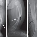
Metaphyseal injuries are common in infant abuse. Therefore, in addition to resection of all sites of suspected injury, the corresponding contralateral metaphysis is usually removed. In addition, because most CMLs in infants involve the distal femurs, the proximal and distal tibias, and the proximal humeri, these regions may also be resected when other osseous injury is encountered (see Chapters 2–4). Removal of the epiphysis, the physis, and 2–3 cm of the adjacent metaphysis is recommended. The bone should be transected with care to avoid any disruption of the osteocartilaginous structures; fractures may be artifactually separated along the plane of injury with vigorous removal. Removing a block of bone extending from the distal femoral metaphysis to the proximal tibial metaphysis simplifies the resection of these important sites, and subsequent division of the knee articulation permits independent radiographic and histologic analysis of the individual metaphyses. Resection of the metaphyses of the distal extremities requires separate skin incisions. The proximal humeral metaphyses can be exposed through extension of the excision that exposes the thorax.
The ribs should be resected with their vertebral articulations. It is advisable to resect the entire vertebral body and both rib arcs as a unit (see Chapter 5). When widespread thoracic injury is present, it is most useful to resect a block of contiguous vertebrae with their costal articulations. In some cases we have removed the entire thorax, permitting analysis of each rib with its vertebral articulation. When the vertebral column is normal radiographically, it is not routinely removed, except at the level of rib fractures. When spinal injuries are present, the fractured vertebrae, as well as the levels above and below it are resected en bloc (see Chapter 21). When fractures are noted involving the long bone shafts, hands, feet, clavicle, and scapula and any other anatomic sites, these regions are removed accordingly.
Specimen radiography
Specimen radiography should be performed with high-detail imaging systems. Digital imaging plates (computed radiography/digital radiography or CR/DR) can be exposed at low kilovoltage and high tube current for high contrast and optimal signal/noise with a conventional radiographic machine. High-resolution images may be acquired in a digital cabinet unit designed for specimen radiography. The extremities are viewed in the frontal and lateral projection, and on occasion oblique/angled projections are required to optimize fracture detection. The rib cage is radiographed in frontal projection, and axial views of the individual ribs and their costovertebral articulations are also obtained. This axial projection is essential for delineating acute rib injuries at any site, and especially healing fractures of the costochondral and costovertebral articulations. The clavicle should be viewed in frontal and angled projections. The vertebral bodies should be viewed in frontal, lateral, and axial projections. It may also be useful to radiograph a resected skull fracture.
Specimen high-resolution CT
If high-resolution or micro-CT is available, it may better define injuries suspected radiographically. Flat-panel detector CT provides substantially higher resolution than customary detector arrays and can elegantly depict metaphyseal injuries, but the resolution still falls below that achieved with micro-CT. Because these techniques entail the acquisition and processing of huge volumes of data and the imaging field size is small with micro-CT, they are at the moment only practical for the assessment of individual fractures and not for global screening. They can confirm injuries suspected radiographically and provide elegant imaging/histopathologic correlations (27).
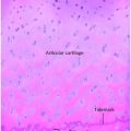
Histologic analysis
As is customary with other forensic pathology specimens, resected material should generally be examined microscopically (10–12). All resected specimens should be immediately placed in formalin and carefully labeled with all identifying information. Once fixation and specimen radiography have been performed, the material can be placed in saline or directly into decalcifying solution. Sectioning should be carried out in conjunction with radiologic analysis. Histologic sections should be obtained in a plane that corresponds best to the radiographic projection that clearly depicts the anatomic alterations. This ensures optimal radiologic–histologic correlation. Slides are generally stained with hematoxylin and eosin; other special stains are used if deemed appropriate.
Data analysis
The SS, specimen radiography, and histologic sections are analyzed and the data carefully recorded. At the author’s institution, we have developed a computerized database that allows tabulation and analysis of the data, region by region and bone by bone. Each fracture is characterized in terms of the portion of bone involved and whether healing is noted radiographically and histologically. This detailed analysis facilitates the construction of a report that encompasses both radiologic and pathologic findings that can be used in the investigation, as well as any subsequent legal proceedings. This aggressive radiologic–histologic assessment to define skeletal injury is most applicable to infants; beyond infancy, the approach can be modified. The SS should be performed in a similar manner, but resection of osseous injury should be restricted to those areas noted to be abnormal or suspicious on SS.
Differential diagnosis of fatal abuse
A variety of conditions may result in sudden unexpected death in infants and children, and skeletal imaging performed initially to evaluate for traumatic injury can also assess for an underlying medical entity. Naturally occurring conditions include various infections, metabolic disorders, cardiac arrhythmias, central nervous system tumors, vascular malformations, endogenous hyperthermia/hypothermia, etc. A discussion of these diseases is beyond the scope of this text, and the reader is referred to other articles and texts for in-depth coverage of these topics (3, 35, 50–53).
SIDS, an apparent life-threatening event (ALTE), and accidental suffocations commonly share the differential diagnosis in cases of SUID (3, 9–12, 40, 54–63). SIDS is defined as the sudden death of an infant less than one year of age that cannot be explained after a thorough investigation is conducted, including a complete autopsy, examination of the death scene, and review of the clinical history. Often previously referred to as “cot” or “crib” death,” it has been the subject of interest throughout history (3, 64–72). SIDS is the leading cause of death between one and six months of age, sharing the peak incidence time period of abuse fatalities (9, 73, 74). The rate of SIDS is higher among black and American Indian/Alaska native populations. A variety of theories have been proposed to explain SIDS, but no single etiologic factor has been identified. It has been strongly linked to prone sleep position and there has been a dramatic reduction in SIDS deaths following recent campaigns aimed at limiting prone sleeping. However, the rates for non-Hispanic black and American Indian/Alaska native infants have shown a slower decline. Because SIDS is a diagnosis of exclusion, skeletal imaging is essential, but as noted above, SSs are often omitted in the forensic assessment of SUID (13).
In the last century, it became evident that some cases of fatal abuse were initially diagnosed as SIDS (9, 51, 65, 66, 70, 75–94). Although there has been progress in making this critical distinction, confusion between these causes of sudden infant fatality persists (9, 36, 40, 41, 95). The subject of SIDS is a complex and interesting one and the reader is referred to the references cited in this text, in particular the discussion by Reece and Christian, which focuses on differentiation of abuse from SIDS (96).
The radiographic SS is designed to identify those osseous alterations that point to inflicted injury. Cases of abuse may initially be mistakenly attributed to SIDS, and the SS plays an important role in detecting findings characteristic of inflicted injury. We found that SIDS and/or apnea was initially suspected in 50% of cases subsequently shown with the aid of postmortem radiography to be abuse related (5). Indications of trauma elucidated by SS are of particular value in cases in which an autopsy fails to identify a cause of death (3). The autopsy findings of SIDS may be indistinguishable from those of suffocation. Valdes-Dapena stated that “it is impossible to distinguish at autopsy between accidental or deliberate asphyxiation with a soft object and SIDS” (93). The presence of new and old inflicted injuries identified on SS before autopsy may allow a more focused postmortem examination and death scene evaluation, as well as law enforcement investigation.
Conclusion
The thorough postmortem imaging analysis of cases of suspected child abuse is an extension of the imaging procedure in living children. This approach, in keeping with the principles of forensic scientific analysis, should be carried out without bias so that the results will not only assist in cases where evidence of abuse is discovered, but also in facilitating the determination of accidental and natural causes of death. The growth of multidisciplinary child fatality review teams has brought the issue of SUID under closer scrutiny and has highlighted the need for thorough and meticulous autopsies and death scene investigations (97, 98). As experts in the diagnostic imaging of abuse and its imitators, pediatric radiologists have an obligation to assist medical examiners and child death review teams with these with challenging cases. All professionals involved with these tragic cases should adhere to high standards of practice to ensure that all necessary forensic data are identified and fully characterized.
References
Related posts:
 Evidence-based radiology and child abuse
Abusive head trauma: clinical, biomechanical, and imaging considerations
Evidence-based radiology and child abuse
Abusive head trauma: clinical, biomechanical, and imaging considerations
 Dating fractures
 Differential diagnosis I: diseases, dysplasias, and syndromes
 The skeleton: structure, growth and development, and basis of skeletal injury
 Epithelioid Hemangioendothelioma
