Post-Transplant Lymphoproliferative Disorder

Published on 19/07/2015 by admin

Filed under Radiology

Last modified 22/04/2025

Print this page

rate 1 star rate 2 star rate 3 star rate 4 star rate 5 star
Your rating: none, Average: 5 (1 votes)

This article have been viewed 1671 times

 Site of presentation may depend partially on transplanted organ

image Extranodal (80%) > nodal involvement (20%)
• Imaging findings of post-transplant lymphoproliferative disorder parallel those of non-Hodgkin lymphoma (NHL) in immunocompetent patients
• GI tract: Imaging findings are similar to NHL, including mass-like bowel wall thickening, aneurysmal dilatation, ulcerated polyploid mass, or submucosal nodules

image Increased prevalence of ulceration and bowel perforation
• Liver: Most frequently involved abdominal solid organ

image Single or multiple poorly enhancing masses, discrete mass in porta hepatis, or diffuse infiltration of liver
• Spleen: Splenomegaly ± discrete lesions (usually multiple, hypoattenuating, and variable in size)
• Kidney: Most common site in renal transplant recipients

image Heterogeneous mass surrounding hilar vessels, parenchymal masses, or diffuse infiltrative disease
• Nodal disease: Abdominal nodal involvement in only 15-20% of cases

image Nodal involvement much less common than in immunocompetent NHL

TOP DIFFERENTIAL DIAGNOSES

• Recurrent or new malignancy
• Opportunistic infections

PATHOLOGY

• Most cases are related to B-lymphocyte proliferation due to Epstein-Barr virus (EBV) infection

CLINICAL ISSUES

• High mortality, with survival rates of only 25-35%
• Treatment: Reduction or cessation of immunosuppression can be effective, although antiviral drugs, chemotherapy, or rituximab may be necessary
image
(Left) Axial CECT in a patient post liver transplant demonstrates a new hypodense mass image 
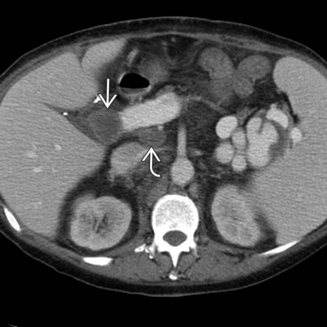
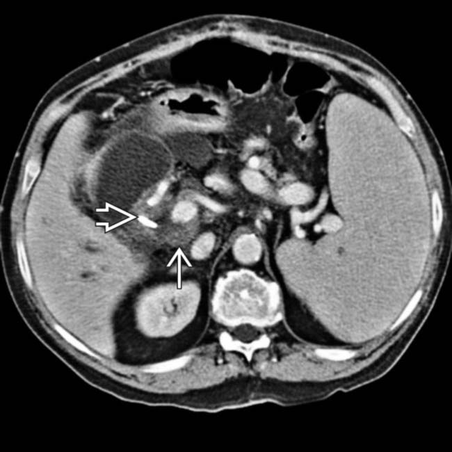
in the porta hepatis, as well as an enlarging portacaval lymph node image.

image
(Right) Axial CECT in the same patient demonstrates extensive retroperitoneal lymphadenopathy image. The findings of post-transplant lymphoproliferative disorder (PTLD) in this case are indistinguishable from traditional non-Hodgkin lymphoma (NHL) in an immunocompetent patient.
image
(Left) Axial NECT demonstrates mass-like wall thickening image 
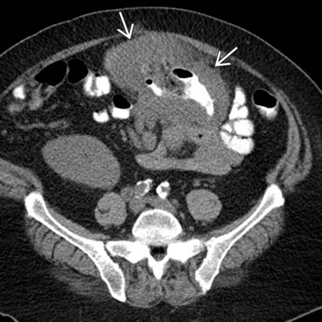
of a segment of colon with aneurysmal dilatation.

image
(Right) Coronal NECT in the same patient again demonstrates the significant wall thickening image 
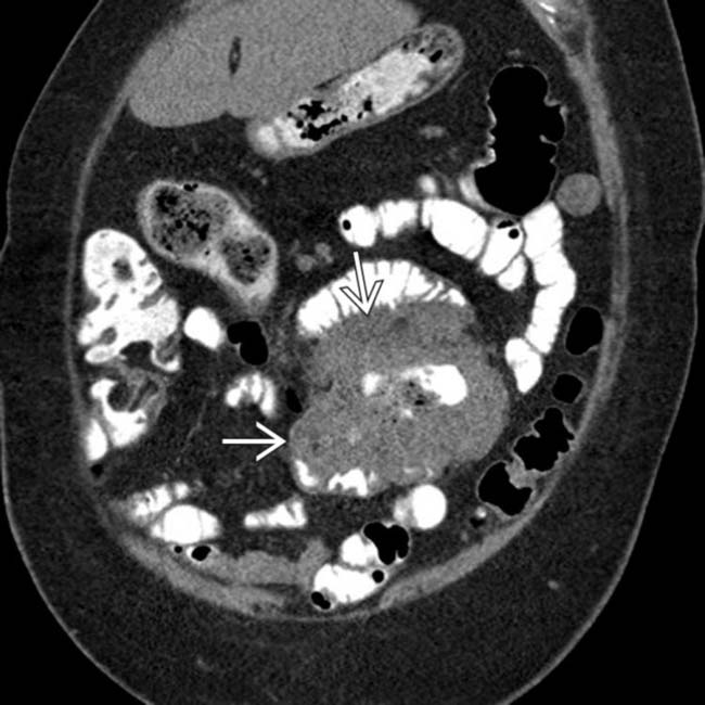
of the bowel segment with dilatation. This is a common appearance for both NHL in immunocompetent patients and PTLD.

TERMINOLOGY

Abbreviations

• Post-transplant lymphoproliferative disorder (PTLD)

Definitions

• Heterogeneous group of lymphoproliferative diseases that occur in post-transplant setting (either solid organ or stem cell transplants), ranging from abnormal lymphoid hyperplasias to frank malignancies

IMAGING

General Features

• Location

image Extranodal involvement (80%) is much more common than nodal involvement (20%)

– Unlike lymphoma in general population where nodal disease predominates
image Can occur nearly anywhere, with common locations including lungs, GI tract, and CNS

– Site of presentation may depend partially on type of transplanted organ
– Abdominal cavity is most frequently involved (up to 50% of all cases)
– May occur within renal and liver allografts

image Some studies have suggested that PTLD may preferentially affect allograft itself
• Size

image Masses and nodes range from < 1 cm to huge masses

Imaging Recommendations

• Best imaging tool

image CECT for initial diagnosis
image PET/CT for staging and follow-up

CT Findings

• Imaging findings of PTLD mostly parallel those of non-Hodgkin lymphoma (NHL) in immunocompetent patients
• GI tract

image Small bowel (distal > proximal) > colon > stomach > duodenum > esophagus
image Imaging findings are similar to NHL in immunocompetent patients

– Mass-like wall thickening (most common) with aneurysmal dilatation of lumen
– Dominant polyploid mass (often with ulceration) or multiple submucosal nodules
– May present with intussusception
image Unlike lymphoma in general population, there is a markedly increased prevalence of ulceration and perforation of bowel

– Spontaneous perforation may be 1st symptom of PTLD
• Liver

image Most frequently involved abdominal solid organ
image Several possible appearances

– Most often single or multiple low attenuation, poorly enhancing masses

image Lesions may vary in size (few mm to few cm)
– Diffuse or geographic infiltration of liver with no discrete lesions (liver appears steatotic)
– Discrete mass in porta hepatis (sometimes with extension into biliary tree or gallbladder)

image Unique manifestation of PTLD (not common with immunocompetent lymphoma)
• Spleen

image Spleen involved in 10-40% of cases (particularly common after liver transplant)
image Possible appearances

– Splenomegaly (most common) ± discrete parenchymal lesions
– Parenchymal lesions are typically multiple, low-attenuation, and variable in size
image Spontaneous rupture is possible complication
• Kidney

image Most commonly involved site in renal transplant recipients and may affect native kidneys or allograft
image Renal allograft involvement

– Heterogeneous mass surrounding hilar vessels
– Multifocal parenchymal masses
image Native kidney involvement

– Almost always unilateral
– Discrete round, hypoenhancing parenchymal lesions
– Diffuse infiltrative disease with nephromegaly
• Adrenal

image Adrenal involvement in 5%
image Diffuse infiltration with adrenal enlargement or discrete homogeneous hypoenhancing mass
• Pancreas

image Rare manifestation of PTLD only described in pancreatic allograft (not native pancreas)
image Diffuse enlargement of pancreas mimicking acute pancreatitis
image Focal hypoenhancing mass in pancreas mimicking pancreatic cancer

– No ductal dilatation or pancreatic atrophy
• Nodal disease

image Abdominal nodal involvement in only 15-20%

– Less common than immunocompetent lymphoma
– Nodal involvement is typically seen along with extranodal disease
image Retroperitoneal nodes > mesenteric nodes
image Discrete homogeneous enlarged lymph nodes or conglomerate mass of confluent enlarged nodes

MR Findings

• Liver

image Low signal intensity on T1WI
image Mildly hyperintense on T2WI
image Variable, usually minimal, enhancement on T1 C+
• Spleen

image Isointense to spleen on T1WI
image Iso- to hypointense on T2WI
image Minimal enhancement on T1 C+
• Kidney

image Isointense to renal parenchyma on T1WI
image Slightly hypointense on T2WI
image Mild enhancement on T1 C+

Fluoroscopic Findings

• Luminal ulceration (“target” or bull’s-eye lesions)

image Indirect signs of larger mass (e.g., displaced bowel loops)
• Aneurysmal dilatation of bowel lumen
• Intraluminal polypoid filling defect

Nuclear Medicine Findings

• PET/CT

image May detect disease that is occult on CT (including activity in normal-sized lymph nodes)
image Useful for follow-up after treatment

– Can differentiate viable tumor after treatment from fibrotic tissue without viable tumor
image Importance of PET/CT partially extrapolated from role in lymphoma in immunocompetent patients
image May be more effective in diagnosing and staging monomorphic PTLD than polymorphic PTLD

DIFFERENTIAL DIAGNOSIS

Recurrent or New Malignancy

• Patients with known prior malignancy are at high risk of recurrent malignancy after transplantation

image e.g., liver transplantation for hepatocellular carcinoma
• Previously indolent or undiscovered malignancies may grow rapidly under immunosuppressive therapy

image Transplant recipients are at markedly increased risk for multiple cancers

– Skin and lips (squamous cell carcinomas): Account for ∼ 40% of malignancies in transplant recipients
– Vulvar, cervical, and uterine cancers
– Kaposi sarcoma (6% incidence)
– Renal cell carcinoma (in native kidneys or allograft)

Intestinal Opportunistic Infection

• May result in bowel wall thickening, but not discrete mass
• May result in modest lymphadenopathy

Hepatic and Splenic Opportunistic Infections

• Serologic markers and clinical findings may be helpful
• Hepatic and splenic microabscesses

image Usually due to fungal infection, such as candidiasis
image Multiple small hypodense nodules on CECT may mimic one of the possible appearances of PTLD
image Usually demonstrate low T1 signal and markedly high T2 signal intensity on MR with variable peripheral enhancement

PATHOLOGY

General Features

• Etiology

image Most cases are related to B-lymphocyte proliferation due to Epstein-Barr virus (EBV) infection

– Immunosuppression (with ↓ T-cell response) results in uncontrolled proliferation of EBV-infected B cells
image Pathogenesis of EBV negative PTLD is unknown

– May account for ∼ 30% of all PTLD cases
image Risk factors

– Primary EBV infection
– Transplant from seropositive donor to seronegative recipient
– Cytomegalovirus infection
– Pediatric age group or age > 60 years
– Greater degrees of immunosuppression (may be associated with certain drug regimens)

image Greatest risk of PTLD in 1st year after transplant when immunosuppression is highest
image Can occur in recipients of either

– Solid organ allografts
– Allogenic hematopoietic stem cell transplantation

Staging, Grading, & Classification

• Hyperplastic (early) lesions

image Polyclonal proliferation of B-lymphocytes without evidence of frank malignancy
image Symptoms similar to acute viral infection
• Polymorphic lesions

image Demonstrate evidence of malignant transformation, but do not meet criteria needed to diagnose traditional lymphomas in immunocompetent patients
• Monomorphic (lymphomatous) lesions

image Frank malignancies that can be described using standard nomenclature for lymphoma subtypes

– B-cell type (most common)
– T-cell type (accounts for 14% of cases)
– NK-cell type
• Other lymphoproliferative disorders (e.g., Hodgkin)

Microscopic Features

• Diffuse polymorphic or monomorphic proliferation of mononuclear cells

CLINICAL ISSUES

Presentation

• Most common signs/symptoms

image Some patients are asymptomatic in early stages
image Fever, night sweats, weight loss, fatigue
image GI symptoms, allograft dysfunction
image Laboratory: Markedly elevated EBV viral load, anemia, thrombocytopenia, leukopenia
• Other signs/symptoms

image Symptoms referable to affected organ

– e.g., headache or seizure with CNS involvement

Demographics

• Epidemiology

image Frequency depends on type of allograft

– Multivisceral transplant recipients (13-33%)
– Intestinal transplant (7-11%)
– Heart-lung transplant (10%)
– Lung transplant (2-8%)
– Heart transplant (3-4%)
– Liver transplant (2%)
– Kidney transplant (1%)
image Highest incidence within 1st year after transplantation (accounts for > 80% of all PTLD cases)

Natural History & Prognosis

• Despite all treatments mortality is still high

image Survival rates of only 25-35%
image Mortality highest with monomorphic PTLD

Treatment

• Reduction or cessation of immunosuppression for early polyclonal lesions

image Return of intact immune system will reject early PTLD as well as allograft in most cases
image Some patients can be weaned off immunosuppressive therapy without rejection
image More likely to work with polymorphic PTLD; unlikely to be effective in monomorphic PTLD
• Other options

image Antiviral medication, such as acyclovir (for early polyclonal disease)
image Chemotherapy (CHOP is most common regimen)
image Rituximab (monoclonal antibody against CD20 receptor)
image Localized radiation therapy
• Surgery for complications

image e.g., obstructed or perforated bowel

DIAGNOSTIC CHECKLIST

Consider

• Any soft tissue density nodal or visceral mass in transplant recipient should be considered PTLD until proven otherwise
• Biopsy will often be required for confirmation

image
(Left) Axial NECT in a patient with a renal transplant shows large lobulated masses image 
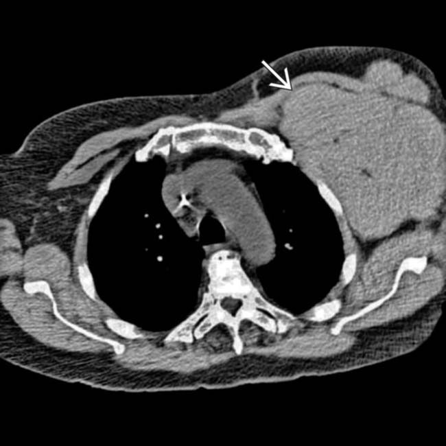
in the left axilla, representing dramatically enlarged left axillary lymph nodes.
image
(Right) Axial NECT in the same patient shows markedly atrophic native kidneys image 
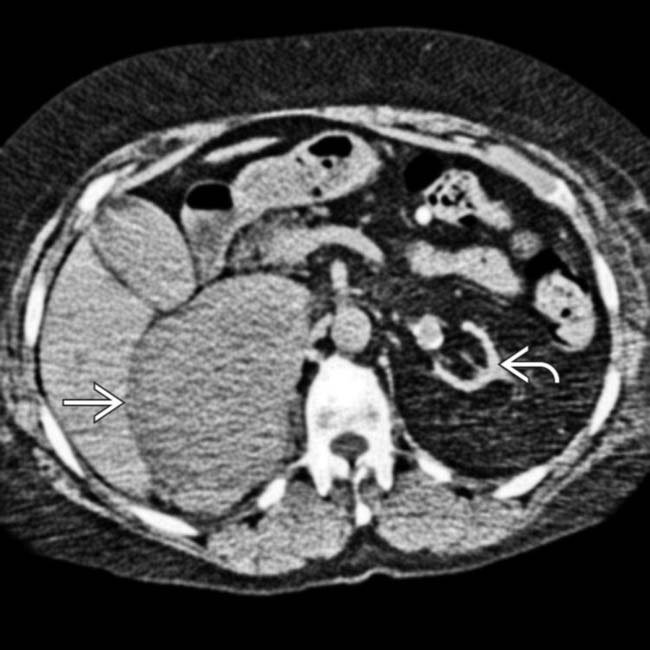
and a right adrenal or retroperitoneal mass image, compatible with the patient’s PTLD. PTLD can affect essentially any organ or lymph node group in the body, and any soft tissue mass in a transplant recipient must be regarded as worrisome for PTLD.
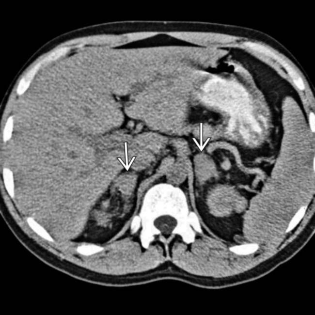
image
(Left) Axial NECT in a patient with a renal transplant shows bilateral adrenal masses image.

image
(Right) More caudal axial NECT section in the same patient shows the renal allograft in the right iliac fossa image
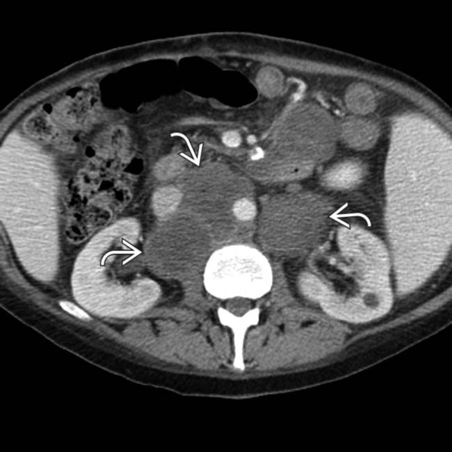
. There is massive pelvic lymphadenopathy image. Both the lymphadenopathy and the adrenal masses represent manifestations of PTLD.
image
(Left) Axial CECT in a liver transplant patient shows a large mass image 
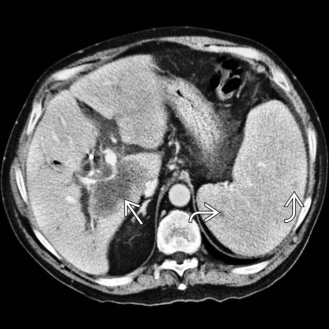
within the liver allograft that envelopes the portal vein and bile ducts. The spleen is also involved, with splenomegaly and dozens of small hypodense nodules image.

image
(Right) Axial CECT in the same patient shows porta hepatis and portacaval adenopathy image present as manifestations of PTLD. Note the biliary stent image placed to treat the extrinsic compression of the common bile duct by enlarged nodes and the liver mass.
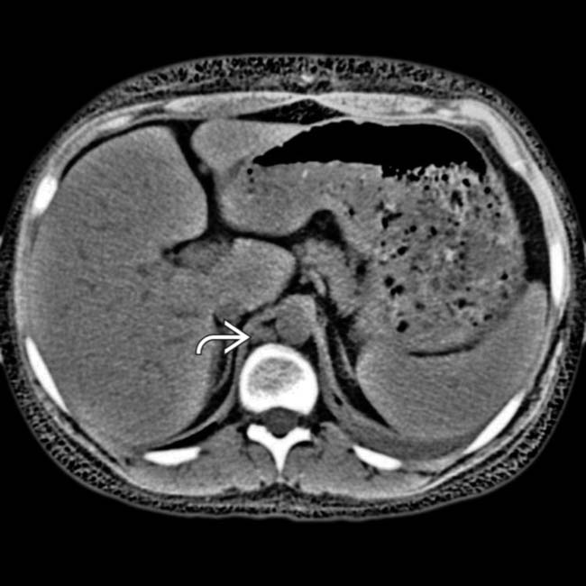
image
Axial NECT in a 20-year-old woman with a heart transplant shows retrocrural lymphadenopathy image.

image
CT in the same patient shows splenomegaly and lymphadenopathy image
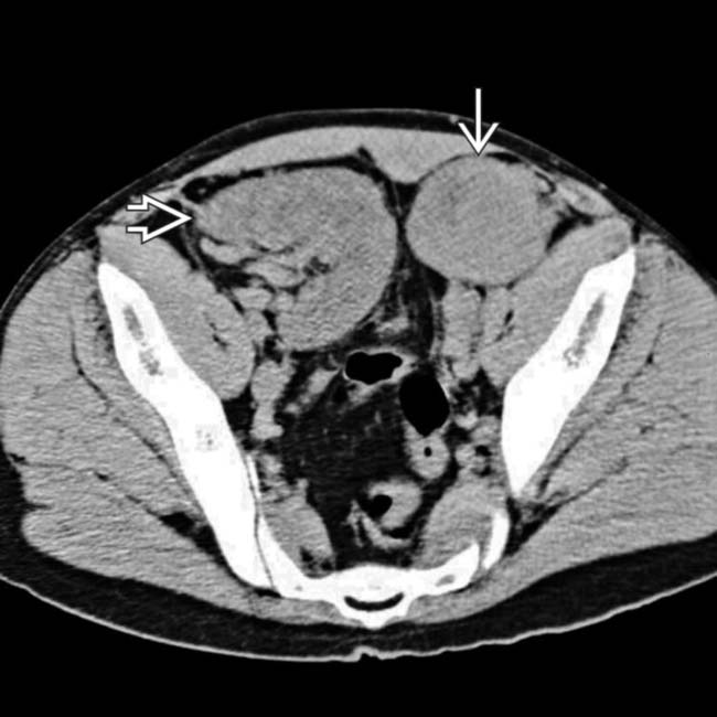
. The lack of oral or IV contrast makes it more difficult to distinguish the mesenteric and retroperitoneal nodes from unopacified vessels and bowel. Patients who have undergone heart transplantation are at markedly increased risk of developing PTLD.
image
Axial NECT shows a cavitated soft tissue mass image 
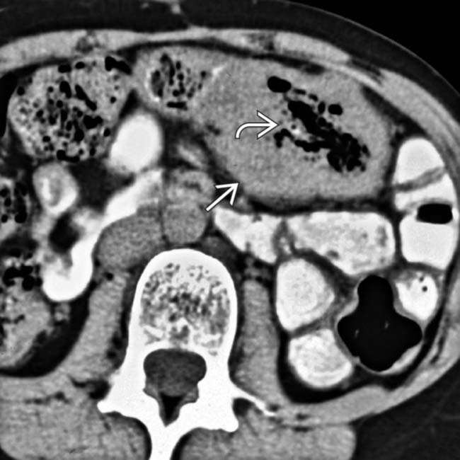
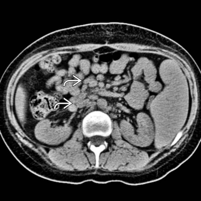
encasing the jejunum. In the center of the mass is an irregular gas-filled lumen image. There is no evidence of bowel obstruction, as more distal segments of bowel fill with enteric contrast medium.
image
Small bowel follow-through shows indirect evidence of the size of the soft tissue mass image 
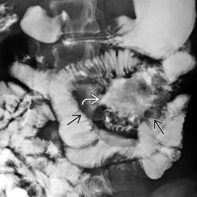
with displaced bowel loops. The cavitated lumen of the involved jejunal segment image is evident as a collection of barium lacking any sign of intact mucosa.

Share this: