Portal Hypertension and Varices

Published on 13/07/2015 by admin

Filed under Radiology

Last modified 22/04/2025

Print this page

rate 1 star rate 2 star rate 3 star rate 4 star rate 5 star
Your rating: none, Average: 0 (0 votes)

This article have been viewed 2379 times

 Ascites: Fluid in dependent recesses of peritoneal cavity

image Splenomegaly: Spleen is enlarged > 500 cc
image Varices: Well-defined, tubular or serpentine collateral vessels with same enhancement as adjacent veins

– Usually opacify on venous phase (not arterial phase)
image Mesenteric edema: ↑ attenuation of mesenteric fat
image Dilation of mesenteric veins : Portal vein > 13 mm
image Slow or reversed flow in portal veins on ultrasound

– Slow flow in portal vein (< 15 cm/sec)
– Portal flow may be biphasic or completely reversed
– ↑ risk of portal vein thrombosis
image Portal gastropathy, enteropathy, and colopathy

– May manifest as thickening of colon (especially right colon), small bowel, and stomach
image Gallbladder wall thickening

PATHOLOGY

• Causes of portal hypertension divided into 3 categories

image Pre sinusoidal: Portal vein/splenic vein thrombosis
image Sinusoidal: Cirrhosis (most common), extensive hepatic tumor, or any severe hepatocellular disorder
image Post sinusoidal: Right-sided heart failure, constrictive pericarditis, Budd-Chiari syndrome
• Varices (portosystemic collaterals) develop when portal pressures increase beyond critical value

image Blood in portal venous system bypasses liver and empties into systemic veins via portosystemic collaterals

CLINICAL ISSUES

• Asymptomatic until varices rupture or ascites are infected
• 30% of patients with esophageal varices experience variceal hemorrhage within 2 years of diagnosis
• Many patients with cirrhotic ascites develop spontaneous bacterial (or rarely tuberculous) peritonitis
image
(Left) Graphic shows esophageal varices image 
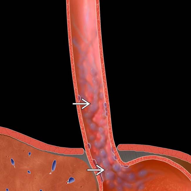
as serpiginous, longitudinally oriented submucosal venous collaterals extending into the gastric fundus.

image
(Right) Esophagram performed after endoscopic sclerosis of varices shows defects image 
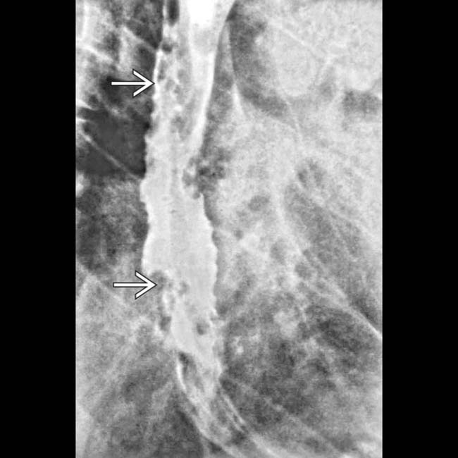
in the esophageal wall. Varices are usually pliable and easily compressed. Varicoid carcinoma could have a similar appearance.
image
(Left) Coronal CECT demonstrates multiple varices image 
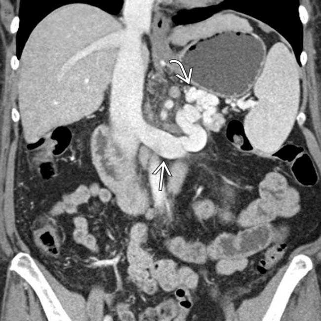
in the left upper quadrant in communication with the splenic vein and the left renal vein image, which appears dilated, forming a splenorenal shunt.

image
(Right) Coronal CECT in a patient with cirrhosis and portal hypertension demonstrates thrombus image 
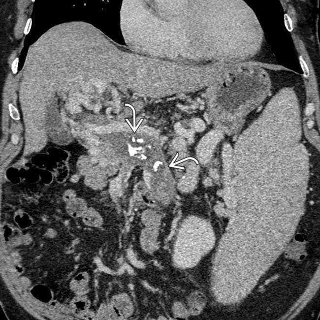
in the portal and superior mesenteric veins, with calcification suggesting chronicity. Portal hypertension increases the risk of portal vein thrombus due to stasis and slow flow.

TERMINOLOGY

Definitions

• Portal hypertension: Elevated portal pressures due to resistance to portal flow, defined as absolute portal venous pressure of > 10 mm Hg or gradient between portal and systemic veins of > 5 mm Hg
• Varices: Abnormally dilated and tortuous veins due to rerouting of blood flow away from liver into lower pressure systemic veins through collateral pathways
• Ascites: Pathologic fluid accumulation in peritoneal cavity

IMAGING

General Features

• Best diagnostic clue
• Location

image Ascites: Dependent recesses of peritoneal cavity
• Common features of portal hypertension

image Ascites: Water density fluid in dependent recesses of peritoneal cavity
image Splenomegaly: Spleen is enlarged > 500 cc

– CT or US can estimate or calculate splenic volume
– Usual upper limits: 13 cm in length and 6 × 8 cm in width and breadth, respectively
image Varices: Well-defined, tubular or serpentine portosystemic collateral vessels with same enhancement as adjacent veins

– Usually opacify on venous phase images (not on arterial phase)
image Mesenteric edema: Stranding and increased attenuation in mesenteric fat
image Dilation of mesenteric veins (portal vein, superior mesenteric vein, and splenic vein)

– Portal vein is often dilated > 13 mm
image Slow or reversed flow in portal veins on Doppler ultrasound

– Normal portal vein flow is hepatopetal (toward liver) with normal flow velocity of 15-40 cm/sec
– As portal hypertension progresses, portal blood flow velocities decrease
– Eventually, portal flow may be biphasic (alternating hepatopetal/hepatofugal flow) or completely reversed
– Stasis of blood flow in portal vein as well as hypercoagulability due to intrinsic liver disease increases risk of portal vein thrombosis
image Portal gastropathy, enteropathy, and colopathy

– May manifest as thickening of colon (especially right colon), small bowel, and stomach
image Gallbladder wall thickening: Due to combination of portal hypertension, hepatocellular dysfunction, and hypoproteinemia
• Varices: Types or locations

image Left gastric venous collateral vessels

– Vascular channels in triangular fatty tissue between medial wall of upper gastric body and posterior margin of left hepatic lobe in lesser omentum
– Coronary (left gastric) vein ≥ 5-6 mm in diameter is considered abnormal
– Usually accompanied by esophageal or paraesophageal varices
image Esophageal varices

– Dilated tortuous submucosal venous plexus of esophagus can be divided into “uphill” and “downhill” varices

image Uphill varices (collateral blood flow into superior vena cava (SVC) from portal vein via azygous vein): Result from portal hypertension and found in distal 1/2 of esophagus
image Downhill varices (collateral flow from SVC into inferior vena cava [IVC] or portal system): Usually due to SVC obstruction and found in proximal 1/3 of esophagus
– Tortuous or serpiginous longitudinal filling defects on esophagography

image Varices may collapse during esophageal peristalsis or with distention of lumen
image Best detected when esophagus is collapsed and esophageal mucosa coated with thick barium
– CECT has limited sensitivity for small esophageal varices, which may not be evident when collapsed

image Can be simulated or hidden by esophagitis or hiatal hernia
– Endoscopy is most sensitive diagnostic tool
– Most clinically significant varices due to risk of bleeding
image Paraesophageal varices

– Collateral vessels in posterior mediastinum behind esophageal wall connect coronary vein with azygos and hemiazygos veins and vertebral plexus
– Can mimic posterior mediastinal mass on chest x-ray
image Recanalized paraumbilical vein

– Dilated collateral vein (≥ 3 mm) arising from left portal vein and coursing between medial and lateral segments of left hepatic lobe in anterior edge of falciform ligament
– Presence is virtually diagnostic of portal hypertension
image Abdominal wall varices

– Prominent collateral veins radiating from umbilicus referred to as “caput medusae” (head of Medusa, a mythological figure who had snakes for hair)
– Connect with superior and inferior epigastric veins and fed by paraumbilical and omental veins
image Perisplenic varices

– Dilated veins in anteroinferior aspect of spleen traversing splenocolic ligament
– May lead to spontaneous splenorenal shunt (communication between left renal vein and splenic vein)
image Retrogastric varices

– Dilated veins in posterior or posteromedial aspect of gastric fundus near cardia
– Fed by coronary (left gastric) vein or gastroepiploic vein; drain into esophageal or paraesophageal veins
– Can mimic tumors on NECT, barium study, or endoscopy
image Omental varices

– Small and numerous varices throughout greater omentum
– Should not be mistaken for metastases or granulomas
image Retroperitoneal-paravertebral varices

– Arise from colic or mesenteric branches
image Mesenteric varices

– Dilated and tortuous branches of superior mesenteric vein in leaves of mesentery ± bowel wall
– Can be cause of lower GI bleeding
image Varices may develop in sites of previous abdominal surgery or intraabdominal inflammation

– e.g., peristomal varices surrounding ostomy in patient with cirrhosis following colectomy
• Ascites

image Cirrhosis present in 80-85% of cases

– Other causes of portal hypertension, e.g., portal or splenic vein thrombosis, massive liver tumors (5-10%)

image May coexist with liver disease, e.g., pancreatitis, hepatocellular carcinoma, alcoholic cardiomyopathy
– Other causes include cardiac ascites (3-5%), peritoneal carcinomatosis (2%)
image Ascites should have near-water attenuation on CT

– Rarely mildly hyperdense due to prior episodes of infection or hemorrhage
image Anechoic on ultrasound unless complicated
image Hypointense on T1WI, hyperintense on T2WI
image Accumulates in dependent recesses of peritoneal cavity

– Morison pouch, subphrenic spaces, paracolic gutters, and pelvis
image Patients with cirrhotic ascites have low protein in serum and ascites fluid

– Leads to more fluid loss from blood vessels
– “Low-protein ascites” more prone to spontaneous bacterial peritonitis

Imaging Recommendations

• Best imaging tool

image CECT is best study to identify varices and other manifestations of portal hypertension
image CT or US can detect and quantify both ascites and splenomegaly

DIFFERENTIAL DIAGNOSIS

Esophageal Carcinoma

• Varicoid carcinoma: Serpiginous filling defects in submucosa that are fixed and unchanged with peristalsis
• Can be simulated by sclerosed or thrombosed varices

Gastric Carcinoma

• Gastric masses may be simulated by gastric varices on either NECT or fluoroscopic barium studies
• Distinction readily made on CECT, which shows characteristic morphology and enhancement of varices

Adrenal Carcinoma

• Perigastric varices can simulate adrenal carcinoma or any mass

Pancreatic Islet Cell Tumors

• Enhanced varices in and around pancreatic head may mimic hypervascular tumor of pancreas

Middle Mediastinal Mass

• Large varices (particularly paraesophageal) mimic mediastinal mass on chest x-ray or NECT, but usually easy to make distinction on CECT
• Lymphadenopathy, aneurysm, and other masses must be considered

Superior Vena Cava Obstruction

• Obstruction of SVC results in downhill varices as collaterals follow esophagus and azygous-hemiazygos system
• Return blood to IVC via collaterals that may traverse liver

Other Cause of Ascites

• May be caused by conditions other than portal hypertension
• Congestive heart failure (cardiac ascites), peritonitis, peritoneal carcinomatosis, renal failure (especially nephrotic syndrome), pancreatitis, peritoneal dialysis, etc.

Other Causes of Gastrointestinal Bleeding

• Not always due to variceal hemorrhage in cirrhotic patients
• Other etiologies include gastritis, gastric ulcers, and Mallory-Weiss tear of distal esophagus

PATHOLOGY

General Features

• Etiology

image Causes of portal hypertension are divided into 3 categories

– Pre sinusoidal: Portal vein thrombosis, splenic vein thrombosis, compression of portal vein by tumor or lymphadenopathy, schistosomiasis
– Sinusoidal : Cirrhosis (most common cause: ∼ 80% of cases), extensive hepatic tumor (metastases, hepatocellular carcinoma), or any other severe hepatocellular disorder
– Post sinusoidal: Right-sided heart failure, constrictive pericarditis, tricuspid insufficiency, Budd-Chiari syndrome, hepatic veno-occlusive disease
image Varices begin to develop when portal pressures increase beyond critical value

– Result from dilation of embryonic channels or redirection of flow within existing veins
image Blood flow always seeks path of least resistance and lowest pressure

– When portal hypertension is severe, blood flows away from liver (hepatofugal flow)
– Blood in portal venous system will bypass liver and empty into adjacent systemic veins via portosystemic collaterals
– Coronary venous collateral vessels are most common varices in portal hypertension (80%)
– Esophageal varices are most important clinically due to risk of bleeding
image Ascites pathophysiology

– Portal hypertension → increased nitric oxide → renal sodium retention → vasodilation → increased intravascular volume → ascites
– Loss of intravascular fluid to ascites stimulates sympathetic nervous activity and more secretion of renin and aldosterone

image Leads to more sodium retention and more ascites

CLINICAL ISSUES

Presentation

• Most common signs/symptoms

image May be asymptomatic in mild cases
image Hematemesis or melena due to bleeding from esophageal or gastric varices
image Abdominal distension due to accumulation of ascites
image Abdominal pain, fever, and ↑ WBC due to spontaneous bacterial peritonitis
image Altered mental status due to hepatic encephalopathy

Demographics

• Gender

image M = F

Natural History & Prognosis

• 30% of patients with esophageal varices experience variceal hemorrhage within 2 years of diagnosis

image Most serious complication of portal hypertension (20-35% of all deaths in cirrhotic patients)
image Mortality rate is 40-70% within 6 weeks
image Highest risk for variceal bleeding is in patients with portosystemic gradient of > 12 mm Hg
• Many patients with cirrhotic ascites develop spontaneous bacterial (or rarely tuberculous) peritonitis

image Diagnosis may not be evident (often causes new onset of encephalopathy, pain, fever)
image Early diagnosis and antibiotic treatment are essential

Treatment

• Primary goal is to prevent/treat symptomatic complications

image Dietary sodium restriction, spironolactone, and diuretics to prevent ascites

– Regular paracentesis for symptomatic relief
image β-blockers and obliteration of varices (endoscopic sclerotherapy, endoscopic variceal ligation, surgical devascularization) to prevent bleeding
image Transjugular intrahepatic portosystemic shunt (TIPS) or surgical portosystemic shunts to reduce portal pressures in patients with intractable ascites or variceal bleeding

DIAGNOSTIC CHECKLIST

Consider

• Visualize and grade size of varices by 3D CECT to estimate danger of hemorrhage

image
(Left) Axial CECT in a cirrhotic patient demonstrates a recanalized paraumbilical vein image 
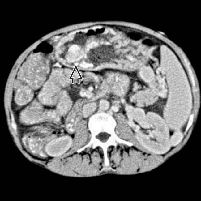
in communication with the left portal vein image, a virtually pathognomonic finding for portal hypertension. Note the multiple periumbilical collaterals image forming a caput medusae.
image
(Right) Sagittal volume-rendered CECT nicely demonstrates the direct communication between a recanalized paraumbilical vein image 
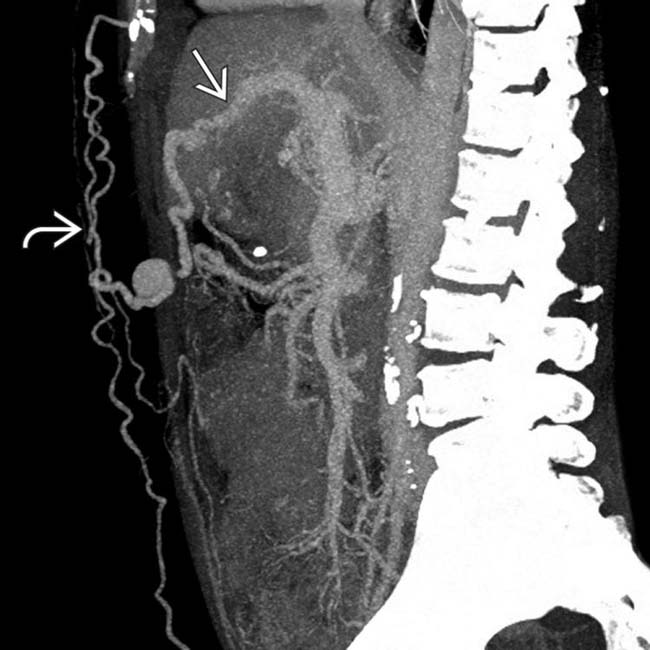
and multiple abdominal wall varices image.
image
(Left) Coronal volume-rendered CECT demonstrates extensive varices image 
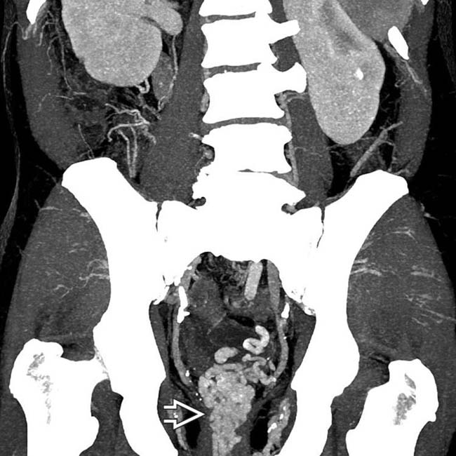
surrounding the rectum in a cirrhotic patient. While often asymptomatic, rectal varices (like varices elsewhere) can bleed and result in gastrointestinal hemorrhage.

image
(Right) Axial CECT demonstrates multiple large gastric varices image 
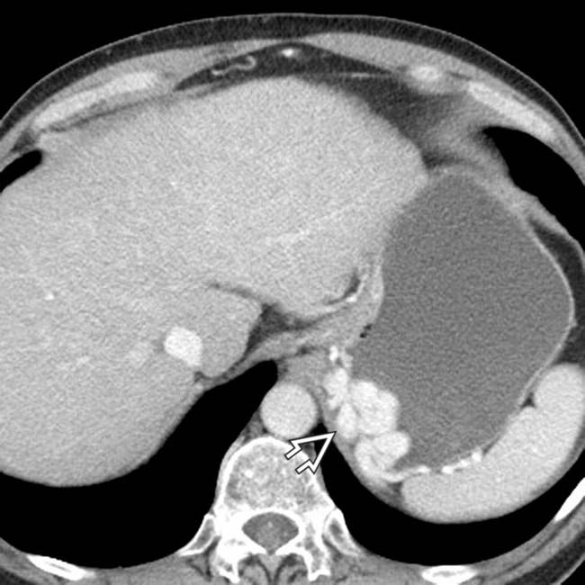
in the left upper quadrant. Although the diagnosis is straightforward on these venous phase images, the varices could mimic an intramural mass on NECT or arterial phase CECT images.
image
(Left) Axial CECT in a middle-aged man with ulcerative colitis and primary sclerosing cholangitis shows a descending colostomy and large varices image 
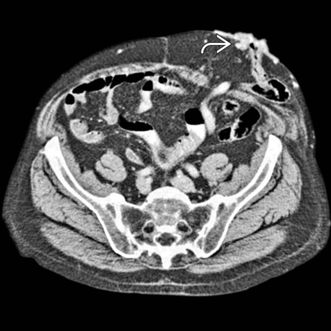
around the stoma. Surgical creation of the stoma creates a new site of portosystemic anastomosis and a potential site for the development of varices.

image
(Right) Axial CECT demonstrates large varices image 
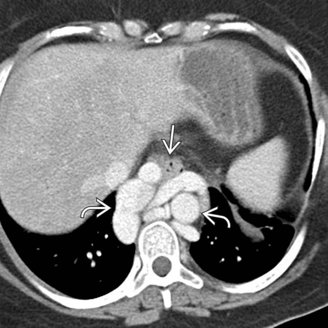
surrounding the esophagus image. Paraesophageal varices are not infrequently mistaken for a mediastinal mass on either CXR or NECT.
image
Axial CECT in a patient with cirrhosis and portal hypertension demonstrates a cluster of dilated mesenteric varices image 
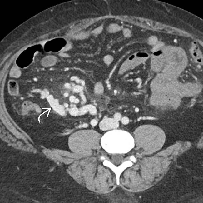
in the right lower quadrant.

image
Coronal volume-rendered CECT demonstrates a cirrhotic morphology of the liver, marked splenomegaly image
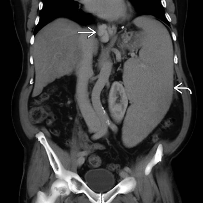
, and paraesophageal varices image, a constellation of findings indicative of portal hypertension.
image
Coronal volume-rendered CECT demonstrates extensive abdominal wall collaterals image 
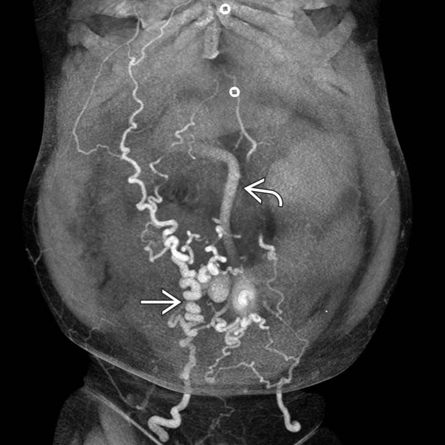
(caput medusae) in communication with a dilated recanalized paraumbilical vein image within the abdomen itself. These abdominal wall varices are frequently directly visible on clinical inspection.
image
Axial CECT demonstrates multiple additional varices image 
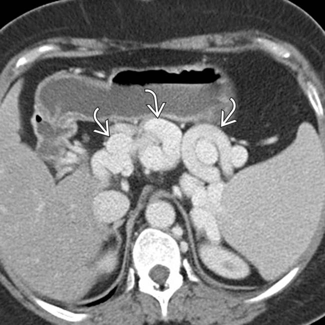
in the upper abdomen adjacent to the spleen and stomach.
image
Coronal volume-rendered CECT demonstrates extensive varices in the anterior abdominal wall, forming a caput medusae.
image
Axial CECT shows a nodular, cirrhotic liver with signs of portal hypertension, including splenomegaly, ascites, and varices image
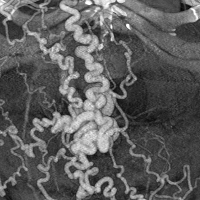
. The intrahepatic ducts image are dilated with an abnormal arborization, suggestive of primary sclerosing cholangitis (PSC). Biopsy showed elements of both PSC and autoimmune hepatitis.
image
Axial CECT shows an occluded portal vein image 
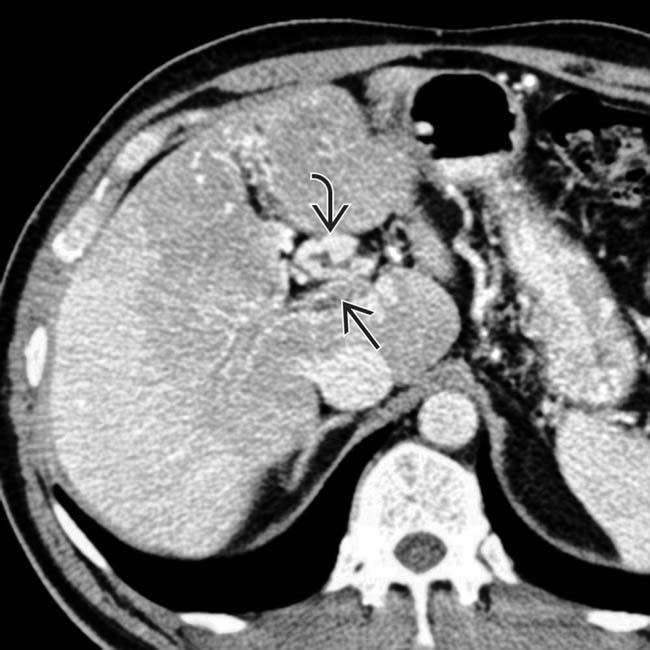
with cavernous transformation image of the portal vein and extensive varices as a result.
image
Axial CT in the same patient shows varices in the wall of the gallbladder image 
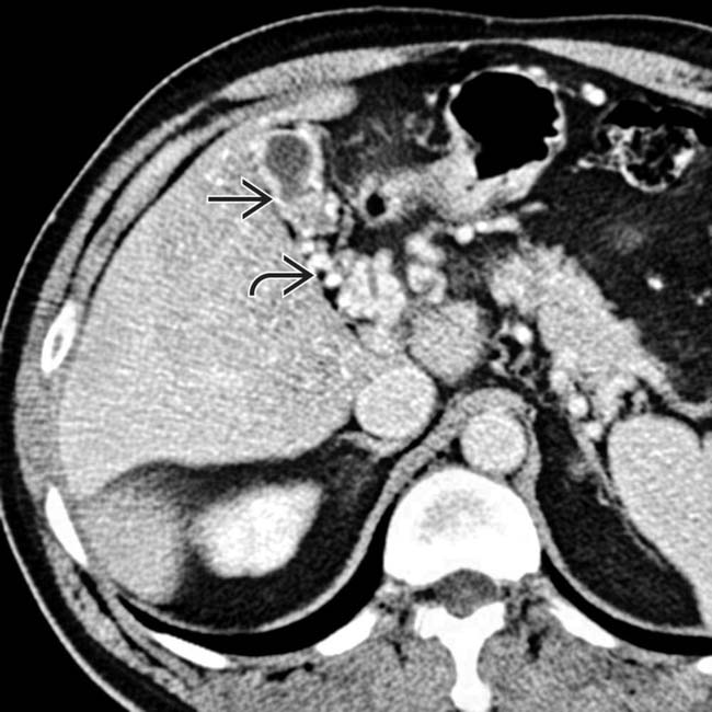
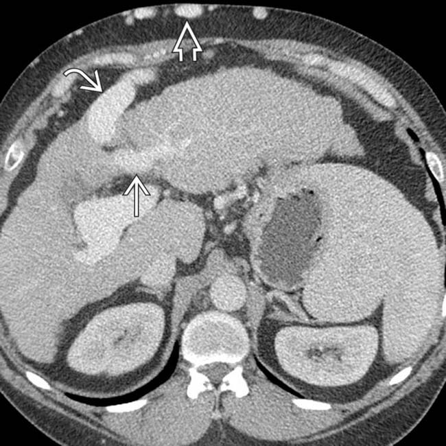
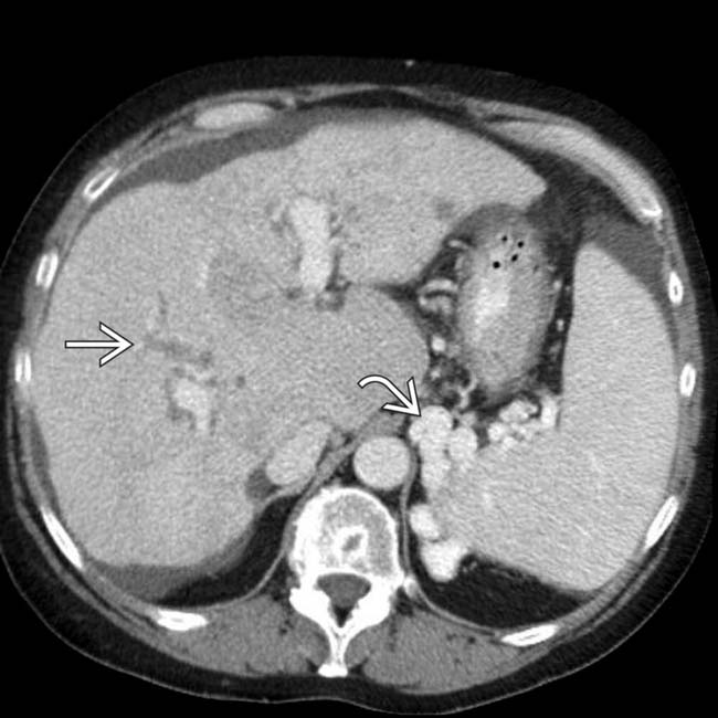
that might mimic primary gallbladder disease and the mass of varices in and around the pancreatic head image that might be mistaken for a hypervascular tumor of the pancreas. Gallbladder wall varices occur almost exclusively in patients with portal vein occlusion.
image
Axial CECT shows typical signs of cirrhosis and portal hypertension, including ascites. Note the thickened wall of the ascending colon image
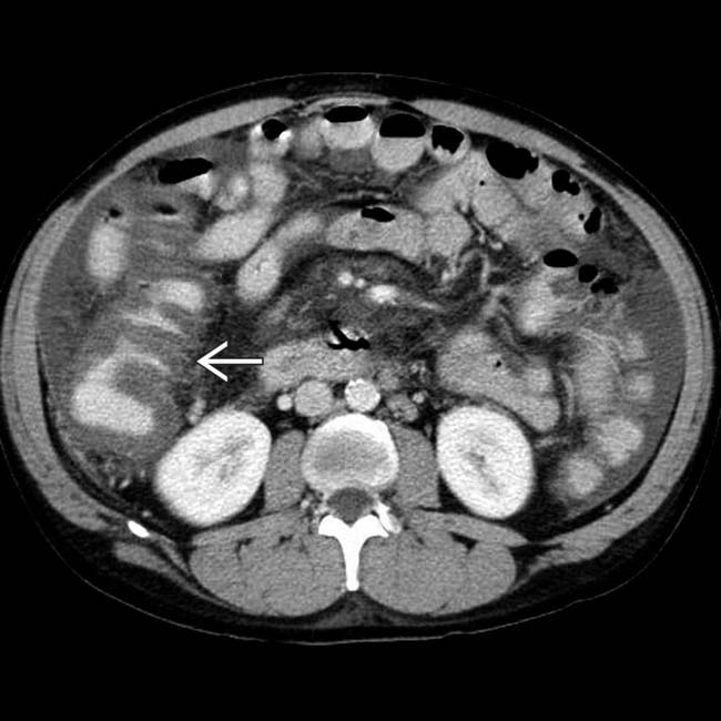
, which simulates findings seen with colitis. On colonoscopy, there was no mucosal inflammation, only venous engorgement, known as portal hypertensive colopathy.
image
Axial NECT shows what appears to be a left adrenal mass image
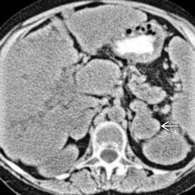
. The liver has a cirrhotic morphology with widened fissures and an increased caudate to right lobe ratio.
image
Axial CECT shows that the “mass” is composed of large varices image 
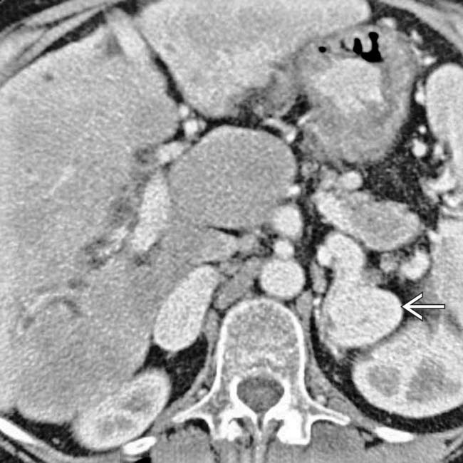
in a patient with cirrhosis and severe portal hypertension.
image
Axial CECT shows thrombosis of the portal vein image
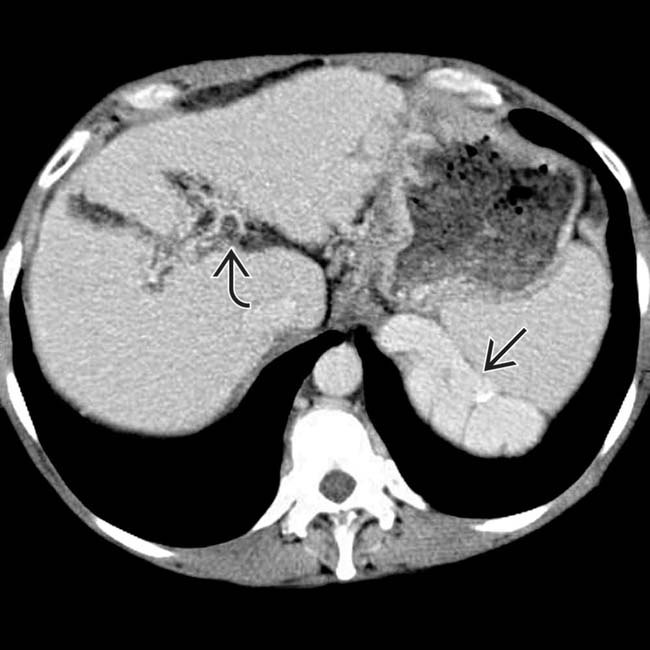
, a cirrhotic liver, and huge upper abdominal varices image.
image
Axial CECT in the same patient shows a hyperdense collection within the stomach image that represents active bleeding from gastric varices (no oral contrast was given). Variceal bleeding is one of the leading causes of death among patients with cirrhosis.
image
Axial CECT shows findings typical of a cirrhotic liver and portal hypertension with ascites, splenomegaly, and a recanalized parumbilical vein or varix image
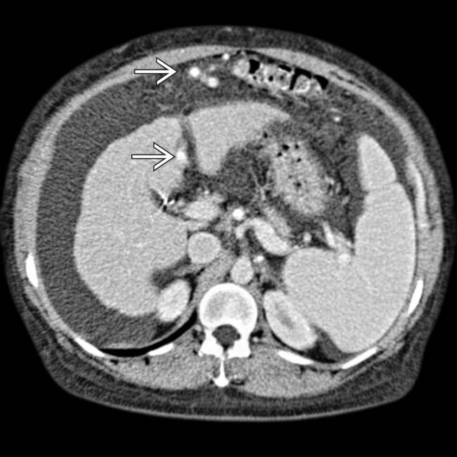
. The collateral vessels continued caudally to surround the umbilicus where parumbilical varices were seen (not shown).

Share this: