16 Popliteal and Saphenous Block
Traditional Block Technique
Placement
Anatomy
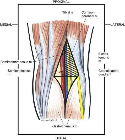
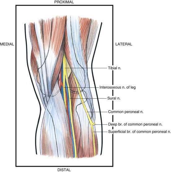
As illustrated in Figure 16-1, the cephalad popliteal fossa is defined by the semimembranosus and semitendinosus muscles medially and the biceps femoris muscle laterally. Its caudad extent is defined by the gastrocnemius muscles both medially and laterally. If this quadrilateral area is bisected, as shown in Figure 16-1, the area of interest to the anesthesiologist is the cephalolateral quadrant (hatched area). Here, both tibial and common peroneal nerve block is possible. The tibial nerve is the larger of these two nerves; it separates from the common peroneal nerve at the upper limit of the popliteal fossa and sometimes higher. The tibial nerve continues the straight course of the sciatic nerve and runs lengthwise through the popliteal fossa immediately under the popliteal fascia. Inferiorly, it passes between the heads of the gastrocnemius muscles. The common peroneal nerve follows the tendon of the biceps femoris muscle along the cephalolateral margin of the popliteal fossa, as illustrated in Figure 16-2. After the common peroneal nerve leaves the popliteal fossa, it travels around the head of the fibula and divides into the superficial peroneal and deep peroneal nerves.
Needle Puncture
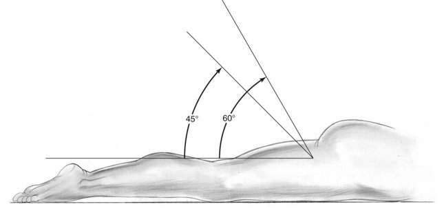
With the patient in the prone position, he or she is asked to flex the leg at the knee, which allows more accurate identification of the popliteal fossa. Once the popliteal fossa has been defined, it is divided into equal medial and lateral triangles, as shown in Figure 16-1. An “X” is placed 5 to 7 cm superior to the skin crease of the popliteal fossa and 1 cm lateral to the midline of the triangles, as shown in Figure 16-1. Through this site, a 22-gauge, 4- to 6-cm needle is advanced at an angle of 45 to 60 degrees to the skin while being directed anterosuperiorly (Fig. 16-3). A paresthesia or motor response is sought; when obtained, 30 to 40 mL of local anesthetic is injected.
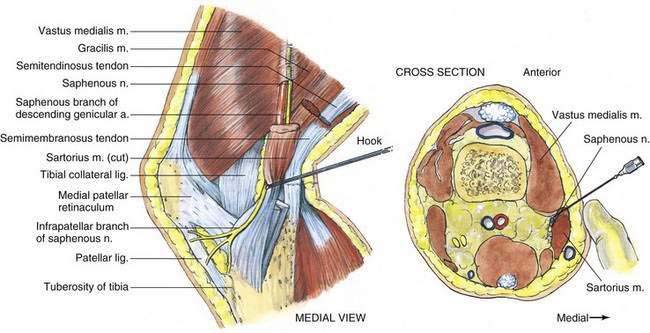
When a saphenous block is added for foot and ankle surgery, the patient’s knee is bent at approximately a 45-degree angle, and the medial aspect of the leg is exposed. Two primary techniques are used for saphenous block. A superficial ring of local anesthetic may be injected just distal to the medial surface of the tibial condyle. Often 5 to 10 mL of local anesthetic is needed. Conversely, a more proximal technique (at the cross-sectional level of the superior border of the patella) is possible (Fig. 16-4). In this case, a 22- to 25-gauge, 3- to 4-cm needle is inserted immediately deep to the sartorius muscle in the plane between the vastus medialis and the sartorius muscles, and 10 mL of local anesthetic is injected.
Ultrasonography-Guided Technique
Popliteal Block
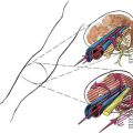
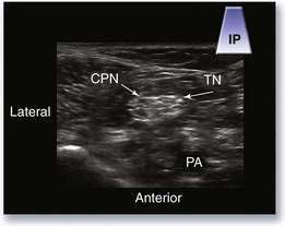
The patient can be placed in either the supine or prone position (Fig. 16-5). The first objective is to define the popliteal artery or the popliteal vein; the former is a pulsatile hypoechoic circle and the latter is a compressible hypoechoic circle. The vein typically lies more posterior than the artery. Immediately posterior to the vessels is the tibial nerve, appearing as a distinct hyperechoic circle with internal hypoechoic fascicles. Moving the transducer in a proximal direction, the operator will soon see a smaller hyperechoic circle joining the tibial component from the lateral aspect of the screen (Fig. 16-6; see Video 8: Popliteal Anatomy on the Expert Consult Website). This second structure is the common peroneal nerve. This is the location where most practitioners perform the block. 
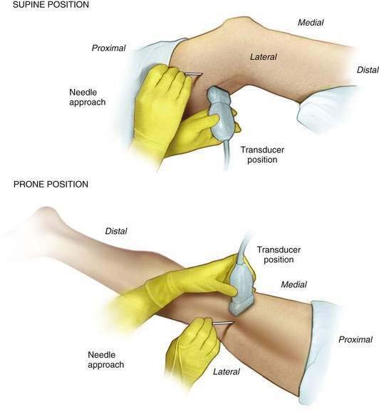
With the patient in the prone position, the needle is inserted using the in-plane needle insertion technique. Typically, the needle is inserted at a roughly 45-degree angle with respect to the skin (see Fig. 16-5). The needle is advanced and contact is made with the outermost hyperechoic layer of the sciatic nerve. There is often a tactile and visual “popping” sensation as the needle penetrates a tissue layer just external to the epineurium. The end result of the injection is the circumferential spread of local anesthetic around the sciatic nerve (see Video 9: Popliteal Nerve Block: In-Plane Technique on the Expert Consult Website). 
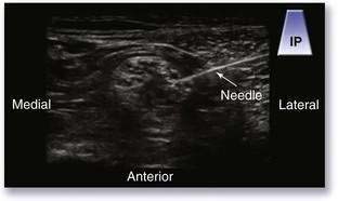
When the patient is in the supine position, the needle is also inserted using the in-plane technique. However, the needle is placed completely parallel with the faceplate of the transducer. With this approach, the needle acts as a strong specular reflector, thus generating the clearest and brightest image of the needle possible (Fig. 16-7).