10 Poisoning, overdose, antidotes
Initial assessment
• The identity of the substance(s) taken.
• The time that has elapsed since ingestion.
Adults may be sufficiently conscious to give some indication of the poison or may have referred to it in a suicide note, or there may be other circumstantial evidence e.g. knowledge of the prescribed drugs that the patient had access to, empty drug containers in pocket or at the scene. The ambulance crew attending to the patient at home may have very valuable information and should be questioned for any clues to the ingested drug. Any family or friends attending with the patient should be similarly questioned.
Many substances used in accidental or self-poisoning produce recognisable symptoms and signs. Some arise from dysfunction of the central or autonomic nervous systems; other agents produce individual effects. They can be useful diagnostically and provide characteristic toxic syndromes or ‘toxidromes’ (Table 10.1).
| Toxidrome | Clinical features | Causative agents |
|---|---|---|
| Antimuscarinic | Tachycardia Dilated pupils Dry, flushed skin Urinary retention Decreased bowel sounds Mild increase in body temperature Confusion Cardiac arrhythmias Seizures |
Antipsychotics Tricyclic antidepressants Antihistamines Antispasmodics Many plant toxins |
| Muscarinic | Salivation Lachrymation Abdominal cramps Urinary and faecal incontinence Vomiting Sweating Miosis Muscle fasciculation and weakness Bradycardia Pulmonary oedema Confusion CNS depression Seizures |
Anticholinesterases Organophosphorus insecticides Carbamate insecticides Galantamine Donepezil |
| Sympathomimetic | Tachycardia Hypertension Hyperthermia Sweating Mydriasis Hyperreflexia Agitation Delusions Paranoia Seizures Cardiac arrhythmias |
Resuscitation
Rapid biochemical ‘screens’ of urine are widely available in hospital emergency departments and will detect a range of drugs (Table 10.2).
Table 10.2 Drugs that can be readily tested for and detected in urine in the emergency department
| Drugs detectable on rapid urine testing |
Supportive treatment
TOXBASE, the primary clinical toxicology database of the UK National Poisons Information Service, is available on the internet to registered users at: http://www.toxbase.org
Special problems introduced by poisoning are as follows:
• Airway maintenance is essential; some patients require a cuffed endotracheal tube but seldom for more than 24 h.
• Ventilation: a mixed respiratory and metabolic acidosis is common; the inspired air is supplemented with oxygen to correct the hypoxia. Mechanical ventilation is necessary if adequate oxygenation cannot be obtained or hypercapnia ensues.
• Hypotension: this is common in poisoning and, in addition to the resuscitative measures indicated above, conventional inotropic support may be required.
• In addition: there is recent interest in the use of high dose insulin infusions with euglycaemic clamping as a positive inotrope in the context of overdose with myocardial depressant agents. The very high insulin doses given (0.5–2 units/kg/h) have so far deterred physicians from the routine use of such therapy. There are, however, a number of case reports that support such an approach. Many of these are in the context of overdosage with non-dihydropyridine calcium channel blockers that are often resistant to conventional inotropic agents.
• Convulsions should be treated if they are persistent or protracted. Intravenous benzodiazepine (diazepam or lorazepam) is the first choice.
• Cardiac arrhythmia frequently accompanies poisoning, e.g. with tricyclic antidepressants, theophylline, β-adrenoceptor blockers.
• Acidosis, hypoxia and electrolyte disturbance are often important contributory factors and it is preferable to observe the effect of correcting these before considering resort to an antiarrhythmic drug. If arrhythmia does lead to persistent peripheral circulatory failure, an appropriate drug may be cautiously justified, e.g. a β-adrenoceptor blocker for poisoning with a sympathomimetic drug.
• Hypothermia may occur if CNS depression impairs temperature regulation. A low-reading rectal thermometer is used to monitor core temperature and the patient is nursed in a heat-retaining ‘space blanket’.
• Immobility may lead to pressure lesions of peripheral nerves, cutaneous blisters, necrosis over bony prominences, and increased risk of thromboembolism warrants prophylaxis.
• Rhabdomyolysis may result from prolonged pressure on muscles from agents that cause muscle spasm or convulsions (phencyclidine, theophylline); may be aggravated by hyperthermia due to muscle contraction, e.g. with MDMA (‘ecstasy’). Aggressive volume repletion and correction of acid–base abnormality are needed; urine alkalinisation and/or diuretic therapy may be helpful in preventing acute tubular necrosis but evidence is not conclusive.
Preventing further absorption of the poison
From the alimentary tract (‘gut decontamination’)1
Oral adsorbents
Activated charcoal (Carbomix) consists of a very fine black powder prepared from vegetable matter, e.g. wood pulp, coconut shell, which is ‘activated’ by an oxidising gas flow at high temperature to create a network of small (10–20 nm) pores with an enormous surface area in relation to weight (1000 m2/g). This binds to, and thus inactivates, a wide variety of compounds in the gut. Indeed, activated charcoal comes nearest to fulfilling the long-sought notion of a ‘universal antidote’.2 Thus it is simpler to list the exceptions, i.e. substances that are poorly adsorbed by charcoal:
• Metal salts (iron, lithium).
• Alcohols (ethanol, methanol, ethylene glycol).
To be most effective, five to ten times as much charcoal as poison, weight for weight, is needed. In the adult an initial dose of 50 g is usual, repeated if necessary. If the patient is vomiting, give the charcoal through a nasogastric tube. Unless a patient has an intact or protected airway its administration is contraindicated.
Accelerating elimination of the poison
• The poison should be present in high concentration in the plasma relative to that in the rest of the body, i.e. it should have a small volume of distribution.
• The poison should dissociate readily from any plasma protein binding sites.
• The effects of the poison should relate to its plasma concentration.
Methods used are:
Alteration of urine pH and diuresis
It is useful to alter the pH of the glomerular filtrate such that a drug that is a weak electrolyte will ionise, become less lipid soluble, remain in the renal tubular fluid, and leave the body in the urine (see p. 80).
Alkalinisation4 may be used for: salicylate (> 500 mg/L + metabolic acidosis, or in any case > 750 mg/L) phenobarbital (75–150 mg/L); phenoxy herbicides, e.g. 2,4-D, mecoprop, dichlorprop; moderately severe salicylate poisoning that does not meet the criteria for haemodialysis.
Specific antidotes5
Specific antidotes reduce or abolish the effects of poisons through a variety of general mechanisms, as indicated in Table 10.3.
| Mechanism | Examples |
|---|---|
| Removal of circulating poison from plasma |
Table 10.4 illustrates these mechanisms with antidotes that are of therapeutic value.
Table 10.4 Specific antidotes useful in clinical practice
| Some specific antidotes, indications and modes of action (see Index for a fuller account of individual drugs) | ||
|---|---|---|
| Antidote | Indication | Mode of action |
| Acetylcysteine | Paracetamol, chloroform, carbon tetrachloride, radiocontrast nephropathy | Replenishes depleted glutathione stores |
| Atropine | Cholinesterase inhibitors, e.g. organophosphorus insecticides | Blocks muscarinic cholinoceptors |
| β-Blocker poisoning | Vagal block accelerates heart rate | |
| Benzatropine | Drug-induced movement disorders | Blocks muscarinic cholinoceptors |
| Calcium gluconate | Hydrofluoric acid, fluorides | Binds or precipitates fluoride ions |
| Desferrioxamine | Iron | Chelates ferrous ions |
| Dicobalt edetate | Cyanide and derivatives, e.g. acrylonitrile | Chelates to form non-toxic cobalti- and cobalto-cyanides |
| Digoxin-specific antibody fragments (FAB) | Digitalis glycosides | Binds free glycoside in plasma, complex excreted in urine |
| Dimercaprol (BAL) | Arsenic, copper, gold, lead, inorganic mercury | Chelates metal ions |
| Ethanol (or fomepizole) | Ethylene glycol, methanol | Competes for alcohol and acetaldehyde dehydrogenases, preventing formation of toxic metabolites |
| Flumazenil | Benzodiazepines | Competes for benzodiazepine receptors |
| Folinic acid | Folic acid antagonists, e.g. methotrexate, trimethoprim | Bypasses block in folate metabolism |
| Glucagon | β-Adrenoceptor antagonists | Bypasses blockade of the β-adrenoceptor; stimulates cyclic AMP formation with positive cardiac inotropic effect |
| Isoprenaline | β-Adrenoceptor antagonists | Competes for and activates β-adrenoceptors |
| Methionine | Paracetamol | Replenishes depleted glutathione stores |
| Naloxone | Opioids | Competes for opioid receptors |
| Neostigmine | Antimuscarinic drugs | Inhibits acetylcholinesterase, causing acetylcholine to accumulate at cholinoceptors |
| Oxygen | Carbon monoxide | Competitively displaces carbon monoxide from binding sites on haemoglobin |
| Penicillamine | Copper, gold, lead, elemental mercury (vapour), zinc | Chelates metal ions |
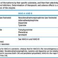
| Phenoxybenzamine | Hypertension due to α-adrenoceptor agonists, e.g. with MAOI, clonidine, ergotamine | Competes for and blocks α-adrenoceptors (long acting) |
| Phentolamine | As above | Competes for and blocks α-adrenoceptors (short acting) |
| Phytomenadione (vitamin K1) | Coumarin (warfarin) and indanedione anticoagulants | Replenishes vitamin K |
| Pralidoxime | Cholinesterase inhibitors, e.g. organophosphorus insecticides | Competitively reactivates cholinesterase |
| Propranolol | β-Adrenoceptor agonists, ephedrine, theophylline, thyroxine | Blocks β-adrenoceptors |
| Protamine | Heparin | Binds ionically to neutralise |
| Prussian blue (potassium ferric hexacyanoferrate) | Thallium (in rodenticides) | Potassium exchanges for thallium |
| Sodium calcium edetate | Lead | Chelates lead ions |
| Unithiol | Lead, elemental and organic mercury | Chelates metal ions |
Poisoning by (non-drug) chemicals
Heavy metal poisoning and use of chelating agents
Acute or chronic exposure to heavy metals can harm the body.6 Treatment is with chelating agents which incorporate the metal ions into an inner ring structure in the molecule (Greek: chele, claw) by means of structural groups called ligands (Latin: ligare, to bind). Effective agents form stable, biologically inert complexes that pass into the urine.
Penicillamine (dimethylcysteine) is a metabolite of penicillin that contains SH-groups; it may be used to chelate lead and copper (see Wilson’s disease, p. 366). Its principal use is for rheumatoid arthritis (see Index).
Desferrioxamine (see Iron, p. 500).
Cyanide
poisoning results in tissue anoxia by chelating the ferric part of the intracellular respiratory enzyme, cytochrome oxidase. It thus uncouples mitochondrial oxidative phosphorylation and inhibits cellular respiration in the presence of adequate oxygenation. Poisoning may occur as a result of: self-administration of hydrocyanic (prussic) acid; accidental exposure in industry; inhaling smoke from burning polyurethane foams in furniture; ingesting amygdalin which is present in the kernels of several fruits including apricots, almonds and peaches (constituents of the unlicensed anticancer agent, laetrile); excessive use of sodium nitroprusside for severe hypertension.7
The principles of specific therapy are as follows:
• Dicobalt edetate. The dose is 300 mg given intravenously over 1 min (5 min if condition is less serious), followed immediately by a 50 mL intravenous infusion of 50% glucose; a further 300 mg dicobalt edetate should be given if recovery is not evident within 1 min.
• Alternatively, a two-stage procedure may be followed by intravenous administration of:
The increasing use of hydroxocobalamin as a first-line treatment is based upon animal studies that have shown a faster improvement of arterial blood pressure compared to sodium nitrate. No benefit in terms of mortality was seen in these studies.
Methanol
• Correcting the metabolic acidosis. Achieving this largely determines the outcome; sodium bicarbonate is given intravenously in doses up to 2 mol in a few hours, carrying an excess of sodium which must be managed. Methanol is metabolised slowly and relapse may accompany too early discontinuation of bicarbonate.
• Inhibiting methanol metabolism. Ethanol, which occupies the dehydrogenase enzymes in preference to methanol, competitively prevents metabolism of methanol to its toxic products. A single oral dose of ethanol 1 mL/kg (as a 50% solution or as the equivalent in gin or whisky) is followed by 0.25 mL/kg/h orally or intravenously, aiming to maintain the blood ethanol at about 100 mg/100 mL until no methanol is detectable in the blood. Fomepizole (4-methylpyrazole), another competitive inhibitor of alcohol dehydrogenase, is effective in severe methanol poisoning and is less likely to cause cerebral depression (it is available in the UK on a named-patient basis).
• Eliminating methanol and its metabolites. Haemodialysis is two to three times more effective than is peritoneal dialysis and is indicated in severe cases.
Folinic acid 30 mg intravenously 6-hourly may protect against retinal damage by enhancing formate metabolism.
Herbicides and pesticides
Rodenticides
include warfarin and thallium (see Table 10.1); for strychnine, which causes convulsions, give diazepam.
Paraquat
is a widely used herbicide that is extremely toxic if ingested; a mouthful of the commercial solution taken and spat out may be sufficient to kill. It is highly corrosive and can be absorbed through the skin. A common sequence is: ulceration and sloughing of8 the oral and oesophageal mucosa, renal tubular necrosis (5–10 days later), pulmonary oedema and pulmonary fibrosis. Whether the patient lives or dies depends largely on the condition of the lung. Treatment is urgent and includes activated charcoal or aluminium silicate (Fuller’s earth) by mouth as adsorbents. Haemodialysis may have a role in the first 24 h, the rationale being to reduce the plasma concentration and protect the kidney, failure of which allows the slow but relentless accumulation of paraquat in the lung.
Incapacitating agents
CS
According to the concentration of CS to which a person is exposed, the effects vary from a slight pricking or peppery sensation in the eyes and nasal passages up to the maximal symptoms of streaming from the eyes and nose, spasm of the eyelids, profuse lachrymation and salivation, retching and sometimes vomiting, burning of the mouth and throat, cough and gripping pain in the chest.9 Exposed subjects absorb small amounts only, and the plasma t½ is about 5 s. There appear to be no long-lasting sequelae but, plainly, it would be prudent to assume that patients with asthma or chronic bronchitis could suffer an exacerbation from high concentrations.
Poisoning by biological substances
Many plants form substances that are important for their survival either by enticing animals, which disperse their spores, or by repelling potential predators. Poisoning occurs when children eat berries or chew flowers, attracted by their colour; adults may mistake non-edible for edible varieties of salad plants and fungi (mushrooms) for they may resemble one another closely and some are greatly prized by epicures. Ingestion of plants is responsible for a significant number of calls to poison information services (10% in US and German surveys) but serious poisonings are rare. Deaths from plant poisoning are thus very rare in industrialised societies. A recent study from the USA covering the period 1983–2000 identified only 30 fatalities over this 18-year period. Plant poisoning is, however, a significant problem in the developing world. Such deaths are almost exclusively deliberate suicide or homicide.10
The range of toxic substances that these plants produce is exhibited in a diversity of symptoms that may be grouped broadly as shown in Table 10.5.
| Symptom complex | Causative agent | Active ingredient |
|---|---|---|
|
Atropenic |
Tropane alkaloids such as
Nicotinic
Muscarinic
Hallucinogenic
Cardiovascular
Cardenolide cardiac glycosides such as
GABA antagonists
Bradbury S., Vale A. Poisons: epidemiology and clinical presentation. Clin. Med. (Northfield Il). 2008;8(1):86–88. (and subsequent papers in this issue)
Body R., Bartram T., Azam F., Mackway-Jones K. Guidelines in Emergency Medicine Network (GeMNet): guideline for the management of tricyclic antidepressant overdose. Emergency Medical Journal. 2011;28:347–368.
Buckley N.A., Juurlink D.N., Isbister G., et al. Hyperbaric oxygen for carbon monoxide poisoning. Cochrane Database Syst. Rev.. 2011;13:4.
Budnitz D.S., Lovegrove M.C., Crosby A.E. Emergency department visits for acetaminophen-containing products. Am. J. Prev. Med.. 2011;40:585–592.
Chyka P.A., Erdman A.R., Christianson G., et al. Salicylate poisoning: an evidence-based consensus guideline for out-of-hospital management. Clin. Toxicol.. 2007;45:95–131.
Evison D., Hinsley D., Rice P. Chemical weapons. Br. Med. J.. 2002;324:332–335.
Gawande A. When law and ethics collide – why physicians participate in executions. N. Engl. J. Med.. 2006;354(12):1221–1229.
Holger J.S., Engebretson K.M., Marini J.J. High dose insulin in toxic cardiogenic shock. Clin. Toxicol.. 2009;47(4):303–307.
Kales S.N., Christiani D.C. Acute chemical emergencies. N. Engl. J. Med.. 2004;350(8):800–808.
Kerins M., Dargan P.I., Jones A.L. Pitfalls in the management of the poisoned patient. J. R. Coll. Physicians Edinb.. 2003;33:90–103.
Ruben Thanacoody H.K., Thomas S.H.L. Antidepressant poisoning. Clin. Med. (Northfield Il). 2003;3(2):114–118.
Skegg K. Self-harm. Lancet. 2005;366:1471–1483.
Volans G., Hartley V., McCrea S., Monaghan J. Non-opioid analgesic poisoning. Clin. Med. (Northfield Il). 2003;3(2):119–123.
Wolf A.D., Erdman A.R., Nelson L.S., et al. Tricyclic antidepressant poisoning: an evidence-based consensus guideline for out-of-hospital management. Clin. Toxicol.. 2007;45:203–233.
1 Joint position statements and guidelines agreed by the American Academy of Clinical Toxicology and the European Association of Poison Centres and Clinical Toxicologists review the therapeutic usefulness of various procedures for gut decontamination. These appear in the Journal of Toxicology, Clinical Toxicology from 1997 onwards, the latest position statements being in 2004 and 2005.
2 For centuries it was supposed not only that there could be, but that there actually was, a single antidote to all poisons. This was Theriaca Andromachi, a formulation of 72 (a magical number) ingredients among which particular importance was attached to the flesh of a snake (viper). The antidote was devised by Andromachus, whose son was physician to the Roman Emperor Nero (AD 37–68).
3 Irrigation with large volumes of a polyethylene glycol–electrolyte solution, e.g. Klean-Prep, by mouth causes minimal fluid and electrolyte disturbance (it was developed for preparation for colonoscopy). Magnesium sulphate may also be used.
4 Proudfoot A T, Krenzelok E P, Vale J A 2004 Position paper on urine alkalinisation. Journal of Toxicology, Clinical Toxicology 42:1–26.
5 Mithridates the Great (?132 BC – 63 bc), king of Pontus (in Asia Minor), was noted for ‘ambition, cruelty and artifice’. ‘He murdered his own mother … and fortified his constitution by drinking antidotes’ to the poisons with which his domestic enemies sought to kill him (Lemprière). When his son also sought to kill him, Mithridates was so disappointed that he compelled his wife to poison herself. He then tried to poison himself, but in vain; the frequent antidotes that he had taken in the early part of his life had so strengthened his constitution that he was immune. He was obliged to stab himself, but had to seek the help of a slave to complete his task. Modern physicians have to be content with less comprehensively effective antidotes, some of which are listed in Table 10.1.
6 Sometimes in unexpected ways; an initiation custom in an artillery regiment involved pouring wine through the barrel of a gun after several shots had been fired. A healthy 19-year-old soldier drank 250 mL of the wine and within 15 min convulsed and became unconscious. His plasma, urine and the wine contained high concentrations of tungsten. He received haemodialysis and recovered. Investigation revealed that the gun barrels had recently been hardened by the addition of tungsten to the steel. Marquet P, François B, Vignon P, Lachâtre G 1996 A soldier who had seizures after drinking a quarter of a litre of wine. Lancet 348:1070.
7 Or in other more bizarre ways. ‘A 23-year-old medical student saw his dog (a puppy) suddenly collapse. He started external cardiac massage and a mouth-to-nose ventilation effort. Moments later the dog died, and the student felt nauseated, vomited and lost consciousness. On the victim’s arrival at hospital, an alert medical officer detected a bitter almonds odour on his breath and administered the accepted treatment for cyanide poisoning after which he recovered. It turned out that the dog had accidentally swallowed cyanide, and the poison eliminated through the lungs had been inhaled by the master during the mouth-to-nose resuscitation.’ Journal of the American Medical Association 1983 249:353.
8 A 19-year-old male was admitted to hospital in Sri Lanka, having ingested 250 mL of paraquat in an episode of deliberate self-harm. He was accompanied by his brother and friend. The unfortunate young man died within 8 h of admission. His brother and friend presented to the same hospital 2 days later with severe swelling and burns to the scrotal skin. They had originally brought the patient to hospital in a three-wheeled taxi with the patient lying across their laps. He had vomited on them several times. They had been wearing sarongs which they had been unable to change out of during their 8-hour vigil before he died. The brother went on to develop evidence of mild systemic toxicity with abnormalities of renal and hepatic function. Both made a complete recovery. The local and systemic toxicity had occurred from prolonged contact with the vomitus-stained clothes. (Premaratna R, Rathnasena B G N, de Silva H J 2008 Accidental scrotal burns from paraquat while handling a patient. Ceylon Medical Journal 53(3):102–103.)
9 Home Office Report (1971) of the enquiry into the medical and toxicological aspects of CS. Part II. HMSO, London: Cmnd 4775.
10 Eddleston M, Rezvi Sheriff M H, Hawton K 1998 Deliberate self-harm in Sri Lanka: an overlooked tragedy in the developing world. British Medical Journal 317:133–135.