[level-membership-for-pediatrics-category]
Chapter 404 Pleurisy, Pleural Effusions, and Empyema
Pleurisy or inflammation of the pleura is often accompanied by an effusion. The most common cause of pleural effusion in children is bacterial pneumonia (Chapter 392); heart failure (Chapter 436), rheumatologic causes, and metastatic intrathoracic malignancy are the next most common causes. A variety of other diseases account for the remaining cases, including tuberculosis (Chapter 207), lupus erythematosus (Chapter 152), aspiration pneumonitis (Chapter 389), uremia, pancreatitis, subdiaphragmatic abscess, and rheumatoid arthritis. Males and females are affected equally.
404.1 Dry or Plastic Pleurisy (Pleural Effusion)
Glenna B. Winnie and Steven V. Lossef
Laboratory Findings
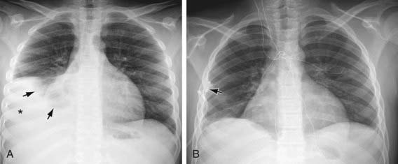
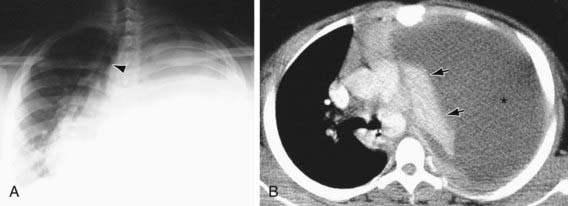
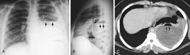
Plastic pleurisy may be detected on radiographs as a diffuse haziness at the pleural surface or a dense, sharply demarcated shadow (Figs. 404-1 and 404-2). The latter finding may be indistinguishable from small amounts of pleural exudate. Chest radiographic findings may be normal, but ultrasonography or CT findings will be positive.
404.2 Serofibrinous or Serosanguineous Pleurisy (Pleural Effusion)
Laboratory Findings
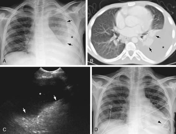
Radiographic examination shows a generally homogeneous density obliterating the normal markings of the underlying lung. Small effusions may cause obliteration of only the costophrenic or cardiophrenic angles or a widening of the interlobar septa. Examinations should be performed with the patient both supine and upright, to demonstrate a shift of the effusion with a change in position; the decubitus position may be helpful. Ultrasonographic examinations are useful and may guide thoracentesis if the effusion is loculated. Examination of the fluid is essential to differentiate exudates from transudates and to determine the type of exudate (Chapter 392). Depending on the clinical scenario, pleural fluid is sent for culture for bacterial, fungal, and mycobacterial cultures; antigen testing; Gram staining; and chemical evaluation of content, including protein, lactic dehydrogenase and glucose, amylase, specific gravity, total cell count and differential, cytologic examination, and pH. Complete blood count and serum chemistry analysis should be obtained; hypoalbuminemia is often present. Exudates usually have at least one of the following features: protein level >3.0 g/dL, with pleural fluid:serum protein ratio >0.5; pleural fluid lactic dehydrogenase values >200 IU/L; or fluid:serum lactic dehydrogenase ratio >0.6. Although systemic acidosis reduces the usefulness of pleural fluid pH measurements, pH <7.20 suggests an exudate. Glucose is usually <60 mg/dL in malignancy, rheumatoid disease, and tuberculosis; the finding of many small lymphocytes and a pH <7.20 suggest tuberculosis. The fluid of serofibrinous pleurisy is clear or slightly cloudy and contains relatively few leukocytes and, occasionally, some erythrocytes. Gram staining may occasionally show bacteria; however, acid-fast staining rarely demonstrates tubercle bacilli.
Treatment
Therapy should address the underlying disease. With a large effusion, draining the fluid makes the patient more comfortable. When a diagnostic thoracentesis is performed, as much fluid as possible should be removed for therapeutic purposes. Rapid removal of ≥1 L of pleural fluid may be associated with the development of reexpansion pulmonary edema (Chapter 388). If the underlying disease is adequately treated, further drainage is usually unnecessary, but if sufficient fluid reaccumulates to cause respiratory embarrassment, chest tube drainage should be performed. In older children with suspected parapneumonic effusion, tube thoracostomy is considered necessary if the pleural fluid pH is <7.20 or the pleural fluid glucose level is <50 mg/dL. If the fluid is clearly purulent, tube drainage with thrombolytic therapy or video-assisted thoracoscopic surgery (VATS) is indicated. Patients with pleural effusions may need analgesia, particularly after thoracentesis or insertion of a chest tube. Those with acute pneumonia may need supplemental oxygen in addition to specific antibiotic treatment.
404.3 Purulent Pleurisy or Empyema
Etiology
Empyema is an accumulation of pus in the pleural space. It is most often associated with pneumonia (Chapter 392) due to Streptococcus pneumoniae, although Staphylococcus aureus is most common in developing nations and Asia as well as in post-traumatic empyema. The relative incidence of Haemophilus influenzae empyema has decreased since the introduction of the Hib vaccination. Group A streptococcus, gram-negative organisms, tuberculosis, fungi, and malignancy are less common causes. The disease can also be produced by rupture of a lung abscess into the pleural space, by contamination introduced from trauma or thoracic surgery, or, rarely, by mediastinitis or the extension of intra-abdominal abscesses.
Laboratory Findings
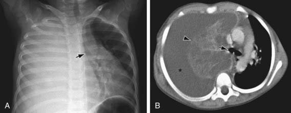
Radiographically, all pleural effusions appear similar, but the absence of a shift of the fluid with a change of position indicates a loculated empyema (Figs. 404-3 to 404-5). Septa may be confirmed by ultrasonography or CT. The maximal amount of fluid obtainable should be withdrawn by thoracentesis and studied as described in Chapter 404.2. The effusion is empyema if bacteria are present on Gram staining, the pH is <7.20, and there are >100,000 neutrophils/µL. The appearance of pus produced by different organisms is not distinctive; cultures of the fluid must always be performed. In pneumococcal empyema, the culture is positive in 58% of cases. In patients with negative culture results for pneumococcus, the pneumococcal polymerase chain reaction (PCR) analysis is most helpful to making a diagnosis. Blood cultures have a high yield, possibly higher than cultures of the pleural fluid. Leukocytosis and an elevated sedimentation rate may be found.
Treatment
Treatment includes systemic antibiotics and thoracentesis and possibly chest tube drainage with or without a fibrinolytic agent, video-assisted thorascopic surgery (VATS), or open decortication (Chapter 392); controlled studies are needed. If empyema is diagnosed early, antibiotic treatment plus thoracentesis achieves a complete cure. The selection of antibiotic should be based on the in vitro sensitivities of the responsible organism. See Chapters 174, 175, and 186 for treatment of infections by Staphylococcus, S. pneumoniae, and H. influenzae, respectively. Clinical response in empyema is slow; even with optimal treatment, there may be little improvement for as long as 2 wk. With staphylococcal infections, resolution is very slow, and systemic antibiotic therapy is required for 3-4 wk. Instillation of antibiotics into the pleural cavity does not improve results.
Akhan O, Ozkan O, Akinci D, et al. Image-guided catheter drainage of infected pleural effusions. Diagn Interv Radiol. 2007;13:204-209.
Aydogan M, Aydogan A, Ozcan A, et al. Intrapleural streptokinase treatment in children with empyema. Eur J Pediatr. 2008;167:739-744.
Aziz A, Healey JM, Qureshi F, et al. Comparative analysis of chest tube thoracostomy and video-assisted thoracoscopic surgery in empyema and parapneumonic effusion associated with pneumonia in children. Surg Infect (Larchmt). 2008;9:317-323.
Cameron R, Davies HR: Intra-pleural fibrinolytic therapy versus conservative management in the treatment of adult parapneumonic effusions and empyema, Cochrane Database Syst Rev (2):CD002312, 2008.
Chiu CY, Wong KS, Huang YC, et al. Echo-guided management of complicated parapneumonic effusion in children. Pediatr Pulmonol. 2006;41:1226-1232.
Kurt BA, Winterhalter KM, Connors RH, et al. Therapy of parapneumonic effusions in children: video-assisted thoracoscopic surgery versus conventional thoracostomy drainage. Pediatrics. 2006;118:e547-e553.
Nyambat B, Kilgore PE, Yong DE, et al. Survey of childhood empyema in Asia. BMC Infect Dis. 2008;8:90.
Sawicki GS, Lu FL, Valim C, et al. Necrotising pneumonia is an increasingly detected complication of pneumonia in children. Eur Respir J. 2008;31:1285-1291.
[/level-membership-for-pediatrics-category][not-level-membership-for-pediatrics-category]
Chapter 404 Pleurisy, Pleural Effusions, and Empyema
Pleurisy or inflammation of the pleura is often accompanied by an effusion. The most common cause of pleural effusion in children is bacterial pneumonia (Chapter 392); heart failure (Chapter 436), rheumatologic causes, and metastatic intrathoracic malignancy are the next most common causes. A variety of other diseases account for the remaining cases, including tuberculosis (Chapter 207), lupus erythematosus (Chapter 152), aspiration pneumonitis (Chapter 389), uremia, pancreatitis, subdiaphragmatic abscess, and rheumatoid arthritis. Males and females are affected equally.
404.1 Dry or Plastic Pleurisy (Pleural Effusion)
Glenna B. Winnie and Steven V. Lossef
Laboratory Findings
Plastic pleurisy may be detected on radiographs as a diffuse haziness at the pleural surface or a dense, sharply demarcated shadow (Figs. 404-1 and 404-2). The latter finding may be indistinguishable from small amounts of pleural exudate. Chest radiographic findings may be normal, but ultrasonography or CT findings will be positive.
