Chapter 20
Percutaneous Coronary Intervention
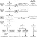
1. What does the term percutaneous coronary intervention mean?
2. Which patients with chronic stable angina benefit from PCI?
The goals of treatment in patients with coronary artery disease are to:
 Prevent adverse outcomes such as cardiovascular death, myocardial infarction (MI), left ventricular (LV) dysfunctions, and arrhythmias.
3. Which patients with unstable angina/non–ST elevation myocardial infarction (UA/NSTEMI) should undergo a strategy of early cardiac catheterization and revascularization?
Two major strategies, conservative (medical therapy without an initial strategy of catheterization and revascularization) and early invasive, are employed in treating patients with UA/NSTEMI. The early invasive approach involves performing diagnostic angiography with intent to perform PCI along with administering the usual antiischemic, antiplatelet, and anticoagulant medications. Evidence from clinical trials suggests that an early invasive approach with UA/NSTEMI leads to a reduction in adverse cardiovascular outcomes, such as death and nonfatal MI, especially in high-risk patients. Several risk-assessment tools are available that assign a score based upon the patient’s clinical characteristics (e.g., TIMI and GRACE scores). Patients who present with UA/NSTEMI should be risk stratified to identify those who would benefit most from an early invasive approach. Patients with the following clinical characteristics indicative of high risk should be taken for early coronary angiography with intent to perform revascularization:
 Recurrent angina or ischemia at rest or with low-level activities
 Elevated cardiac biomarkers (troponin T or I)
 New or presumably new ST segment deviation
 Congestive heart failure [CHF], new or worsening mitral regurgitation, or hypotension
 Reduced LV function (ejection fraction less than 40%)
 Sustained ventricular tachycardia
 High-risk findings from noninvasive testing
 PCI within the last six months or prior coronary artery bypass graft (CABG)
 High risk score as per risk-assessment tools (e.g., TIMI, GRACE)
4. What are the contraindications to PCI and the predictors of adverse outcomes?
 Bleeding diathesis or other conditions that predispose to bleeding during antiplatelet therapy
 Severe renal insufficiency unless the patient is on hemodialysis, or severe electrolyte abnormalities
 Poor patient compliance with medications
 A terminal condition that indicates short life expectancy such as advanced or metastatic malignancy
 Other indications for open heart surgery
 Failure of previous PCI or not amenable to PCI based upon previous angiograms
 Patients with severe cognitive dysfunction or advanced physical limitations
Patients generally should not undergo PCI if the following conditions are present:
 There is only a very small area of myocardium at risk.
 There is no objective evidence of ischemia (unless patient has clear anginal symptoms and has not had a stress test).
 There is a low likelihood of technical success.
 The patient has left-main or multivessel CAD with high SYNTAX score and is a candidate for CABG.
 There is insignificant stenosis (less than 50% luminal narrowing).
 The patient has end-stage cirrhosis with portal hypertension resulting in encephalopathy or visceral bleeding.
Angiographic predictors of poor outcomes include the presence of thrombus, degenerated bypass graft, unprotected left main disease, long lesions (more than 20 mm), excessive tortuosity of proximal segment, extremely angulated lesions (more than 90 degrees), a bifurcation lesion with involvement of major side branches, or chronic total occlusion.
5. What are the major complications related to PCI?
Death: The overall in-hospital mortality rate is 1.27% ranging from 0.65% in elective PCI to 4.81% in ST elevation myocardial infarction (STEMI) (based on the National Cardiac Data Registry [NCDR] CathPCI database of patients undergoing PCI between 2004 and 2007).
MI: The incidence of PCI-related MI is 0.4% to 4.9%; the incidence varies depending on the acuity of symptoms, lesion morphology, definition of MI, and frequency of measurement of biomarkers.
Stroke: The incidence of PCI-related stroke is 0.22%. In-hospital mortality in patients with PCI-related stroke is 25% to 30% (based on a contemporary analysis from the NCDR).
Emergency CABG: The need to perform emergency CABG in the stent era is extremely low (between 0.1% and 0.4%).
Vascular complications: The incidence of vascular complications ranges from 2% to 6%. These include access-site hematoma, retroperitoneal hematoma, pseudoaneurysm, arteriovenous fistula, and arterial dissection. In randomized trials, closure devices were only beneficial in reducing time to hemostasis but did not reduce the incidence of vascular complications.
Radial artery access complications: There is significant reduction of vascular complications with the use of radial arterial access as compared to femoral artery access. Complications include loss of the radial pulse in fewer than 5% of cases, compartment syndrome, pseudoaneurysm in fewer than 0.01% of cases, sterile abscess, and radial artery spasm.
Other complications associated with PCI: Other complications include transient ischemic attack (TIA), renal insufficiency, and anaphylactoid reactions to contrast agents.
6. When should cardiac biomarkers be assessed in patients undergoing PCI?
7. What is abrupt vessel closure?
Stent thrombosis is when there is complete occlusion of the artery due to thrombus formation in the stent. This may occur in the first 24 hours after stent deployment (acute stent thrombosis); in the first month after implantation (subacute stent thrombosis); between 1 and 12 months after implantation (late stent thrombosis); or even more than 1 year after implantation, most notably after drug-eluting stent (DES) implantation (very late stent thrombosis). Most instances of stent thrombosis occur in the first thirty days after implantation, at a rate of less than 1% per year. Beyond 30 days, the incidence is 0.2% to 0.6% per year depending on patient characteristics and type of stent used. Stent thrombosis is a potentially catastrophic event and often presents as STEMI, requiring emergency revascularization. Stent thrombosis carries a mortality rate of 20% to 45%. The primary factors contributing to stent thrombosis are inadequate stent deployment, incomplete stent apposition, residual stenosis, unrecognized dissection impairing blood flow, and noncompliance with dual antiplatelet therapy (DAPT). Noncompliance to DAPT is the most common cause of stent thrombosis. Resistance to aspirin and clopidogrel, and hypercoagulable states, such as those associated with malignancy, are additional, less common causes of stent thrombosis.
9. What do the terms slow-flow and no-reflow mean?
10. Are bleeding complications related to PCI clinically important?
11. What are the important complications that can occur at the access site?
12. What are some treatment options for various vascular complications?
Pseudoaneurysm: For small pseudoaneurysms, observation is recommended. For larger pseudoaneurysms, ultrasound-guided compression or percutaneous thrombin injection under ultrasound guidance is the treatment of choice. For pseudoaneurysms with a large neck, simultaneous balloon inflation to occlude the entry site can be helpful. In cases of thrombin failure, surgical repair should be considered. Endovascular repair with stent-graft implantation can also be used in the treatment of pseudoaneurysms.
Arteriovenous fistula: For small arteriovenous fistulae, observation is recommended; most close spontaneously or remain stable. For a large fistula or when significant shunting is present, options include ultrasound-guided compression, covered stent, or surgical repair.
Dissection: If there is no effect on blood flow, a conservative approach is indicated. In the presence of flow impairment (distal limb ischemia), angioplasty, stenting, and surgical repair are the treatment options.
Retroperitoneal bleeding: This should always be suspected with unexplained hypotension, marked decrease in hematocrit, flank/abdominal or back pain, and high arterial sticks. Treatment includes intravascular volume replacement, reversal of anticoagulation, blood transfusion, and, occasionally, vasopressor agents and monitoring in the intensive care unit with serial hemoglobin and hematocrit checks. Endovascular management with covered stents, prolonged balloon inflation, or surgical repair are options, but are rarely necessary.
13. What is contrast nephropathy?
 Neointimal hyperplasia caused by smooth muscle cell migration and proliferation and extracellular matrix production
 Platelet deposition and thrombus formation
 Vessel elastic recoil after balloon inflation and vessel negative remodeling over time
Coronary stents prevent vessel elastic recoil and negative remodeling, and significantly reduce both angiographic and clinical restenosis rates. The main factor leading to restenosis in coronary arteries treated with bare metal stents (BMS) is neointimal hyperplasia as a result of smooth muscle cell proliferation and extracellular matrix production. Angiographic restenosis rates with BMS range from 16% to 32% with a target lesion revascularization rate of 12% and target-vessel revascularization rate of 14% at 1 year. Similar to PTCA, restenosis after BMS typically occurs within the first 6 months.
15. What are the recommendations regarding antiplatelet therapy after PCI?
 Patients who are already on aspirin should continue with 81 to 325 mg of aspirin. Those who are not on aspirin should receive 325 mg of non–enteric-coated aspirin preferably 24 hours prior to PCI, after which it should be continued indefinitely at a dose of 81 mg daily.
Recommendations for P2Y12 receptor inhibitors:
 A loading dose of a P2Y12 inhibitor should be given prior to PCI with stent placement. The loading doses for the three recommended drugs are clopidogrel 600 mg, prasugrel 60 mg, and ticagrelor 180 mg.
 Following a loading dose of a P2Y12 inhibitor, a maintenance dose is continued. The recommendations for dose and duration are as follows:
 For patients undergoing stent BMS or DES implantation in the setting of ACS, clopidogrel 75 mg daily or prasugrel 10 mg daily or ticagrelor 90 mg twice daily should be continued for at least 12 months.
 For patients undergoing elective DES implantation, the duration of P2Y12 inhibitor therapy should be at least 12 months. Prasugrel and ticagrelor are more effective at reducing stent thrombosis in patients with ACS or STEMI.
 In patients undergoing elective BMS implantation, the duration of P2Y12 inhibitor therapy should be a minimum of one month, and preferably up to 12 months. In patients who are at high risk of bleeding, a minimum duration of 2 weeks is recommended.
16. What steps should be taken to prevent premature discontinuation of dual antiplatelet therapy?
 Patients should be clearly educated about the rationale for not stopping antiplatelet therapy and the potential consequences of stopping such therapy. Each should be instructed to call their cardiologist if bleeding develops or if another physician advises them to stop antiplatelet therapy.
 Health care providers who perform invasive or surgical procedures and are concerned about periprocedural bleeding must be made aware of the potentially catastrophic risks of premature discontinuation of P2Y12 therapy. The professionals who perform these procedures should contact the patient’s cardiologist to discuss optimal patient management strategy.
 Any elective procedure for which there is significant risk of perioperative bleeding should be deferred until patients have completed an appropriate course of P2Y12 therapy (12 months after DES implantation if they are not at high risk of bleeding, and a minimum of 1 month for BMS implantation).
 For patients treated with DES who are to undergo procedures that mandate discontinuation of P2Y12 therapy, aspirin should be continued if at all possible, and thienopyridine restarted as soon as possible after the procedure.
17. What should be the management of a patient with a DES who requires urgent noncardiac surgery?
Bibliography, Suggested Readings, and Websites
1. Investigators, A.C.T. Acetylcysteine for prevention of renal outcomes in patients undergoing coronary and peripheral vascular angiography. main results from the randomized Acetylcysteine for Contrast-Induced Nephropathy Trial (ACT). Circulation. 2011;124:1250–1259.
2. Anderson, J.L., Adams, C.D., Antman, E.M., et al. ACCF/AHA Focused Update Incorporated Into the ACC/AHA 2007 Guidelines for the Management of Patients With Unstable Angina/Non–ST-Elevation Myocardial Infarction A Report of the American College of Cardiology Foundation/American Heart Association Task Force on Practice Guidelines. Circulation. 2011;123:e426–e579. 2011
3. Bavry, A.A., Kumbhani, D.J., Rassi, A.N., Bhatt, D.L., Askari, A.T. Benefit of early invasive therapy in acute coronary syndromes a meta-analysis of contemporary randomized clinical trials. J Am Coll Cardiol. 2006;48:1319–1325.
4. Boden, W.E., O’Rourke, R.A., Teo, K.K., et al. Optimal medical therapy with or without PCI for stable coronary disease. N Engl J Med. 2007;356:1503–1516.
5. Gonzales, D.A., Norsworthy, K.J., Kern, S.J., et al. A meta-analysis of N-acetylcysteine in contrast-induced nephrotoxicity: unsupervised clustering to resolve heterogeneity. BMC Med. 2007;5:32.
6. Grines, C.L., Bonow, R.O., Casey, D.E., et al. Prevention of premature discontinuation of dual antiplatelet therapy in patients with coronary artery stents. J Am Coll Cardiol. 2007;49:734–739.
7. Keeley, E.C., Boura, J.A., Grines, C.L. Comparison of primary and facilitated percutaneous coronary interventions for ST-elevation myocardial infarction: quantitative review of randomised trials. Lancet. 2006;367:579–588.
8. Levine, G.N., Bates, E.R., Blankenship, J.C., et al. ACCF/AHA/SCAI Guideline for Percutaneous Coronary Intervention. A report of the American College of Cardiology Foundation/American Heart Association Task Force on Practice Guidelines and the Society for Cardiovascular Angiography and Interventions. J Am Coll Cardiol. 2011;58:e44–e122.
9. Levine, G.N., Kern, M.J., Berger, P.B., et al. For the American Heart Association Diagnostic and Interventional Cardiac Catheterization Committee: management of patients undergoing percutaneous coronary revascularization. Ann Intern Med. 2003;139:123–136.
10. Patel, M.R., Dehmer, G.J., Hirshfeld, J.W., Smith, P.K., Spertus, J.A. ACCF/SCAI/STS/AATS/AHA/ASNC/HFSA/SCCT 2012 Appropriate use criteria for coronary revascularization focused update: a report of the American College of Cardiology Foundation Appropriate Use Criteria Task Force, Society for Cardiovascular Angiography and Interventions, Society of Thoracic Surgeons, American Association for Thoracic Surgery, American Heart Association, American Society of Nuclear Cardiology, and the Society of Cardiovascular Computed Tomography. J Am Coll Cardiol. 2012;59:857–881.
11. Shaw, L.J., Berman, D.S., Maron, D.J., et al. Optimal Medical Therapy With or Without Percutaneous Coronary Intervention to Reduce Ischemic Burden Results From the Clinical Outcomes Utilizing Revascularization and Aggressive Drug Evaluation (COURAGE) Trial Nuclear Substudy. Circulation. 2008;117:1283–1291.
12. Serruys, P.W., Morice, M.C., Kappetein, A.P., et al. Percutaneous Coronary Intervention versus Coronary-Artery Bypass Grafting for Severe Coronary Artery Disease. N Engl J Med. 2009;360:961–972.
13. Stergiopoulos, K., Brown, D.L. Initial coronary stent implantation with medical therapy vs medical therapy alone for stable coronary artery disease: meta-analysis of randomized controlled trials. Arch Intern Med. 2012;172:312–319.


