Patient Positioning for Spinal Surgery

Published on 13/03/2015 by admin

Filed under Neurosurgery

Last modified 22/04/2025

Print this page

rate 1 star rate 2 star rate 3 star rate 4 star rate 5 star
Your rating: none, Average: 1 (1 votes)

This article have been viewed 13109 times

CHAPTER 27 Patient Positioning for Spinal Surgery

To obtain optimal outcomes from spinal surgery, any operation must be performed effectively and safely. Achieving the surgical objectives depends, in part, on the surgical field being positioned in a way that facilitates the procedure. The surgeon must select the appropriate surgical approach and then position the patient properly to ensure a safe surgical corridor. At the same time, the surgeon must ensure the patient’s safety during the procedure. Particularly during long procedures and operations performed with the patient prone or lateral, morbidity may occur as a result of the patient’s position. Appropriate positioning and attention to detail will minimize the probability of complications and facilitate the surgical procedure.

Equipment

Table

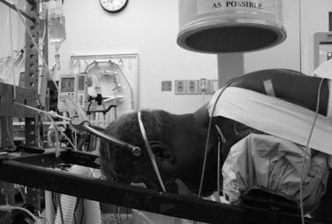
A basic electric operating table may be used for many spinal procedures (Fig. 27-1). Operations in the supine position, such as anterior cervical procedures and anterior lumbar fusions in the distal lumbar spine (L3-S1), are easily performed with such a table. Reversing the table may improve clearance under the table for the fluoroscopic unit. A lateral approach for thoracic, thoracolumbar, and lumbar procedures may also be performed on an electric table. For thoracoabdominal and retroperitoneal flank approaches, it is often helpful to place the level of pathology at the table break and flex the patient laterally.

A posterior approach for thoracic and lumbar procedures may also be performed on a standard operating table with either a Wilson frame or padded bolsters. If an instrumented lumbosacral arthrodesis is planned, however, care must be taken to ensure adequate lumbar lordosis. We generally do not use a Wilson frame for posterior thoracolumbar procedures that include instrumentation and fusion because of the possibility of inadvertently causing an iatrogenic flat-back deformity.

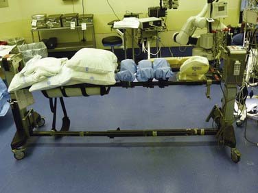
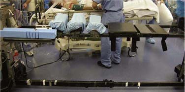
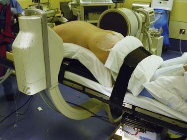
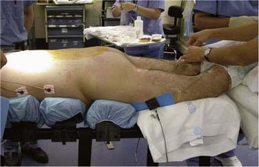
There are many advantages to modular spine-specific operating tables such as the Jackson Spinal table (Mizuho OSI, Union City, CA) (Fig. 27-2). The carbon fiber frame is radiolucent and low profile to allow 360-degree fluoroscopy and the use of intraoperative CT scanners such as the O-arm (Medtronic Navigation, Louisville, CO). Modular components allow customization for each procedure and for a wide range of patient phenotypes. The head may be secured in a foam headrest, in a rigid fixator, or with traction. The legs may be supported in a sling to allow lumbar flexion or on a rigid tabletop to enhance lordosis (Fig. 27-3). Finally, intraoperative repositioning for anterior-posterior or posterior-anterior surgery is facilitated with a rotational capability that obviates the need for moving the patient from one table to another.

Principles of Positioning

Patient Safety and Protection

Patient safety and avoidance of morbidity are important secondary considerations in operative positioning. Although the overall approach (posterior, anterior, lateral) is selected to allow achievement of the surgical objective, it is meticulous attention to the specific details of positioning that ensures that patients do not suffer adverse sequelae from their position during the procedure. Properly padding all areas that may be exposed to pressure and placing extremity joints in relaxed, natural positions are basic preventive measures. Other important considerations may include head positioning and facial pressure and the relationship of the operative field to the level of the heart.

Neuropathies and Prevention

Ulnar neuropathy, one of the most common postoperative neuropathies, accounts for a third of all nerve injury claims in the American Society of Anesthesiologists Closed Claims Study database.1 Although the etiology of postoperative peripheral nerve injury is not entirely known, it is thought to be related to intraneural capillary ischemia resulting from nerve overstretch or compression, perhaps exacerbated by prolonged intraoperative hypotension. The ulnar nerve appears to be more vulnerable to ischemia than the median and radial nerves, with a reported incidence of 0.04% after noncardiac surgery to 37% in one series of cardiac patients who underwent detailed postoperative sensory testing. The time of onset of ulnar nerve symptoms varies from immediately after surgery to 3 days postoperatively. The duration of symptoms tends to vary across reports, with some completely resolving spontaneously in days and others persisting for years after the initial insult.2 Risk factors for postoperative ulnar neuropathy include diabetes, increased age, and male gender.3

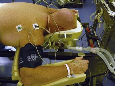
Anatomically, the ulnar nerve appears to be particularly susceptible to direct compression as it courses through the superficial condylar groove at the elbow. Elbow flexion, especially to greater than 110 degrees, can tighten the cubital tunnel retinaculum and directly compress the nerve,4 and external compression in the absence of flexion may compromise the nerve. With the patient in the supine position, direct pressure on the ulnar nerve at the elbow is significantly higher if both forearms are pronated than if they are in a neutral and supinated position (Fig. 27-4).5

Brachial plexus neuropathy may have findings similar to ulnar neuropathy but may additionally be characterized by symptoms such as shoulder pain, scapular winging, and shoulder weakness. The incidence of brachial plexopathy during posterior spinal surgery has been estimated to be between 3.6% and 15%, as compared with 0.02% in a large study of 15,000 general surgical patients. Most patients achieve partial or full functional recovery, although some have persistent symptoms at late (1- to 3-year) follow-up.6,7

The majority of brachial plexus injuries are manifested as upper trunk injuries after surgery in the supine position and as lower trunk injuries after procedures in the prone position. The brachial plexus may be especially vulnerable to stretch in a prone-positioned patient with elbow flexion and shoulder abduction. Patients with congenital anomalies such as cervical ribs and shoulder contractures may have an increased susceptibility to stretch injury.

Some investigators have suggested intraoperative somatosensory evoked potential (SSEP) monitoring as a way to detect impending nerve injury. One retrospective study of 1000 spinal surgeries determined that the overall incidence of position-related upper extremity SSEP changes was 6.1%, with the lateral decubitus position (7.5%) and prone “superman” position (7.0%) having the highest incidence of position-related upper extremity SSEP changes. In this study, postoperative deficits did not develop in any patient who had positionally reversible SSEP changes.8

Lower extremity neuropathies have not been well studied in spinal surgery because they typically occur in patients undergoing surgery in the lithotomy position and after lower extremity orthopedic procedures. Injury to the common peroneal nerve has been reported more frequently than injury to any other lower extremity peripheral nerve, probably because of its vulnerable anatomic location. The common peroneal nerve is fixed in a superficial location as it traverses the head of the fibula, which leaves it susceptible to direct compression injury by devices that hold the legs in place. The legs should be padded and well protected at the level of the fibular head, particularly for procedures in the lateral decubitus position.9

In the event of a new postoperative neurological deficit, it is important to distinguish peroneal nerve injury from an acute L5 radiculopathy. A peroneal neuropathy is characterized by complete plegia of dorsiflexion and eversion without significant pain complaints, whereas an L5 radiculopathy usually results in dermatomal pain and sensory deficit accompanied by weakness of dorsiflexion, toe extension, and foot inversion.10

The lateral femoral cutaneous nerve (LFCN) originates from the L2-3 nerve roots and travels along the lateral border of the psoas major muscle and across the ilium toward the anterior superior iliac spine (ASIS). Because of its anatomic exit below the ASIS, compression neuropathy of the LFCN by posts or pads that support the pelvis may develop in patients who are placed in the prone position. In patients who sustain perioperative injury to the LFCN, hypoesthesia of the anterolateral aspect of the thigh usually develops, but some experience pain and dysesthesia as well. Few studies have been conducted to assess the incidence of positioning-related LFCN injury, although most estimates are around 20%. One study in particular estimated the incidence of LFCN injury to be 23.8% in patients who underwent prone spinal surgery with use of the Relton-Hall frame. All these patients experienced resolution of symptoms within 1 week to 2 months postoperatively.11

Soft Tissue Injuries

The patient’s soft tissues must also be assiduously protected. Prolonged pressure leads to local ischemia and, in severe cases, tissue necrosis. The risk for injury may be minimized by, first, ensuring that sensitive structures do not bear significant pressure and, second, by distributing the pressure over a wide surface with careful padding. Materials that contact the skin should be permeable to prevent moisture buildup. Before placing the patient in the final position, an inspection should be performed to ensure that there are no electrocardiographic leads, intravenous line connectors, or other devices located in areas that will rest on the supporting pads.

With the patient in either the lateral or the prone position, the abdomen should be as free as possible. In the lateral position, the free abdomen will tend to fall anteriorly away from the spine, thereby facilitating a thoracoabdominal or retroperitoneal flank approach. With the patient positioned prone, having the abdomen free decreases intra-abdominal pressure. This both facilitates ventilation of the patient and decreases pressure in the valveless epidural venous plexus and thus reduces epidural bleeding.

Proper positioning of the breasts, particularly for relatively large women placed in the prone position, can be difficult. Adequate support of the upper thoracic region is necessary to achieve neutral cervical alignment and a stable operative platform. In general, the breasts are positioned so that they are medial and caudal to the supporting pads. Particular care is taken to ensure that direct pressure on the nipples is avoided, if possible.

Breast implants may pose a difficult positioning problem as well. The implants tend to be less compressible and less mobile than natural breast tissue, and it may be difficult to avoid direct pressure on the implants. It is important to discuss this potential problem with the patient beforehand and explain the risk for soft tissue injury from pressure and the rare possibility of implant rupture.

Head Positioning

Secure, neutral positioning of the cervical spine is a fundamental principle of patient positioning for all spinal operations, not just those directly involving the cervical region. Patients with degenerative disease in the thoracolumbar region frequently have concomitant cervical spondylosis and may therefore be at risk for postoperative cervical myeloradiculopathy if improperly positioned.

There are three main methods for providing head support and maintaining neutral cervical alignment. For lateral and supine cases, soft supports such as doughnut-shaped foam or gel pads or pillows may be used. Appropriately sized pads should be selected to avoid hyperextension, hyperflexion, or excessive lateral flexion. A specialized foam head holder with or without a custom rigid support may also be used to support the head for prone procedures. These are most appropriate for relatively short operations that do not involve the cervical or upper thoracic region.

Rigid head-holding devices may also be used. Three-point pin fixation devices with a table-mounted holder, such as the Mayfield system (Integra, Plainsboro, NJ), are familiar to most neurological surgeons, but perhaps less so to orthopedic surgeons. Proper positioning of the pins is necessary to prevent slippage of the head in the holder and to minimize the likelihood of perforating the skull. One benefit of rigid head fixation is that the occipitocervicothoracic region can be precisely aligned and the position maintained throughout the operation. A military prone position to reduce a malaligned dens fracture, for example, can be readily achieved, confirmed with fluoroscopy, and securely held during surgery. If a long instrumented fusion is planned, care must be taken during positioning to ensure proper alignment in all three planes and avoid creating an iatrogenic deformity.

Finally, traction systems can be used to secure the head. They allow some movement of the head and neck during surgery, which can have at least two benefits. First, by setting up dual vectors for traction, alignment of the spine can be altered during surgery by the surgeon while still scrubbed (Fig. 27-5). Second, the small amount of movement produced by the placement of upper thoracic pedicle screws might cause dislodgment of the head from a rigid fixator; a properly adjusted traction system allows a safe amount of movement and eliminates this potential complication.

Visual Loss and Its Prevention

Postoperative visual loss (POVL) is an infrequently recognized but devastating complication of spinal surgery. Given that the estimated incidence of POVL has varied considerably across the surgical literature and was thought to be escalating in the mid-1990s, the American Society of Anesthesiologists created a POVL registry in 1999. Interim analyses of the POVL registry suggested a frequency of 0.0008% in noncardiac surgical patients undergoing procedures as disparate as hip arthroplasty, thoracotomy, and neck dissection. POVL in spinal surgery appears to be up to 100 times more frequent, with an incidence of roughly 0.08%.12

The most common cause of POVL in spinal surgery is ischemic optic neuropathy (ION) (89% of registry cases), which is more frequently unilateral than bilateral. POVL may also be attributed to central retinal artery occlusion (11% of registry cases). Although the precise etiology of ION in spinal surgery remains unclear, the leading hypothesis attributes ION to compromised blood flow in the optic nerve as a result of increased venous pressure and interstitial edema. Seventy-two percent of all ION cases were associated with prone spinal surgery; it occurred both in patients whose heads were maintained in facial supporters and in those for whom Mayfield pins alone were used for the entirety of the procedure. These data demonstrate that ION occurs independent of external pressure on the globe. Early data also suggested a relationship between POVL and prolonged anesthetic duration (94% of procedures had an anesthetic duration of 6 hours or longer), as well as between POVL and significant blood loss (82% of patients had estimated blood loss of 1 L or greater).13

Spinal Alignment

As increasing numbers of instrumented fusions are performed, spinal surgeons are recognizing the relationship between achieving and maintaining proper spinal alignment and good clinical outcome. For procedures in which no arthrodesis is performed, such as lumbar microdiskectomy or cervical foraminotomy from a posterior approach, the patient’s intraoperative position may be optimized to facilitate safe, thorough neural decompression. This usually involves a moderate amount of regional flexion. If the spine is instrumented as an adjunct to fusion, however, care should be taken to place the spine in anatomic alignment to avoid creating an iatrogenic deformity such as lumbar hypolordosis (“flat back”). There are important considerations specific to each spinal region that the surgeon should address when checking the patient’s position before surgery.

Proper alignment of the occipitocervical region is essential for good patient outcomes after instrumentation and arthrodesis of the region from the occiput to C2. Improper positioning can lead to an overly extended position and an inability of patients to see their body. Excessive flexion or retraction can make swallowing difficult. Finally, coronal or axial (rotational) malalignment will require patients to compensate for head tilt or rotation to maintain level, forward gaze.

One option to ensure proper occipitocervical alignment is to place the patient in a halo and vest preoperatively. Adjustments can then be made to the patient’s position before surgery. This strategy may be appropriate for patients who will require halo-vest immobilization postoperatively. It is less useful for procedures in which repositioning during the procedure is necessary or advantageous, such as combined transoral decompression and posterior occipitocervical fixation and arthrodesis. There can also be practical issues in accommodating the halo and vest on the operating table.

Estimating the chin-brow angle from fluoroscopy or radiographs can be difficult. We generally use a combination of low-magnification fluoroscopy, which maximizes the field of view and the ability to judge the relationship between the occipitocervical region and the subaxial cervical spine, and direct inspection of the relationship between the head and the torso. In some cases this does require the surgeon to scrub out of the sterile field to look under the surgical drapes.

When performing instrumentation and arthrodesis of the subaxial cervical spine, the surgeon must attend to the restoration or preservation of normal cervical lordosis. Patients who are fixed in a straight alignment are likely to complain about their head and neck position or pain, or both. To facilitate laminectomies, foraminotomies, and placement of lateral mass fixation, we prefer an intraoperative position of relative neck flexion. By using the dual-vector traction system described earlier, we can easily place the patient into cervical lordosis before rod placement and grafting. Although well-placed lateral mass screws can tolerate modest amounts of corrective force during rod placement, their relatively low pullout strength and the lack of a good salvage fixation option in the event of pullout has led us to try to achieve the final alignment through patient positioning and to use the fixation to maintain rather than achieve the final lordotic alignment (see Fig. 27-5).

Alignment of the cervicothoracic region deserves special mention. Without careful attention, it is easy to position the patient so that there is relative cervicothoracic kyphosis and a straight subaxial cervical spine. This is a particularly debilitating position in that the patient’s head juts forward from the upper thoracic region and forward gaze is maintained only through hyperextension between the occiput and the upper cervical spine. Direct inspection is often the best method to detect whether this problem is in fact occurring intraoperatively. Again, we try to minimize the amount of force placed on implants and prefer to obtain optimal alignment through patient positioning.

Finally, the importance of restoring or maintaining adequate lumbar lordosis has received much attention—and rightly so. Lumbar flexion facilitates decompressive lumbar laminectomy and lumbar microdiskectomy but should be assiduously avoided if an arthrodesis is to be performed (Fig. 27-6). Adequate lumbar lordosis can most easily be achieved with maximal hip extension. Use of the Wilson frame and a leg sling should generally be avoided in procedures that involve lumbar arthrodesis. Instead, we use modular hip and thigh pads on a spine table and a flat leg rest padded with pillows (Fig. 27-7).

Surgeon Ergonomics

An important and often overlooked aspect of patient positioning is to optimize the working environment for the surgeon. Particularly for long or difficult procedures, surgeon comfort should be considered carefully, although it should never assume priority over patient safety. Working for a long period in an uncomfortable position or being unable to optimally visualize the surgical field may compromise the surgeon’s ability to achieve the operative objectives. The operative field should be at a comfortable height for the surgeon. Tables designed for spine surgery are often adjustable through a greater vertical range than possible with standard electric operating room tables. This is advantageous in that it may obviate the need for the surgeons and assistants to use standing stools.

In general, we try to position the operative field in as close to a horizontal plane as possible. For lower cervical and cervicothoracic procedures, this often means that the patient must be placed in a considerable amount of reverse Trendelenburg positioning. In the thoracic and lumbar regions, the table is usually fairly level.

The operating microscope can also improve surgeon comfort, as well as provide excellent illumination and magnification. Particularly in the cervical region, its use can reduce surgeon discomfort during both anterior and posterior procedures. An additional benefit is that the assistant has an excellent view of the procedure and can assist, if necessary.

Specific Procedures

Anterolateral, Retropleural Thoracic, Lateral Lumbar

Lateral positioning follows the same principles as for the more common anterior and posterior approaches. There is significant potential, however, for soft tissue or peripheral nerve injury secondary to focal pressure. Attention is therefore meticulously paid to these areas to ensure that adequate padding is used. The patient is placed on a beanbag covered by a sheet. A soft roll is placed under the dependent axilla to prevent excessive shoulder abduction and to distribute the pressure over a greater area. The dependent arm is externally rotated and the elbow is flexed to approximately 90 degrees. The upper part of the arm is also gently flexed. A folded pillow may be placed between the arms to keep them roughly parallel to the floor while allowing adequate access to the face. The dependent leg is flexed gently and the upper part of the leg is at most flexed slightly. Addition flexion may be helpful for lumbar flank approaches because it will tend to relax the iliopsoas and aid exposure. The common peroneal is susceptible to pressure injury as it crosses the proximal portion of the fibula just distal to the knee. This area must therefore be amply padded.

It is also helpful to have the patient in a true lateral position. The resultant orthogonal approach to the spine allows the surgeon to remain oriented to the location of the canal during decompression and placement of grafts and instrumentation. We also prefer to have the patient’s back as close to the edge of the table as possible because we tend to operate mainly from that side, which reduces the effective depth of the wound.

Intraoperative Repositioning

In some cases it is necessary or advantageous to perform “circumferential” surgery via different approaches on the same day. This requires intraoperative repositioning from a supine to a prone position or vice versa and may be accomplished with the use of two standard electric operating tables. We have found, however, that the patient must usually be moved to a stretcher before repositioning on the second table because the bulky table bases prevent them from being moved sufficiently close together to allow direct transfer of the patient from one table to the other.

It is more efficient to use a modular spine table that allows the placement of a second tabletop and rotating the patient on the table. Meticulous attention to detail is necessary to ensure safe repositioning. Adequate assistance must be available, but one individual should be in charge and delegate responsibility. We generally secure the head with Gardner-Wells tongs and 10 lb of in-line traction. For cervical procedures or if there is a concern about the cervical spine, we often also place a hard cervical collar on the patient.

Perhaps it seems obvious, but it is important to remember that what is on top of the patient before rotation will be underneath the patient afterward. The surgeon should therefore ensure that adequate smooth padding is in place and that the patient will not be lying on intravenous lines or other noncompressible objects. Before rotating the patient, a final check should be made that the tabletops are secure, that all lines are disconnected and secured, and that everyone understands the direction of the rotation. The ventilator is disconnected, the patient is rotated, the table is secured, the ventilator is reconnected, and the upper tabletop is removed.

Suggested Readings

Cooper DE, Jenkins RS, Bready L, et al. The prevention of injuries of the brachial plexus secondary to malposition of the patient during surgery. Clin Orthop Relat Res. 1988;228:33-41.

Heitz JW, Audu PB. Asymmetric postoperative visual loss after spine surgery in the lateral decubitus position. Br J Anaesth. 2008;101:380-382.

Kamel IR, Drum ET, Koch SA, et al. The use of somatosensory evoked potentials to determine the relationship between patient positioning and impending upper extremity nerve injury during spine surgery: a retrospective analysis. Anesth Analg. 2006;102:1538-1542.

Lee LA, Roth S, Posner KL, et al. The American Society of Anesthesiologists Postoperative Visual Loss Registry: analysis of 93 spine surgery cases with postoperative visual loss. Anesthesiology. 2006;105:652-659.

O’Driscoll SW, Horii E, Carmichael SW, et al. The cubital tunnel and ulnar neuropathy. J Bone Joint Surg Br. 1991;73:613-617.

Prielipp RC, Morell RC, Butterworth J. Ulnar nerve injury and perioperative arm positioning. Anesthesiol Clin North Am. 2002;20:589-603.

Prielipp RC, Morell RC, Walker FO, et al. Influence of arm position and relationship to somatosensory evoked potential. Anesthesiology. 1999;91:345-354.

Schwartz DM, Drummond DS, Hahn M, et al. Prevention of positional brachial plexopathy during surgical correction of scoliosis. J Spinal Disord. 2000;13:178-182.

Seyfer AE, Grammer NY, Bogumill GP, et al. Upper extremity neuropathies after cardiac surgery. J Hand Surg Am. 1985;10:16-19.

Vazquez-Jimenez JF, Krebs G, Schiefer J, et al. Injury of the common peroneal nerve after cardiothoracic operations. Ann Thorac Surg. 2002;73:119-122.

Warner MA. Perioperative neuropathies. Mayo Clin Proc. 1998;73:567-574.

Warner MA, Warner ME, Martin JT. Ulnar neuropathy: incidence, outcome, and risk factors in sedated or anesthetized patients. Anesthesiology. 1994;81:1332-1340.

Yang SH, Wu CC, Chen PQ. Postoperative meralgia paresthetica after posterior spine surgery: incidence, risk factors, and clinical outcomes. Spine. 2005;30:E547-E550.

References

1 Prielipp RC, Morell RC, Walker FO, et al. Influence of arm position and relationship to somatosensory evoked potential. Anesthesiology. 1999;91:345-354.

2 Seyfer AE, Grammer NY, Bogumill GP, et al. Upper extremity neuropathies after cardiac surgery. J Hand Surg Am. 1985;10:16-19.

3 Warner MA, Warner ME, Martin JT. Ulnar neuropathy: incidence, outcome, and risk factors in sedated or anesthetized patients. Anesthesiology. 1994;81:1332-1340.

4 O’Driscoll SW, Horii E, Carmichael SW, et al. The cubital tunnel and ulnar neuropathy. J Bone Joint Surg Br. 1991;73:613-617.

5 Prielipp RC, Morell RC, Butterworth J. Ulnar nerve injury and perioperative arm positioning. Anesthesiol Clin North Am. 2002;20:589-603.

6 Cooper DE, Jenkins RS, Bready L, et al. The prevention of injuries of the brachial plexus secondary to malposition of the patient during surgery. Clin Orthop Relat Res. 1988;228:33-41.

7 Schwartz DM, Drummond DS, Hahn M, et al. Prevention of positional brachial plexopathy during surgical correction of scoliosis. J Spinal Disord. 2000;13:178-182.

8 Kamel IR, Drum ET, Koch SA, et al. The use of somatosensory evoked potentials to determine the relationship between patient positioning and impending upper extremity nerve injury during spine surgery: a retrospective analysis. Anesth Analg. 2006;102:1538-1542.

9 Warner MA. Perioperative neuropathies. Mayo Clin Proc. 1998;73:567-574.

10 Vazquez-Jimenez JF, Krebs G, Schiefer J, et al. Injury of the common peroneal nerve after cardiothoracic operations. Ann Thorac Surg. 2002;73:119-122.

11 Yang SH, Wu CC, Chen PQ. Postoperative meralgia paresthetica after posterior spine surgery: incidence, risk factors, and clinical outcomes. Spine. 2005;30:E547-E550.

12 Heitz JW, Audu PB. Asymmetric postoperative visual loss after spine surgery in the lateral decubitus position. Br J Anaesth. 2008;101:380-382.

13 Lee LA, Roth S, Posner KL, et al. The American Society of Anesthesiologists Postoperative Visual Loss Registry: analysis of 93 spine surgery cases with postoperative visual loss. Anesthesiology. 2006;105:652-659.