This chapter covers several important pathological processes that used to be classified as ‘tissue degenerations’. They are unified by the fact that they all involve abnormal accumulations of pigments or other substances that build up as a result of a variety of different pathological processes. These materials can be classified as endogenous if they are produced In the body, or exogenous if they are derived from outside.
13.1. Abnormal endogenous accumulations
You should:
• state the main characteristics and pathological significance of fatty change, lipofuscin, haemosiderin and amyloid
• distinguish metastatic from dystrophic calcification
• recognise abnormal melanin accumulation
• understand the term hyaline
• discuss the principles of the inherited storage disorders.
Haemosiderin
Haemosiderin is a golden yellow to brown pigment found in lysosomes within the cell cytoplasm. It is composed of aggregates of partially degraded ferritin, which is protein-covered ferric oxide and phosphate. It can be visualised using the Prussian blue reaction, when haemosiderin appears dark blue.
Haemosiderin represents iron deposition and accumulates in tissues in two main circumstances. First, if there is haemorrhage into a tissue the haemoglobin is broken down and haemosiderin is deposited in macrophages locally. This process occurs in many chronic inflammatory conditions; the extravasated red blood cells are broken down and haemosiderin is produced from the iron they contain. The identification of haemosiderin in a tissue sample can be a clue to previous haemorrhage or inflammation.
Second, an excess of circulating iron can result in systemic haemosiderin accumulation. For example, primary haemochromatosis is an inherited disease in which there is excessive absorption and widespread deposition of haemosiderin in the tissues, especially the liver, pancreas, heart and skin. The iron is toxic to the tissues and leads to fibrosis of the liver (cirrhosis), fibrosis of the pancreas (leading to diabetes mellitus), and heart failure.
Other causes of systemic iron deposition include increased absorption of iron from the intestine, haemolytic anaemia and recurrent blood transfusions. The presence of iron in the tissues is termed haemosiderosis, which is not be confused with the disease haemochromatosis as described previously.
Melanin
Melanin is the brown/black pigment normally present in the cytoplasm of cells in the basal layer of the epidermis, called melanocytes. Melanin is derived from tyrosine, stored in melanosomes and distributed to the other epidermal cells. The function of melanin is to block harmful UV rays from reaching the epidermal nuclei. It may accumulate in excessive quantities in benign or malignant melanocytic neoplasms and its presence is a useful diagnostic feature for such lesions. In inflammatory skin lesions, where the epidermis is damaged, melanin may be released from injured basal cells and taken up by dermal macrophages. This gives rise to post-inflammatory pigmentation of the skin. Melanin can be identified in tissue sections by the use of the Masson–Fontana stain.
Excessive melanin pigmentation of the skin is a feature of certain diseases, e.g. Addison’s disease and neurofibromatosis.
Lipofuscin
This is the yellow/brown ‘wear-and-tear’ pigment seen in atrophic tissues (see atrophy, Ch. 3). It also accumulates in ageing cells of the liver, myocardium and elsewhere. Lipofuscin does not seem to damage cells and does not cause clinical problems.
Calcification
Calcification is the deposition of calcium salts within tissues, often causing them to become chalky, hard or brittle. There are two main types of calcification:
• dystrophic
• metastatic.
Dystrophic calcification
This type of calcification occurs within diseased tissues. The plasma calcium and phosphate levels are normal. The exact mechanism by which dystrophic calcification occurs is not known. Examples include calcification within areas of necrosis, foci of old tuberculosis, atheromatous plaques, and in neoplasms. The calcification can often be identified on radiographs.
Metastatic calcification
Metastatic calcification occurs in normal tissues as a consequence of raised plasma calcium concentrations (hypercalcaemia). Common causes of hypercalcaemia include widespread metastatic cancer in the bones, hyperparathyroidism and multiple myeloma. Metastatic calcification does not often cause clinical problems, although occasionally renal failure can follow calcification of the kidneys.
Hyaline
Hyaline simply means ‘glassy’ and is used as a descriptive term by pathologists for a variety of materials that have a uniform pink (eosinophilic) appearance under the microscope. Deposits of protein in renal tubules and Mallory bodies in the liver can be described as exhibiting a hyaline appearance. In longstanding diabetes and hypertension, accumulation of proteins in the walls of arterioles causes them to become hyalinised.
Amyloidosis
The term amyloid means ‘starch-like’ and as such is misleading because amyloid is not a carbohydrate. Amyloid is a descriptive term used for a group of proteinaceous substances that may be deposited in tissues and organs to give characteristic naked eye, microscopic and ultrastructural appearances.
What is amyloid?
Amyloid is an abnormal protein characterised by a β-pleated sheet configuration deposited in the extracellular matrix. Many different proteins can produce amyloid, and the type of protein found in the amyloid deposits depends on the underlying disease. For example, in patients with multiple myeloma (a neoplastic proliferation of antibody-producing plasma cells), the amyloid is composed of antibody fragments. The β-pleated sheets form long, non-branching fibrils with a diameter of about 8nm; they can be observed under the electron microscope. Once the β-pleated sheets have formed, the body’s intra- and extracellular proteolytic enzyme systems find it almost impossible to digest; therefore, it accumulates inexorably.
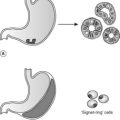
Almost all types of amyloid also contain a second substance, a glycoprotein known as the P component (or P protein). The P component is a doughnut-shaped pentamer.
When amyloid is deposited in large amounts, the tissue becomes pale, smooth and waxy in texture. Histologically, amyloid stains pink with Congo red dye; the pink colour turns apple green under polarised light, a property unique to amyloid.
Where does amyloid accumulate?
Amyloid can accumulate within any tissue or organ. The disease may affect just one tissue or organ, i.e. localised amyloidosis, or several, i.e. systemic amyloidosis. It always accumulates outside cells and has a predilection for basement membranes and interstitial connective tissues.
Where there is abundant amyloid deposition, cells become ‘strangled’ and organ failure occurs. There is also evidence that some types of amyloid have a direct toxic effect on cells. For example, β-amyloid in the brain damages neurones, possibly by free radical production.
When does amyloid accumulation occur?
It is thought that defective proteolysis of the precursor protein results in amyloid production. The two most common examples of systemic amyloidosis are:
• AL amyloid: Patients with multiple myeloma typically have excessive amounts of circulating intact and fragmented immunoglobulin light chains. Defective proteolysis of these light chains gives an amyloid protein called AL amyloid.
• AA amyloid: This type of amyloid complicates long-term inflammatory conditions. These can be suppurative processes, such as bronchiectasis, or autoimmune diseases, such as rheumatoid disease. In these conditions, the liver produces a number of acute-phase proteins that have various functions in the inflammatory process. Among these is serum amyloid A protein (SAA). This normal protein can be converted into amyloid A (AA amyloid).
Examples of localised amyloid are:
• β-amyloid: Derived from amyloid precursor protein (APP) in the brain, this is a component of the plaques of Alzheimer’s disease.
What are the effects of amyloid on tissues/organs?
The effects of amyloid deposition depend on the organs involved and the amount of amyloid deposited. The most serious complications of systemic amyloidosis are usually seen in the kidney and heart. Renal involvement can lead to the loss of large amounts of protein in the urine and even renal failure. Myocardial amyloid may stop the heart contracting properly and cause heart failure, or, if deposited in the conducting system, cause a lethal arrhythmia.
The storage disorders
These conditions are rare inborn errors of metabolism that cause accumulation of a macromolecule in cells. They are all due to a genetic disorder and generally show recessive inheritance. The genetic defect results in the massive accumulation of a substance within tissues, causing secondary cell injury. The majority of these conditions can be divided into two main groups: the glycogen storage diseases and lysosome storage diseases.
The glycogen storage diseases, or glycogenoses, are due to defects in an enzyme involved in glycogen metabolism. For example, deficiency of glucose 6-phosphatase causes glycogen to accumulate particularly in the liver and kidney. Other glycogen storage diseases may primarily affect muscle, or cause generalised systemic deposition of glycogen.
The lysosome storage diseases are due to a failure in lysosomal digestion. The defect is found in the activity of an enzyme that breaks down a macromolecule into its component subunits or in a transport protein that is required for moving the digested material out of the lysosome. Consequently, the macromolecule accumulates within lysosomes that become massively distended. For example, deficiency of glucocerebrosidase causes Gaucher’s disease, in which gangliosides accumulate in macrophages in the bone marrow, spleen and liver, causing reduction in blood cell production (pancytopenia) and hepatosplenomegaly.
13.2. Abnormal exogenous accumulations
If insoluble substances that cannot be adequately eliminated enter the body, they will accumulate. A variety of pigments and other materials can do this. They may be toxic and produce inflammatory tissue reactions or they may be relatively inert. There are two main routes by which such materials enter the body: through the skin and the lungs.
Exogenous skin pigmentation is due to tattooing. The pigments are engulfed by dermal macrophages that become immobilised, retaining the pigments in the skin indefinitely.
Inhaled dusts entering the respiratory tract can accumulate in the lungs. They are taken up by pulmonary macrophages that remain in the walls of the airways. Carbon derived from soot is found in virtually everyone living where there is air pollution, and its accumulation is called anthracosis. Pure carbon is relatively inert, but other substances are toxic and can cause significant disease. The general term for a disease of the lungs caused by inhaled dust is pneumoconiosis. Coal dust, asbestos, silica and other dusts cause fibrosis of the lung that may compromise lung function and cause breathlessness and reduced exercise tolerance. By interfering with normal blood flow through the lungs, they can also cause cor pulmonale and right ventricular failure. In addition, some inhaled dusts are carcinogenic and can cause cancer.
Self-assessment: questions
One best answer questions
2. A 25-year-old man has an appendicectomy for abdominal pain. The pathologist finds no evidence of acute inflammation in the appendix, but the deposition of a pigment suggests that the appendix has been inflamed in the past. Which one of the following is most likely to have led to this conclusion?
a. carbon
b. haematoxylin
c. haemosiderin
d. lipofuscin
e. melanin
3. Which one of these materials shows apple-green birefringence when stained with Congo red?
a. amyloid
b. glycogen
c. lipofuscin
d. Mallory bodies
e. melanin
True-false questions
1. The following are correctly paired:
a. lipofuscin pigment – tattoos
b. haemosiderin pigment – skin bruising
c. asbestosis – pneumoconiosis
d. AL amyloid protein – bronchiectasis
e. parathyroid adenoma – metastatic calcification
2. The following statements are true:
a. fatty change caused by alcoholic liver damage is reversible
b. lipofuscin accumulation is associated with ageing
c. calcification of atherosclerotic plaques is rare
d. anthracosis is caused by silica
e. Gaucher’s disease is inherited as an autosomal dominant disorder
Extended matching items questions (EMIs)
EMI 1
Theme: Abnormal accumulations
A. AA amyloid
B. AL amyloid
C. β-amyloid
D. asbestos
E. calcium
F. glycogen
G. haemosiderin
H. intracellular lipid
I. lipofuscin
J. melanin
For each of the following scenarios, select the substance from the list that is most likely to be responsible for the clinical and pathological features.
1. A 95-year-old woman dies and an autopsy is performed. The pathologist finds the heart to be atrophic and to have a uniform brown appearance.
2. A 75-year-old woman with longstanding rheumatoid arthritis presents with proteinuria. A renal biopsy shows deposition of eosinophilic material in the glomeruli.
3. A 50-year-old man with type 2 diabetes mellitus has a routine blood test that shows abnormal levels of liver enzymes. Ultrasound of the liver shows diffuse increased echogenicity.
4. A 6-month-old baby boy dies of heart failure. Histological sections of the heart show the accumulation of large amounts of periodic acid-Schiff (PAS)-positive material in the cytoplasm of the myocytes. A section of liver shows similar material in the hepatocytes.
5. A 65-year-old retired docker has a history of poor lung function associated with extensive lung fibrosis. He presents with a lung mass, and squamous cell carcinoma is diagnosed by biopsy of the mass.
Case history questions
Case history 1
A 56-year-old woman presented with generalised symptoms of feeling unwell. She had noticed swelling of her ankles and face and was breathless. On examination, she had signs of heart failure and oedema. Her urine, collected over 24 hours, contained 11g of protein (normal is less than 150mg) and her serum albumin level was 20g/L (normal is about 40g/L).
A kidney biopsy was performed and showed amyloid deposition. Further examination of the urine and serum proteins showed increased amounts of λ light chain proteins. Electrophoresis of the blood revealed a monoclonal band of immunoglobulin. Bone marrow biopsy was performed.
2. What type of amyloid would you expect to find in this case?
3. State two physical characteristics of the amyloid in this case.
4. Why might the loss of large amounts of protein in the urine lead to ankle swelling?
Case history 2
A 58-year-old accountant was noticed by his family and firm to have become unreliable, mentally slow, confused and have a poor memory. Over the next few years, he became progressively unable to walk and lead an independent existence. He died at the age of 67. Clinically, a diagnosis of Alzheimer’s disease was made during his illness.
1. State the type of amyloid found in the plaques of Alzheimer’s disease, and the normal protein from which it is derived.
2. Would you expect to find the amyloid inside or outside cells?
3. Why does the study of Down’s syndrome help us in the understanding of Alzheimer’s disease?
Self-assessment: answers
One best answer
2. c. Haemosiderin can be found in macrophages for some time after the inflammation has died down. Perl’s stain is often used by pathologists to demonstrate it in tissue sections; this stain uses the Prussian blue reaction. Note that haematoxylin is a dye used in routine histological stains.
3. a. This staining reaction is a useful histological test for amyloid.
True-false answers
1.
a. False. Lipofuscin is the brown intracellular pigment which accumulates in cells with age. It is present in autophagic vacuoles within cells and is often known as wear-and-tear pigment. Organs which are very atrophic may actually look brown to the naked eye from excessive amounts of this pigment. Tattoos gain their colour and permanence from the intradermal injection of inks and dyes, often based on carbon or mercury pigments. These are ingested by local macrophages which cannot digest them and become immobilised at the site of the tattoo, the colour remaining visible through the skin.
b. True. Haemosiderin is the iron-based pigment resulting from the breakdown of haemoglobin. When haemorrhage occurs into tissues, red cell haemoglobin is degraded and ingested by macrophages. Haemosiderin is a brownish yellow pigment which gives the bruise its characteristic colour.
c. True. Asbestosis is characterised by fibrosis of the lungs and reduced respiratory function. It is acquired by inhaling asbestos dust, and so it is a type of pneumoconiosis. Asbestos also causes other diseases, such as lung carcinoma and pleural malignant mesothelioma.
d. False. AL amyloid is composed of κ or λ immunoglobulin light chains and is found in amyloid derived from immunoglobulin, most commonly in multiple myeloma. Multiple myeloma is a neoplastic monoclonal proliferation of plasma cells which produces abnormal amounts of a single immunoglobulin. Bronchiectasis leads to chronic infection of the lungs which can lead to AA protein amyloid formation.
e. True. Parathyroid adenoma is a benign tumour of the parathyroid glands which can produce abnormal amounts of parathyroid hormone. This leads to increased levels of calcium in the bloodstream (hypercalcaemia), which causes fits, vomiting and excessive urine production (polyuria), and sometimes metastatic calcification.
2.
a. True. Fatty change represents reversible tissue injury. In the liver, intracellular triglycerides must be complexed with protein in order to transport them through the cell. Alcohol interferes with this metabolic process and hence causes fatty change. The liver is the most common site for fatty change to be clinically apparent.
b. True. Lipofuscin is associated with ageing and can be seen accumulating in many major organs in elderly people. Lipofuscin itself is not injurious to cells.
c. False. Dystrophic calcification of atherosclerotic plaques is common.
d. False. Anthracosis is caused by the accumulation of carbon pigment in lung macrophages. It is seen in coal miners and city dwellers.
e. False. If only one allele is abnormal, the cell can produce enough normal enzyme. Only if both alleles are abnormal does the cell accumulate glucocerebroside. Gaucher’s disease is inherited as an autosomal recessive condition; storage diseases in general show recessive rather than dominant inheritance.
EMI answers
EMI 1
Theme: Abnormal accumulations
1. I. The heart shows ‘brown atrophy’, i.e. the accumulation of ‘wear and tear’ lipofuscin pigment in the atrophic tissue has imparted a brown appearance.
2. A. AA amyloid complicates chronic inflammatory conditions such as systemic autoimmune diseases, tuberculosis and chronic inflammatory bowel disease. The nephrotic syndrome (see Ch. 23) can follow renal involvement. The eosinophilic material in the glomeruli is the amyloid; it will be positive with Congo red.
3. H. This patient has non-alcoholic fatty liver due to type 2 diabetes. The ultrasound findings are not specific but point to a diffuse abnormality of the liver, in this case accumulation of fat in hepatocytes.
4. F. This description is of Pompe’s disease, a type of glycogen storage disease. Glycogen is positive with the PAS stain.
Case history answers
Case history 1
1. This patient has multiple myeloma, which is an uncontrolled proliferation of plasma cells. Therefore, the type of cell expected to be present in excess in the bone marrow is the plasma cell. Bone marrow biopsy is a useful diagnostic test in this condition. Comment: The specific clues to the diagnosis in the scenario are the light chain proteinuria and the monoclonal band in the plasma. They represent the excessive amounts of abnormal immunoglobulin being produced by the plasma cells.
2. AL amyloid (derived from the light chains).
3. Comment: A good response would be:
• β-pleated sheet
• fibrillary structure.
4. Protein loss in the urine leads to a reduced intravascular colloid osmotic pressure and hence hydrostatic pressure forces fluid out of the blood vessels into the interstitium. This is clinically seen as tissue oedema.
Case history 2
1. β-amyloid, derived from APP.
2. Outside: amyloid is an extracellular deposit.
3. Individuals with Down’s syndrome (trisomy 21) who survive to middle age show similar neuritic plaques and neurofibrillary tangles in their cerebral cortex. Therefore, it has been postulated that genes on chromosome 21 have an important role in the development of these degenerative changes in Alzheimer’s disease.