Pancreatobiliary Parasites

Published on 19/07/2015 by admin

Filed under Radiology

Last modified 22/04/2025

Print this page

rate 1 star rate 2 star rate 3 star rate 4 star rate 5 star
Your rating: none, Average: 0 (0 votes)

This article have been viewed 2315 times

 CT: Linear high-density foci within dilated biliary tree

image US: Tubular echogenic structure, 3-6 mm in diameter, with central anechoic area (digestive tract of worm)
• Clonorchiasis: Flukes typically involve peripheral intrahepatic ducts, not GB, CBD, or PD

image CT/MR: Preferential dilatation of small peripheral intrahepatic ducts with intraductal high-density foci
image US: Flukes appear as echogenic filling defects in bile ducts, which float with changes in position
• Fascioliasis: Flukes usually involve large intrahepatic ducts, extrahepatic duct, and GB (after liver involvement)

image CT: Low-density abscesses forming tract from entry site at Glisson capsule into liver parenchyma

– High-density foci within duct lumen represent trematodes with associated mild ductal dilatation
• Echinococcosis: Communication of hepatic hydatid cyst with small biliary radicles or rupture of cyst into bile ducts

image CT: High-attenuation material within dilated bile ducts, often contiguous with wall defect in hepatic hydatid cyst 

– Air-fluid level within adjacent hydatid cyst may indicate cyst infection or biliary communication
image US: Linear/round filling defects within dilated bile ducts

CLINICAL ISSUES

• Ascariasis most prevalent helminth infection worldwide
• Clonorchiasis endemic in Asia and present in Western world secondary to travel and immigration
• Echinococcosis endemic in underdeveloped grazing regions (Mediterranean, Africa, South America)
• Fascioliasis affects temperate sheep-rearing areas including South America, Europe, China, Africa, and Middle East
image
(Left) Specimen photograph shows an Ascaris worm retrieved from a bile duct with multiple stones. These biliary parasites may grow up to 30 cm in length. If multiple, they may fill the entire bile duct and produce the so-called spaghetti sign.

image
(Right) Liver flukes are short, flat, somewhat transparent, and taper anteriorly. They have prominent oral and ventral suckers. (Courtesy J. Doss, MD.)
image
(Left) T-tube cholangiogram of a woman who recently immigrated from China and presented with RUQ pain and symptoms of cholangitis demonstrates a tubular filling defect in the proximal common bile duct image 
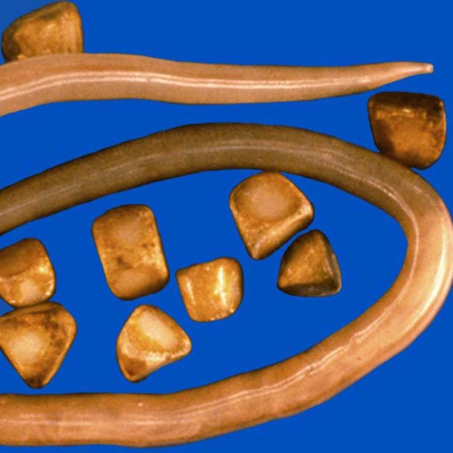
from a biliary Ascaris worm.

image
(Right) Ultrasound of the porta hepatis shows an Ascaris worm within the dilated common duct image
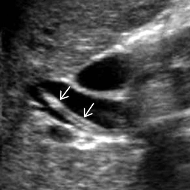
. The worm was mobile at real-time examination. Central hypoechogenicity is thought to represent the digestive tract of the worm. (Courtesy A. Dasyam, MD.)

TERMINOLOGY

Synonyms

• Biliary ascariasis, biliary clonorchiasis, biliary fascioliasis, biliary echinococcosis

Definitions

• Biliary and pancreatic duct (PD) involvement by parasitic infections (e.g., ascariasis, clonorchiasis, echinococcosis, fascioliasis)

IMAGING

General Features

• Best diagnostic clue

image Ascariasis/clonorchiasis: Longitudinal filling defect in bile ducts or PD on ERCP/MRCP
image Echinococcosis: Dilated, debris-filled biliary ducts adjacent to ruptured hepatic hydatid cyst on CT, US, MR
image Fascioliasis: Clustered low-density hepatic lesions forming tract from liver capsule into parenchyma on CT/MR
• Location

image Ascariasis may involve entire biliary tract and PD
image Clonorchiasis typically involves peripheral intrahepatic ducts, not gallbladder (GB), common bile duct (CBD), or PD (except in heavy infections)

– Small or medium-sized peripheral ducts typically diffusely dilated, while more central intrahepatic or extrahepatic ducts are normal in caliber
image Echinococcosis: Any portion of biliary tree can potentially communicate with hydatid cyst
image Fascioliasis: Usually large intrahepatic ducts, extrahepatic duct, and GB
• Size

image Ascaris: 2-30 cm (3-6 mm thick)
image Other parasites (Fasciola, Clonorchis) are smaller
• Morphology

image Linear or rounded

CT Findings

• Ascariasis/clonorchiasis: Intraductal high-density foci within dilated biliary tree due to biliary worms/flukes or debris

image Imaging evidence of complications, including peripancreatic inflammation due to pancreatitis, intrahepatic abscess, and abnormal biliary tree wall enhancement or heterogeneous parenchymal enhancement due to cholangitis
image Characteristic pattern of biliary dilatation in clonorchiasis with preferential dilatation of small peripheral intrahepatic ducts (with sparing of more central ducts)

– Bile duct walls may be thickened due to parasite-related infection/inflammation
• Echinococcosis: High-attenuation material within dilated bile ducts, often continuous with wall defect in hepatic hydatid cyst

image Air-fluid level within adjacent hydatid cyst may indicate cyst infection or biliary communication
• Fascioliasis: Clustered low-density hepatic abscesses forming a tract from entry site at Glisson capsule into parenchyma

image High-density foci within duct lumen represent trematodes, usually with associated mild ductal dilatation and ductal wall thickening/hyperenhancement

MR Findings

• Ascariasis: MRCP or T2WI demonstrate dilated ducts with linear low-signal filling defects

image Ascaris worms have characteristic 3-lines appearance on T2WI/MRCP with central high-signal intensity line between 2 low-signal intensity lines
• Clonorchiasis: Preferential peripheral biliary dilatation with low-signal filling defects on T2WI or MRCP
• Echinococcosis: Low-signal linear or rounded filling defects within dilated ducts, often with adjacent deformed hydatid cyst

image Direct communication between adjacent irregular hydatid cyst and biliary tree may be demonstrated
• Fascioliasis: Liver lesions are low signal on T1WI and high signal on T2WI with extension from liver capsule into deeper liver

image T2WI and MRCP demonstrate mild ductal dilatation with low-signal filling defects

Ultrasonographic Findings

• Grayscale ultrasound

image Ascariasis: Ultrasound very sensitive for worms in biliary system, but insensitive for worms in duodenum or ampulla (sensitivity for pancreatobiliary ascariasis only 50%)

– “Bull’s-eye” appearance due to echogenic filling defect
– Tubular echogenic structure, 3-6 mm in diameter, with central anechoic area (digestive tract of worm)
– Motility of worms may be evident, and worms that have not moved for 10 days are usually dead
– May fill entire bile duct when multiple, producing spaghetti or railway track sign
– No acoustic shadowing
image Clonorchiasis: Flukes appear as echogenic filling defects (without shadowing) in bile ducts which float with changes in position

– Often associated with bile duct stones (including hepatolithiasis), which are echogenic and demonstrate posterior acoustic shadowing
image Echinococcosis: Linear or round filling defects within dilated bile ducts without shadowing
image Fascioliasis :  Abscesses in liver can show variable echogenicity (hypoechoic > hyperechoic)

– Ductal thickening and dilatation with mobile fluke sometimes seen within duct lumen

Radiographic Findings

• ERCP: Linear, elliptical, or rounded filling defects within dilated biliary tree or PD

Nonvascular Interventions

• ERCP

image Biliary stent and liver resection for hepatic Echinococcus complicated by biliary rupture

Nuclear Medicine Findings

• Hepatobiliary scan: Lack of GB filling in ascariasis-related cholecystitis

Imaging Recommendations

• Best imaging tool

image US as initial imaging modality
image MRCP may better illustrate parasite within biliary tree or communication of bile duct with hydatid cyst

DIFFERENTIAL DIAGNOSIS

Bacterial Cholangitis

• Pyogenic infection of biliary tree due to biliary obstruction
• Dilated biliary tree with bile duct wall thickening and hyperenhancement, as well as heterogeneous parenchymal liver enhancement

Recurrent Pyogenic Cholangitis

• Disease associated with formation of pigment stones throughout biliary tree, as well as multiple biliary strictures and repeated bouts of cholangitis
• Almost always in patients of Southeast Asian origin
• Dilation of biliary tree with disproportionate dilatation of central intrahepatic and extrahepatic ducts, as well as stones in both intrahepatic and extrahepatic ducts
• May be related to parasitic (ascariasis, clonorchiasis) or bacterial infection of biliary tree

Biliary Tract Stones

• Echogenic nodules within biliary tree with posterior acoustic shadowing on US
• Posterior acoustic shadowing uncommon with pancreaticobiliary parasites

Cholangiocarcinoma

• Tumor arising from bile duct with characteristic delayed enhancement on multiphase imaging
• Mass typically causes biliary dilatation, parenchymal atrophy peripheral to tumor, and capsular retraction
• Tumors with predominantly intraductal growth appear more mass-like and infiltrative than parasites

PATHOLOGY

General Features

• Etiology

image Ascariasis: Ova ingested, larvae hatch in small bowel

– Invade through small bowel mucosa → migrate through circulatory system to lungs
– Invade alveoli → ascend tracheobronchial tree → swallowed → mature into adult worms in small bowel
image Clonorchiasis :  Flukes ingested with uncooked freshwater fish → metacercariae excyst in duodenum → move to ampulla of Vater and ascend biliary tree

– May live in biliary tree for 15-20 years (usually small- or medium-sized ducts)
image Echinococcosis: Intermediate host is sheep, while definitive host is dog

– Ova ingested → reach liver through portal veins
– Hydatid cyst is larval stage infection of Echinococcus
– Communication of hepatic hydatid cyst with small biliary radicles is common
– Frank rupture of cyst into biliary tree occurs in 5-15%
image Fascioliasis: Humans ingest infected watercress or contaminated water with encysted larva → larva penetrates wall of duodenum → enters peritoneal cavity and penetrates Glisson capsule to enter liver

– Eventually penetrates biliary tract and can survive in bile duct lumen for many years
• Associated abnormalities

image Cholangiocarcinoma with clonorchiasis and ascariasis (may be associated with 6-8x increased risk)
image Clonorchiasis has strong relationship with recurrent pyogenic cholangitis (RPC)
• Cholangitis, periductal fibrosis, acute pancreatitis

Gross Pathologic & Surgical Features

• Worms in biliary tree, cholangitis, acute cholecystitis, acute pancreatitis
• Hydatid “sand” within dilated biliary tree, biliary strictures

Microscopic Features

• Periductal round cell infiltrate and fibrosis; pancreatic edema and infiltration

CLINICAL ISSUES

Presentation

• Most common signs/symptoms

image Patients with low disease burdens usually asymptomatic
image RUQ pain (symptoms similar to biliary colic)

– Fever, elevated WBC, or sepsis should suggest acute cholangitis, acute cholecystitis, or hepatic abscess
image Weight loss, diarrhea, jaundice, anorexia, palpitations
image Small bowel obstruction common with ascariasis (usually due to worms lodged in distal ileum)
• Other signs/symptoms

image Ascariasis: Peripheral eosinophilia, ascariasis eggs in stool samples
• Clinical profile

image Patient with poor nutrition

Demographics

• Age

image Ascariasis more common in children, but hepatobiliary involvement more common in adults (pediatric biliary tree may be too small for parasites to enter)
image Clonorchis can colonize bile ducts for 25 years, and symptomatic infections are more common in older adults as disease burden can increase over time
image Echinococcosis can occur at any age, but involvement of liver/lungs is more common in elderly patients
image Fascioliasis most often affects young children
• Gender

image Hepatobiliary ascariasis has female predominance (3:1)
image Female predominance
• Ethnicity

image Higher incidence of clonorchiasis in Asian patients
• Epidemiology

image Ascariasis most prevalent helminth infection worldwide

– 33% of world’s population estimated to be infected
– Most common in tropical countries (south/southeast Asia) with malnutrition and where untreated sewage is released into drinking water or used for fertilizer
image Clonorchiasis endemic in Asia and present in Western world secondary to travel and immigration

– > 80% of cases in China
image Echinococcus granulosus  endemic in underdeveloped grazing regions (Mediterranean, Africa, South America)
image Fascioliasis affects sheep-rearing areas (usually temperate climates), including South America, Europe, China, Africa, and Middle East

Natural History & Prognosis

• May be asymptomatic with early infection, as clinical symptoms often require heavy parasite load
• Complications usually with larger disease burdens, and include acute cholangitis, acute cholecystitis, hepatic abscess, or acute pancreatitis
• Biliary parasites increase risk for cholangiocarcinoma
• Medication therapy generally effective for biliary flukes

Treatment

• Antihelminthic drugs are primary treatment: Praziquantel is moderately effective for clonorchiasis (20% cure rate with single dose), albendazole is very effective for ascariasis, and triclabendazole is very effective for fascioliasis
• Patients with poor response to medications and evidence of acute cholangitis may require endoscopic extraction of parasites or decompression/stenting of biliary system

image GB involvement generally requires cholecystectomy
• Hepatic resection and biliary stenting for hydatid cyst with biliary invasion/rupture

DIAGNOSTIC CHECKLIST

Consider

• Consider parasitic diseases

Image Interpretation Pearls

• Linear or rounded intraductal filling defects in PD or bile ducts
image
(Left) ERCP shows ascariasis image 
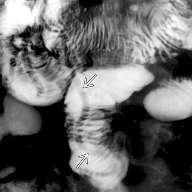
in the main pancreatic duct. Pancreatic duct involvement is much less common than biliary invasion, likely because of the relatively small caliber of the pancreatic duct. Ascariasis-induced pancreatitis is due to either invasion of the pancreatic or biliary ducts.

image
(Right) ERCP demonstrates left duct dilatation image 
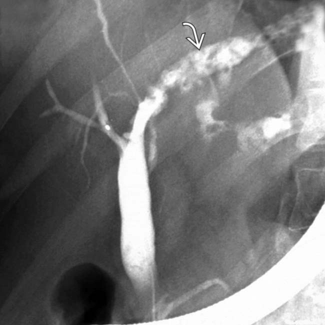
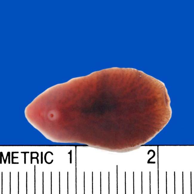
and filling defects from intrahepatic flukes. The elongated configuration of the defects suggests flukes rather than intrahepatic biliary calculi.
image
(Left) Coronal MRCP of a young man with right upper quadrant pain and jaundice shows debris within a dilated extrahepatic duct image
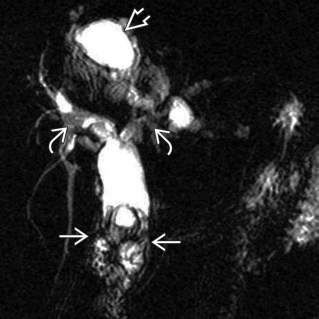
, irregular right and left hepatic ducts image, and an adjacent hydatid cyst image. (Courtesy A. Dasyam, MD.)

image
(Right) ERCP in the same patient shows distal duct debris image
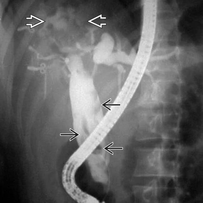
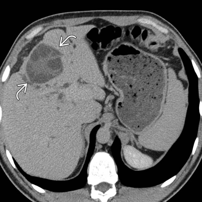
, biliary ductal dilatation, and communication with a partially opacified, ruptured hydatid cyst image. Stents may relieve biliary obstruction, but surgical resection of the cyst is ultimately needed. (Courtesy A. Dasyam, MD.)
image
(Left) Color Doppler ultrasound in a patient with fever and jaundice shows an elongated echogenic filling defect image 
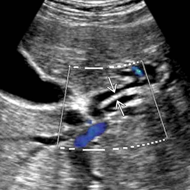
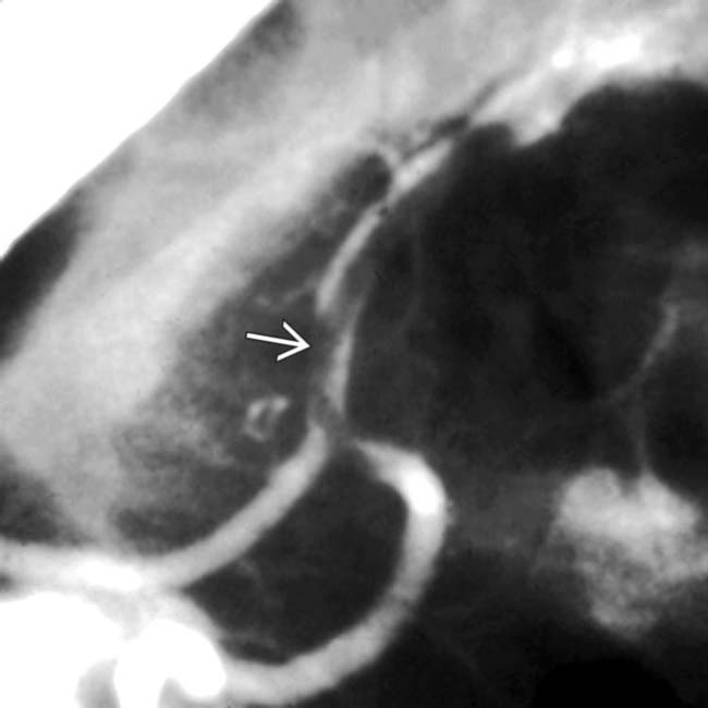
within a dilated left bile duct representing a worm. (Courtesy A. Dasyam, MD.)

image
(Right) Coronal MRCP in the same patient shows intra- and extrahepatic biliary ductal dilatation and Ascaris worm extending from the left bile duct to the distal common bile duct image
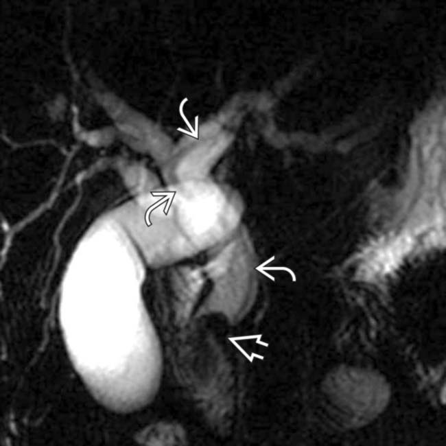
. The distal duct filling defect image was ultimately shown to be cholangiocarcinoma, a known complication of ascariasis. (Courtesy A. Dasyam, MD.)
image
Frontal ERCP in a child from South Africa who presented with biliary colic demonstrates a linear filling defect image
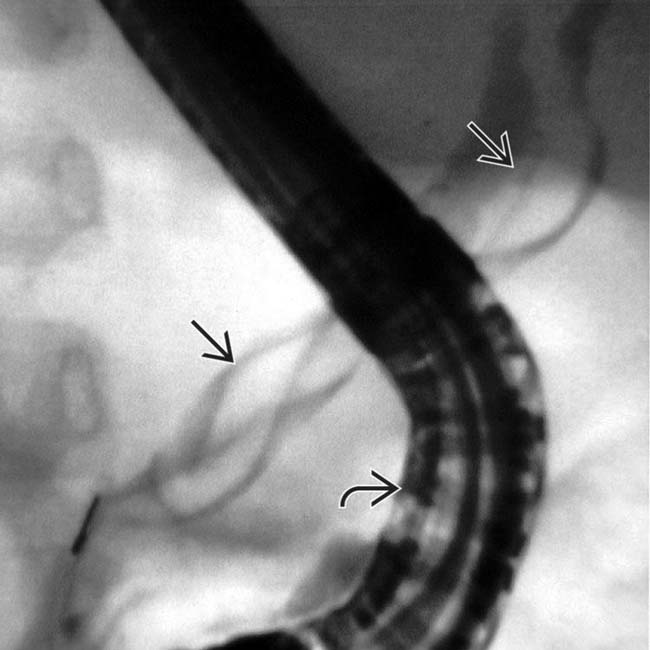
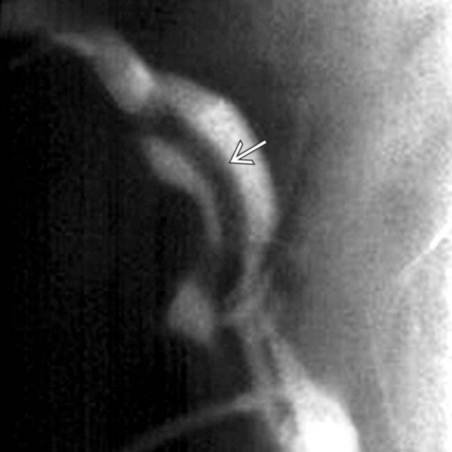
, which represents the adult worm lodged in the common bile duct. Note the overlying endoscope image.

image
Axial CECT shows a classic echinococcal cyst image within the medial segment and mild intrahepatic biliary ductal dilatation.
image
Coronal MRCP in the same patient with a ruptured hydatid cyst shows debris within the mid common duct image 
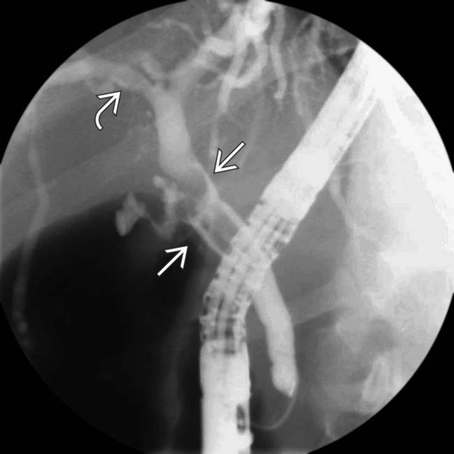
and proximal right duct image. (Courtesy M. Harisinghani, MD.)
image
Supine anteroposterior image from a small bowel follow demonstrates a tubular filling defect in the terminal ileum from an ascaris worm image.

Share this: