Pancreatic Trauma

Published on 19/07/2015 by admin

Filed under Radiology

Last modified 22/04/2025

Print this page

rate 1 star rate 2 star rate 3 star rate 4 star rate 5 star
Your rating: none, Average: 0 (0 votes)

This article have been viewed 2626 times

 Secondary signs of pancreatic injury are usually present (peripancreatic fat stranding and fluid, thickening of pararenal fascia, peripancreatic hematoma)

– Peripancreatic signs of traumatic pancreatitis often subtle; more evident in 24-48 hours
image 
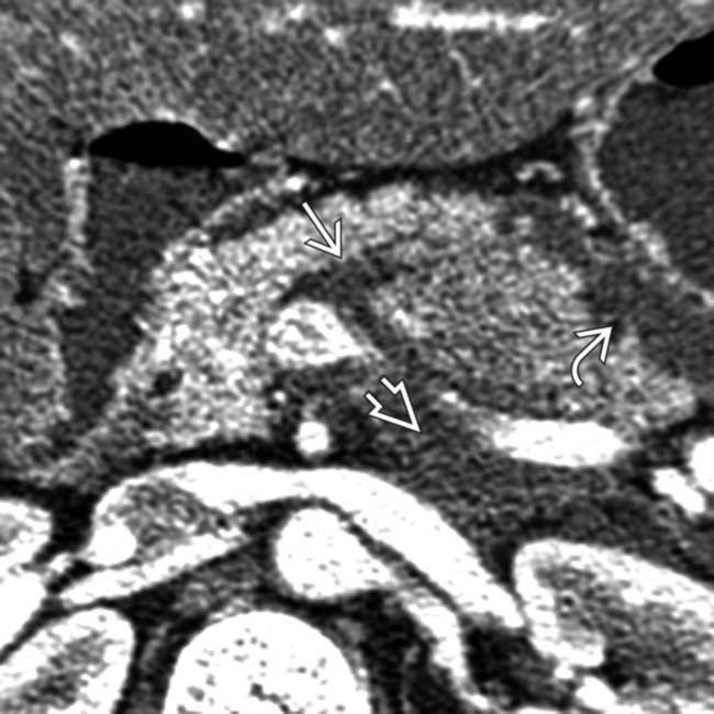
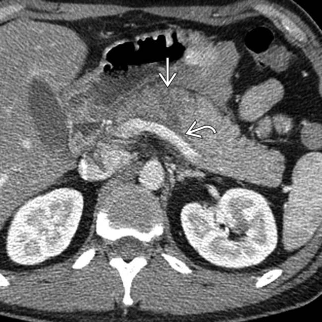
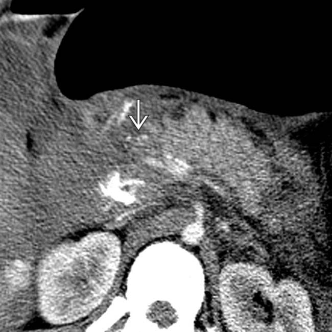
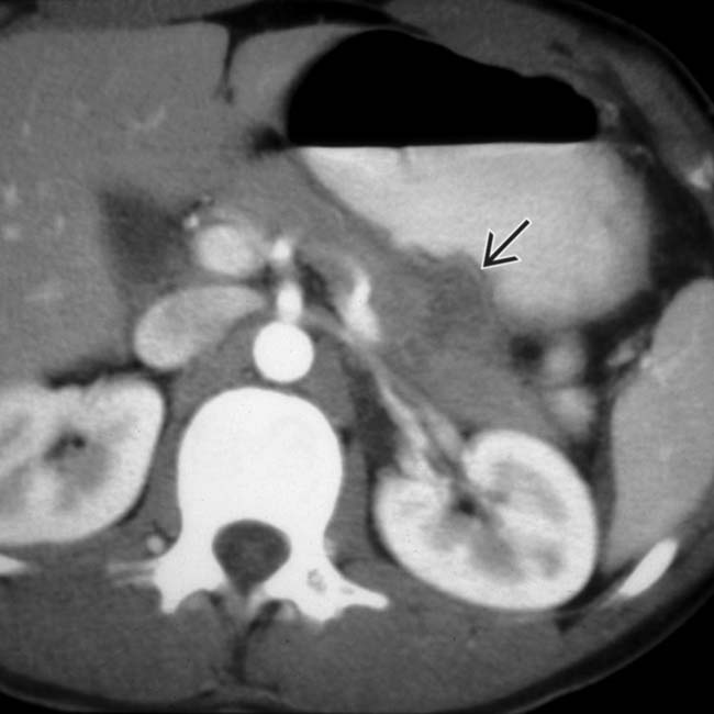
Pancreatic contusion: Ill-defined focal hypoattenuation at site of injury (not linear like laceration)
image Pancreatic laceration: Discrete linear hypodense cleft through pancreas
image Pancreatic fracture: Large laceration with clear separation of 2 ends of gland
image CT insensitive for pancreatic duct injury (usually inferred by laceration extending through duct)
• MR: MRCP (± secretin) is a useful tool in determining presence of pancreatic ductal disruption
• ERCP: Best modality for pancreatic ductal injury

image Transection of pancreatic duct: Abrupt duct termination or contrast extravasation

PATHOLOGY

• May result from either penetrating or blunt trauma

image Blunt traumatic injury usually results from anterior/posterior compression force to abdomen
• Pancreatic injuries almost never isolated and usually associated with polytrauma

CLINICAL ISSUES

• Blunt pancreatic injuries often clinically occult and unrecognized on initial evaluation
• Clinical presentation often due to traumatic pancreatitis: Upper abdominal pain, abdominal distention
• Serum amylase/lipase levels: Elevated in 90% of patients, but may be normal immediately after trauma
• Treatment: Penetrating trauma generally requires immediate laparotomy

image AAST grades I and II: Conservative management
image AAST grades III, IV, and V: Typically require surgery (including possible pancreatic resection)
image
(Left) Axial CECT shows subtle laceration of the pancreas image with fluid in the lesser sac image as well as retropancreatic fluid image.

image
(Right) Axial CECT in the same patient reveals fluid image tracking posterior to the pancreas along the splenic vein from extravasated pancreatic juice. Secondary signs of injury, such as peripancreatic fluid, hematoma, or fat stranding, are almost always present as a clue to the diagnosis.
image
(Left) Axial CECT in a patient with pancreatic fracture shows a fracture plane image 
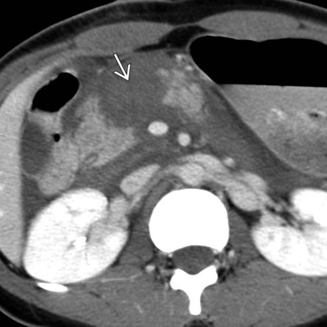
through the neck of the pancreas. The pancreatic duct was disrupted, and the body and tail of the pancreas were resected at surgery.

image
(Right) Axial CECT 48 hours after trauma shows a pseudocyst image 
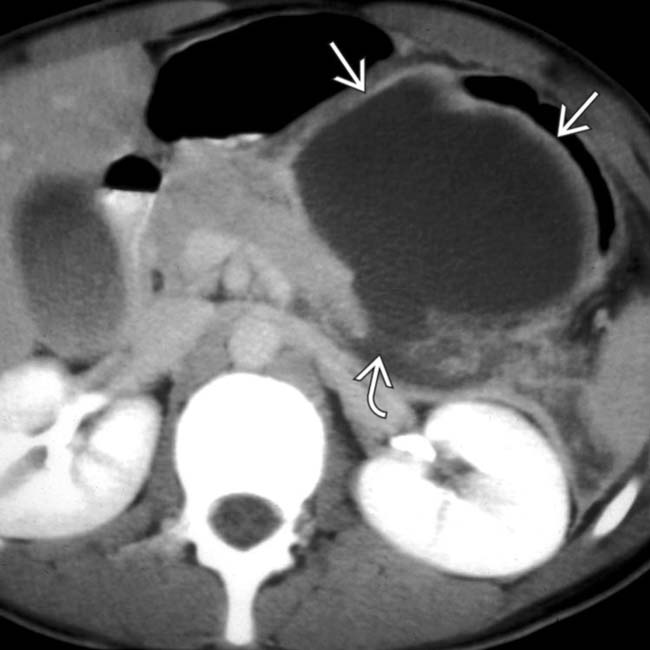
in the lesser sac in this pancreatic transection image.The fluid collection developed as a result of leakage of fluid from the site of the transected pancreatic duct.

TERMINOLOGY

Synonyms

• Traumatic pancreatic injury

Definitions

• Inflammatory disease of pancreas secondary to trauma

IMAGING

General Features

• Best diagnostic clue

image Enlarged, heterogeneous pancreas with peripancreatic fluid or hematoma in patient with history of trauma
• Location

image Most commonly involves pancreatic body > head > tail
• Morphology

image Spectrum of injury: Acute pancreatitis, contusions, deep lacerations, fractures with ductal disruption

Radiographic Findings

• ERCP

image Normal in cases of pancreatic contusion
image Best modality to identify pancreatic duct (PD) injury

– Transection of PD: Abrupt duct termination or contrast extravasation
– Communication of pseudocyst with PD
image May cause pancreatitis

CT Findings

• Secondary signs of pancreatic injury or post-traumatic pancreatitis usually present even in absence of discrete contusion/laceration

image Peripancreatic fat stranding and fluid with loss of normal peripancreatic fat planes almost always present

– Fluid separating pancreas from splenic vein is sensitive (60-90%) for pancreatic injury
– Fluid or hematoma is often seen in lesser sac, left anterior pararenal space, transverse mesocolon, adjacent to spleen, and mesenteric root
image Thickening of anterior pararenal fascia
image Peripancreatic or intrapancreatic hematoma: Intrapancreatic hematoma is more specific for pancreatic injury
image Peripancreatic signs of traumatic pancreatitis are often subtle: May be more evident in 24-48 hours
• Pancreatic contusion: Ill-defined focal hypoattenuation at site of injury 

image Appearance ranges from subtle contour deformity of pancreas to rounded mass-like enlargement of pancreas several cm in diameter
image Often associated with focal or diffuse pancreatic enlargement
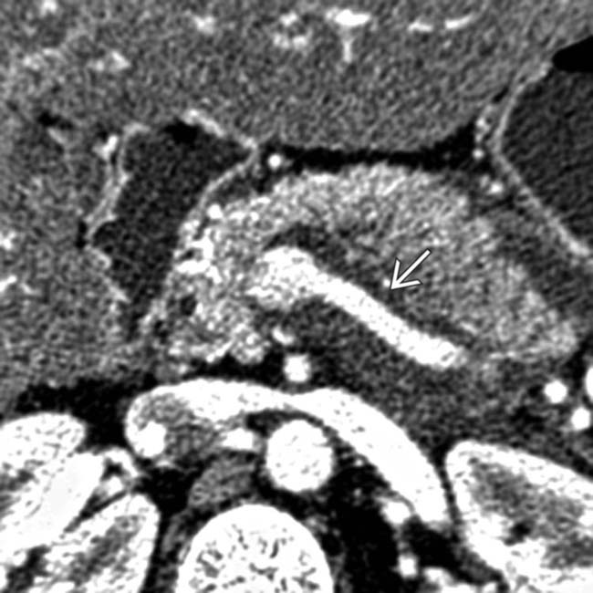
• Pancreatic laceration: Discrete linear cleft of hypoattenuation running through pancreas (usually perpendicular to long-axis of gland)

image Much more likely to be associated with PD injuries than contusion
image Often associated with distortion or irregularity of contour of pancreas and hypoenhancement of gland upstream from laceration
image Lacerations may produce subtle parenchymal density changes and may be undetectable on CT in some cases 

– 20-40% of pancreatic injuries not visible on initial imaging
– May only be faintly visible on initial imaging, and become more conspicuous on follow-up imaging
image CT is not sensitive for detection of PD injury (∼ 40%):  Inferred by presence of laceration extending through duct (> 50% of pancreatic thickness)
• Pancreatic fracture: Linear low attenuation running through pancreatic parenchyma with clear separation of 2 ends of gland

image Most often through pancreatic neck
• Pancreatitis secondary to ERCP (± papillotomy, etc.) usually more severe in/around pancreatic head

MR Findings

• Variably decreased signal on T1WI at sites of contusion or laceration ± high T1 signal related to hematoma
• High signal on T2WI at sites of contusion or laceration
• Heterogeneous enhancement on T1WI C+ images with areas of nonenhancement related to fluid collections, pseudocysts, necrosis, laceration, or severe contusion
• MRCP useful tool to determine PD disruption

image Secretin stimulation may improve diagnostic sensitivity
image Ductal injury suggested by discontinuity in PD, along with direct communication to adjacent pseudocyst or fluid collection
image 

Ultrasonographic Findings

• Not sensitive for pancreatic injury or complications
• Findings similar to pancreatitis (enlarged, hypoechoic gland)

Imaging Recommendations

• Best imaging tool

image CECT for initial evaluation after trauma
image Emergency ERCP: Investigate pancreatic injuries when CT positive and status of PD uncertain
• Protocol advice
• Repeat CT at 24-48 hours may identify pancreatic injuries not appreciated on original examination

DIFFERENTIAL DIAGNOSIS

Shock Pancreas

• Part of hypoperfusion complex seen in severe traumatic injuries or in setting of severe hypotension
• Abnormally intense enhancement of pancreas, bowel wall, and kidneys, with decreased caliber of aorta and inferior vena cava, and diffuse dilatation of intestine with fluid

image Findings resolve spontaneously after fluid resuscitation
• Moderate to large peritoneal fluid collections
• Pancreas appears edematous, enlarged, and hyperenhancing with surrounding fluid and fat stranding, mimicking post-traumatic pancreatitis or injury
• Differentiate from direct traumatic injury by looking for other imaging features of hypoperfusion complex

Duodenal Injury Without Pancreatic Injury

• Duodenal injury (including rupture or hematoma) may simulate or coexist with pancreatic injury
• Duodenal hematoma appears as focal high-attenuation thickening of duodenal wall

image Picket-fence appearance on fluoroscopy from hemorrhage
image Smooth intramural mass causing incomplete bowel obstruction
• Duodenal rupture results in gas or fluid tracking into anterior pararenal space ± oral contrast extravasation

image Air-fluid level in adjacent extraperitoneal space

Acute Pancreatitis

• Pancreatic edema with peripancreatic fluid and stranding in lesser sac and anterior pararenal space

image ± areas of nonenhancement in setting of necrosis
• No history of trauma

PATHOLOGY

General Features

• Etiology

image May result from either penetrating or blunt trauma

– Penetrating injuries include gunshot wounds (45% of cases) and stab wounds (18% of cases)

image Pancreatic injuries in 20-30% of penetrating trauma
– Mechanism in blunt trauma

image Most commonly the result of motor vehicle collisions, but also falls, crush injuries, and assaults (with direct blow to abdomen)
image Pancreatic injuries in children often due to blunt trauma, including trauma from bicycle handlebar, motor vehicle crash, or child abuse
image Usually result from anterior/posterior compression force to abdomen (more rarely lateral force vector)
image Pancreas is susceptible to injury, as it is relatively fixed anterior to spine and commonly compressed against vertebral column
image Lacerations to other structures usually accompany midline compression injury, including left hepatic lobe, duodenum, and central renal vascular pedicle
– Iatrogenic injuries to pancreas can result from endoscopic procedures or surgery

image Endoscopic procedures: ERCP, especially papillotomy, stone extraction, stent placement
image Surgery: Billroth II resections, splenectomy, biliary surgery, aortic graft surgery
• Associated abnormalities

image Pancreatic injuries are almost never isolated and are usually associated with polytrauma
image Duodenum (19-50% of cases) and liver (∼ 20% of cases) frequently also injured
image Vascular injuries also commonly associated (aorta, IVC, left renal vein, right renal artery)

Staging, Grading, & Classification

• Grade of pancreatic injury independent predictor of pancreatic complications and mortality
• American Association for Surgery in Trauma (AAST) grading system

image Grade I: Minor contusion or superficial laceration (PD intact)
image Grade II: Major contusion or deep laceration (PD intact)
image Grade III: Distal laceration either with PD injury or complete transection of gland
image Grade IV: Proximal (right of superior mesenteric artery) laceration or transection involving ampulla or bile duct
image Grade V: Massive disruption of pancreatic head (usually requiring Whipple procedure)
• CT grading system

image Grade A: Superficial (< 50% of gland thickness) laceration or mild pancreatitis
image Grade B: Deep (> 50% of gland thickness) or complete laceration of pancreatic tail
image Grade C: Deep or complete laceration of pancreatic head
• ERCP Grading System

image Grade I: Normal MPD
image Grade IIa: Injury to branches of MPD with contrast extravasation inside parenchyma
image Grade IIb: Injury to branches of MPD with contrast extravasation into retroperitoneum
image Grade IIIa: Injury to MPD at body or tail
image Grade IIIb: Injury to MPD at head

CLINICAL ISSUES

Presentation

• Most common signs/symptoms

image History of trauma (either blunt or penetrating) and multiple other traumatic injuries
image Blunt pancreatic injuries not uncommonly clinically occult and unrecognized on initial evaluation
image Presentation often reflects traumatic pancreatitis: Abdominal pain, postprandial vomiting, abdominal distention
• Clinical profile

image Serum amylase/lipase levels: Elevated in 90% of patients, but may be normal immediately after trauma

– Not specific, as amylase levels may be elevated due to trauma to liver, duodenum, or salivary glands
– Persistently rising amylase levels over time more specific (particularly 3 hours after injury)
– Elevated amylase in diagnostic peritoneal lavage (DPL) fluid much more sensitive/specific than serum amylase
image Leukocytosis
image Diagnosis: Exploratory laparotomy

Demographics

• Gender

image M > F
• Epidemiology

image Uncommon; 3-12% of all abdominal injuries

– Acute post-traumatic pancreatitis represents only 0.4% of acute pancreatitis with pseudocyst formation
– Incidence is much higher with penetrating injuries compared to blunt trauma
image Combined injury of other organs in 80% of patients
image Accounts for 5% of all abdominal injuries in children and is most common cause of pseudocyst in children (often related to child abuse)

Natural History & Prognosis

• Mortality from pancreatic injuries between 9-34% (only 5% due to pancreatic injury itself)

image Most deaths occur within first 48 hours after trauma
• Major complications occur in 50% of patients

image Pancreatic fistula (11%), pancreatic pseudocyst (30%), pancreatitis (17%), intraabdominal abscesses (18%), associated liver or intestinal injuries (> 80%), abdominal hemorrhage, pseudoaneurysm
• Subcutaneous fat necrosis/polyarthritis secondary to post-traumatic pancreatitis reported in < 1% of patients
• Cerebral fat embolism: Rare possible complication of traumatic pancreatitis
• Morbidity higher with external drainage compared to exploration without drainage

Treatment

• Penetrating trauma generally requires immediate laparotomy
• AAST grades I and II (blunt trauma): Conservative nonoperative management

image Superficial lesions not affecting PD can be managed nonoperatively
image Total parenteral nutrition ± somatostatin or octreotide
image Endoscopy with pancreatic stenting is increasingly common practice
• AAST grades III, IV, and V (blunt trauma): Require surgery within 24 hours

image Grade III: Usually distal pancreatectomy and splenectomy (with spleen preservation in children), although pancreas may be preserved with “clean” pancreatic lacerations
image Grades IV and V: Pancreatic resection may be required, but approach is variable depending on concomitant duodenal and ampullary injuries

– Surgical options include Whipple, extended distal pancreatectomy, central pancreatectomy, or surgical drainage
image Surgical indications: Persistently elevated serum amylase levels, increasing cyst size

DIAGNOSTIC CHECKLIST

Consider

• Pancreatic injuries may be occult on initial imaging, but can become more evident after 24-48 hours
• Thickening of anterior pararenal fascia on CT in trauma patient should prompt careful evaluation of pancreas
image
(Left) Axial CECT shows a low-attenuation fracture plane traversing the mid body of the pancreas image
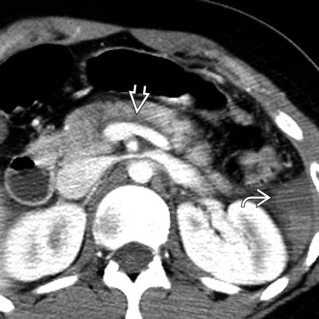
. Note the subtle retropancreatic fluid image adjacent to the splenic vein.

image
(Right) Axial CECT in the same patient at a more caudal level again shows the fracture plane image 
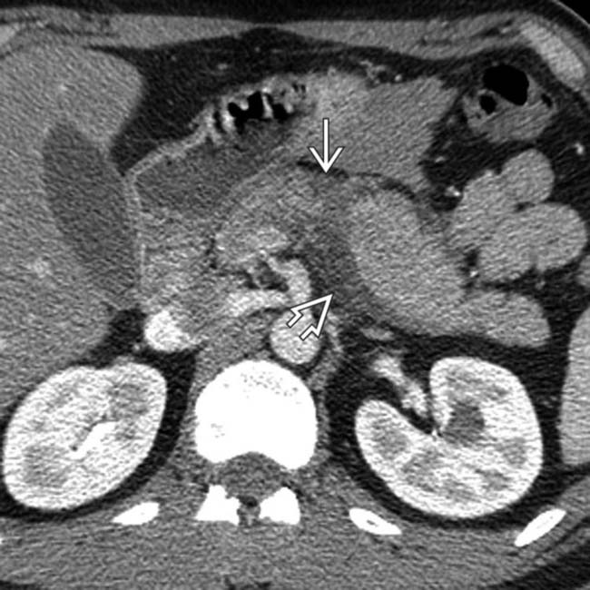
and more obvious adjacent peripancreatic fluid image. In some patients, retropancreatic fluid is the most conspicuous sign of a pancreatic fracture.
image
(Left) Axial CECT 24 hours after blunt trauma reveals ischemic necrosis in the body of the pancreas along the fracture plane image 
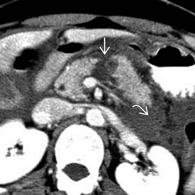
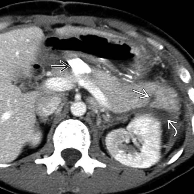
and a perirenal fluid collection image.

image
(Right) Axial CECT in the same patient shows a subtle fracture of the head of the pancreas image with fluid in the pancreatic groove image. Note the adjacent shock bowel image, which is thickened and hyperenhancing.
image
(Left) Axial CECT in a trauma patient demonstrates a focal hematoma at the pancreatic head/neck junction anteriorly image
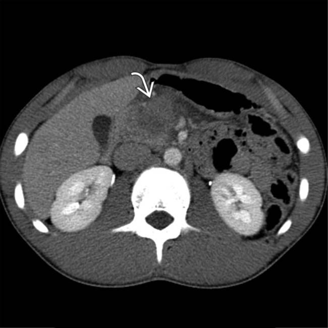
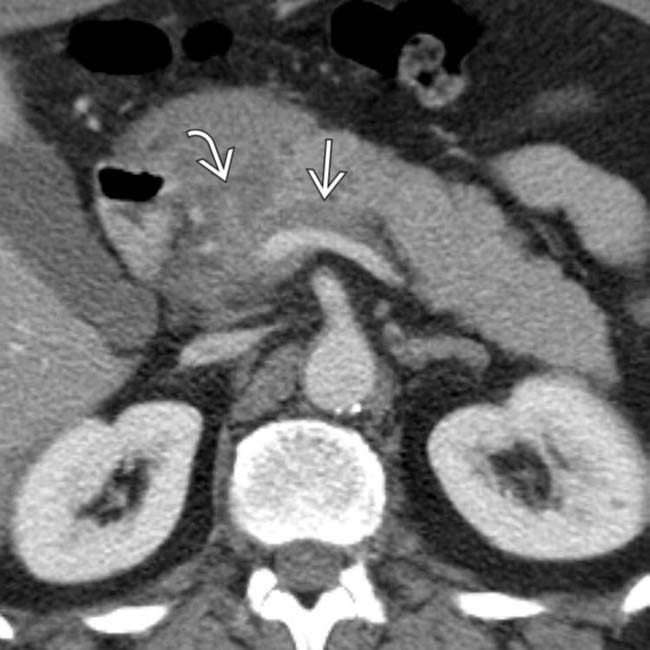
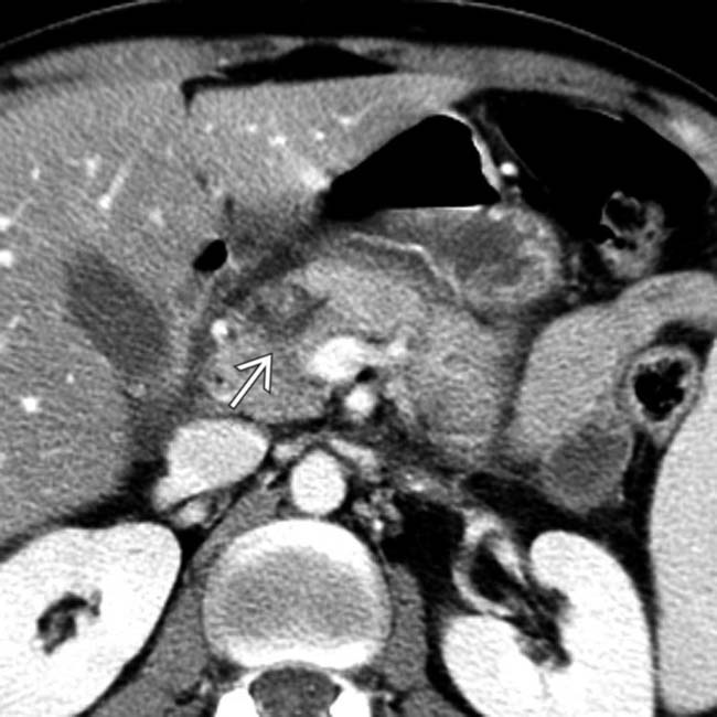
, in keeping with this patient’s pancreatic contusion. No discrete laceration was visualized.

image
(Right) Axial CECT shows fluid surrounding the pancreas image
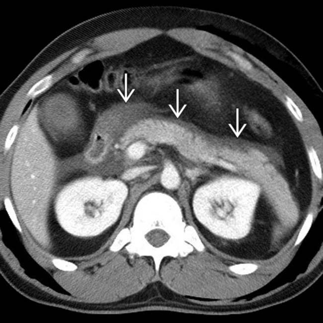
. While no parenchyma laceration was detected by CT, the isolated peripancreatic fluid, particularly in the presence of elevated serum lipase and amylase, is highly suggestive of a pancreatic injury.
image
Axial CECT shows a pancreatic fracture following blunt trauma. Note the fracture image 
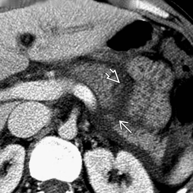
in the mid body of the pancreas and adjacent fluid in the lesser sac image.

image
Axial CECT at a lower plane of section in the same patient demonstrates fluid dissection image posterior to the pancreas and anterior to the splenic vein.
image
Axial CECT shows a patient with a missed pancreatic fracture. Scan performed 48 hours after blunt trauma reveals a linear fracture image 
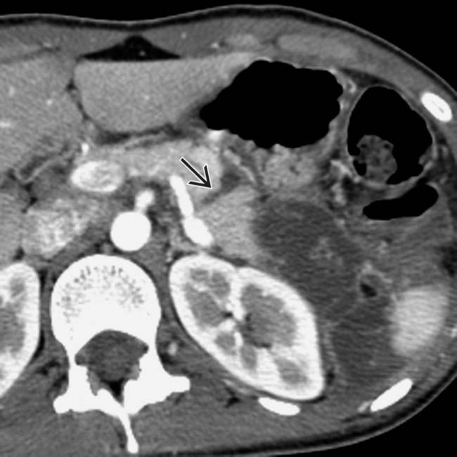
in the mid body of the pancreas.
image
Axial CECT in the same patient more caudally demonstrates an acute fluid collection image
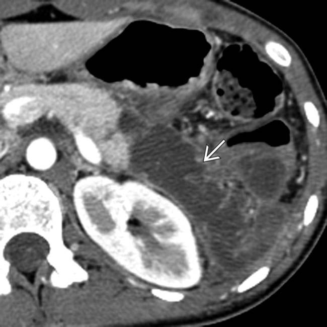
. in the anterior pararenal space.
image
Axial CECT shows laceration to the pancreatic head following blunt trauma. Note the fracture plane image 
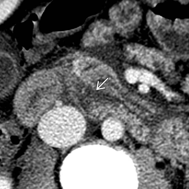
in the head of the pancreas.
image
Plane of section through the uncinate process demonstrates a hematoma with an ill-defined, low-attenuation mass image
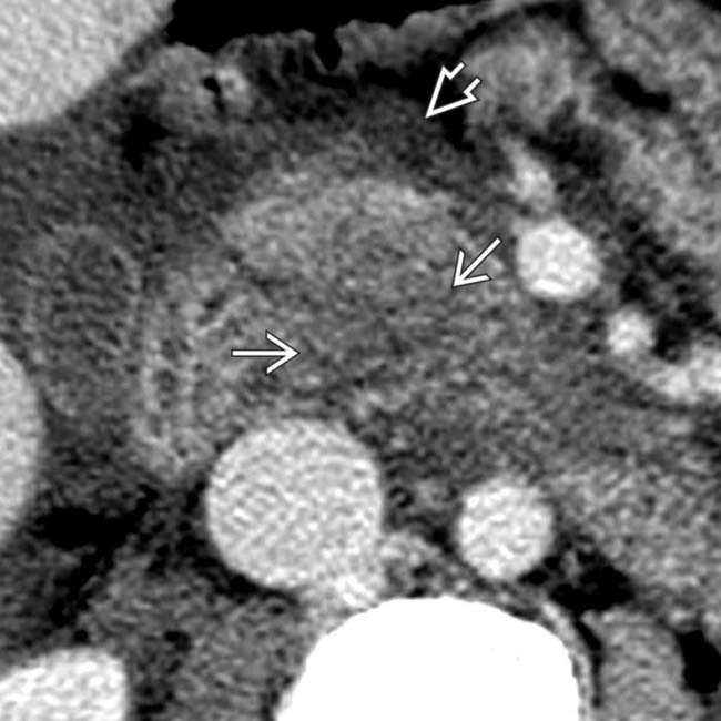
. Note the surrounding peripancreatic fluid and blood image.
image
Axial CECT of a patient with combined duodenal and pancreatic trauma from blunt injury. Note the multiple high-attenuation areas of peripancreatic active arterial bleed image and a large surrounding hematoma image.
image
Axial CECT in the same patient at a lower plane of section shows a fracture plane image through the neck and body of the pancreas.
image
Axial CECT in a patient with pancreatic transection shows a fracture plane through the neck of the pancreas and peripancreatic edema and hemorrhage.
image
Axial CECT in a patient with pancreatic contusion shows a heterogeneous mass effect image 
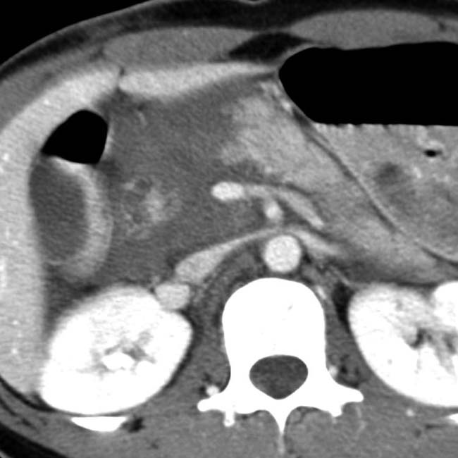
at the pancreatic neck as well as fluid image between the splenic vein and pancreatic body.
image
Axial CECT in a patient with pancreatic contusion shows infiltration of the fat planes anterior and posterior to the pancreatic neck.
image
Initial axial CECT in a patient with pancreatic transection shows a subtle fracture plane image 
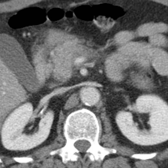
and hematoma in the body of the pancreas.
image
In this case of pancreatic transection, axial CECT shows a fracture plane image 
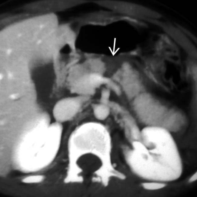
completely through the pancreatic neck.
image
Axial CECT shows extensive infiltration of the fat planes and spaces around the pancreatic head, while the pancreatic body and tail are uninvolved in this case of iatrogenic (post-ERCP) pancreatitis.
image
Axial CECT shows a splenic fracture image
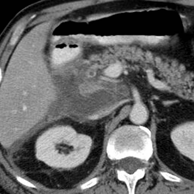
. Note the small amount of fluid posterior to the stomach in the lesser sac image. There are subtle linear areas of decreased enhancement within the body of the pancreas representing a laceration image.
image
Axial CECT in the same patient at a more caudal level demonstrates retropancreatic fluid image anterior to the splenic vein indicating a pancreatic injury. Note also the hemoperitoneum in the left paracolic gutter from the splenic fracture image.
image
Axial CECT shows a linear laceration image in the tail of the pancreas that was not detected at the time of initial surgical exploration. Note the surgically placed drain image in the pancreatic bed and a small amount of peripancreatic fluid image near the pancreatic tail.
image
Axial CECT in the same patient at more caudal level demonstrates that the laceration image 
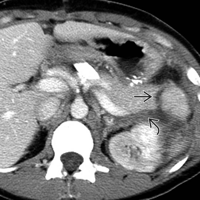
extends through the entire tail of the pancreas thus representing a pancreatic fracture. Note also the peripancreatic fluid image.
image
Axial CECT shows an area of parenchymal laceration image 
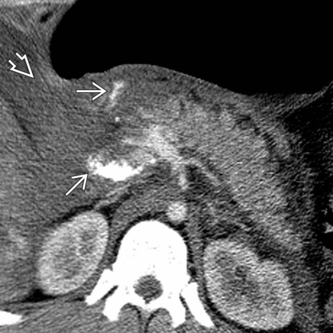
to the neck of the pancreas with an adjacent high-density hematoma image in the pancreatic groove.
image
Axial CECT in the same patient at a more caudal level demonstrates that the hematoma involves the retropancreatic space image 
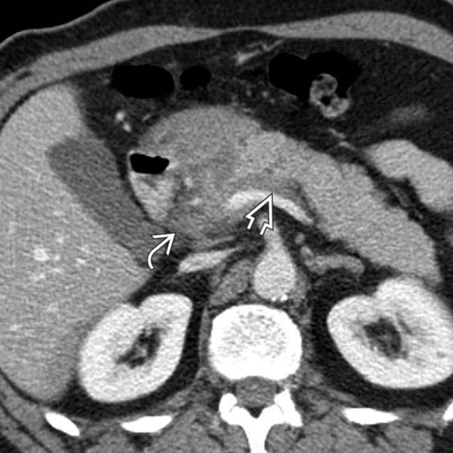
as well as the pancreatic groove image.
image
Axial CECT shows hypodense pancreatic laceration image involving the head and neck of the pancreas.
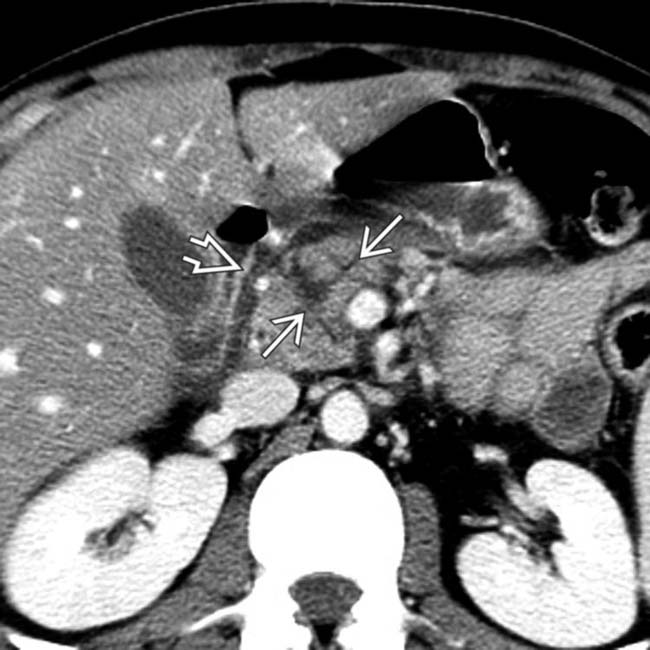
image
Axial CECT in the same patient at a more caudal level shows the stellate nature of the pancreatic laceration image and adjacent fluid image in the groove between the head of the pancreas and the 2nd duodenum.
image
Axial CECT shows obvious liver laceration image 
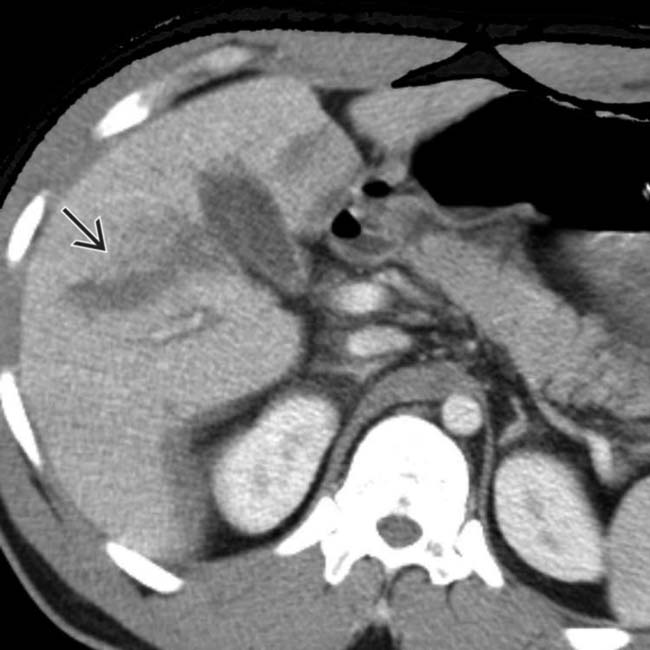
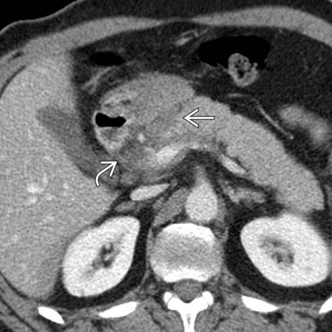
evident as irregular hypodense plane through right lobe.
image
Axial CECT in the same patient demonstrates more subtle signs of pancreatic injury. Note less evident infiltration image 
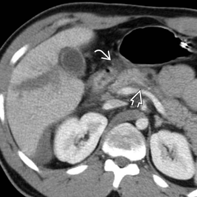
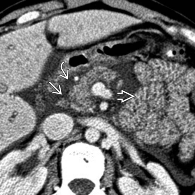
of retroperitoneal and mesenteric fat planes surrounding the pancreas, not explainable by liver injury. In addition, note the hypodense fluid image between the pancreas and splenic vein. The patient had several days of abdominal pain and elevation of serum amylase and lipase. However, there was no development of a pancreatic pseudocyst or sign of disruption of the pancreatic duct. The patient was managed without surgery and was sent home in good condition after a few days.
image
Axial CECT wedge-shaped area of low attenuation image 
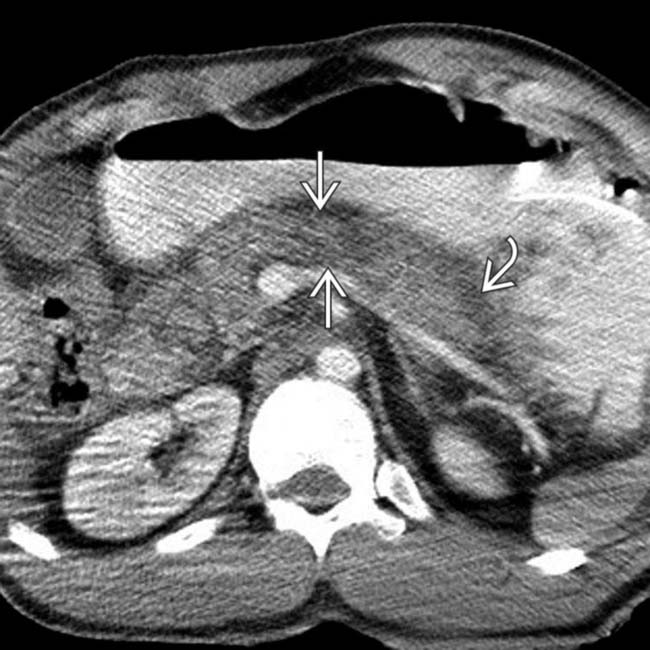
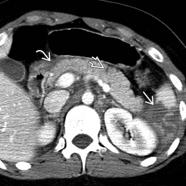
in pancreatic body, indicative of pancreatic contusion, possibly laceration. Note peripancreatic stranding image consistent with pancreatitis.
image
Axial CECT in same patient as left demonstrates flattened cava image 
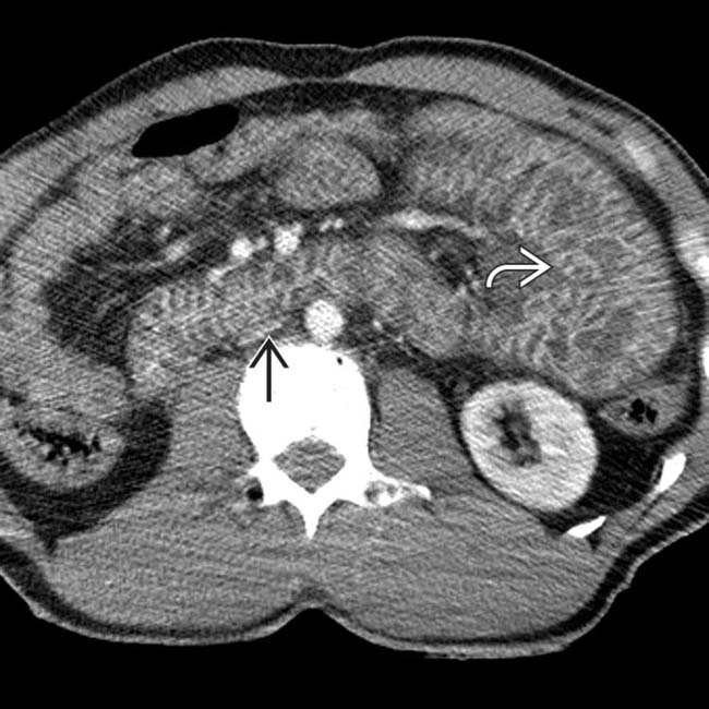
and thick-walled, vividly enhancing small bowel image, consistent with shock bowel.
image
Axial CECT shows fracture plane image 
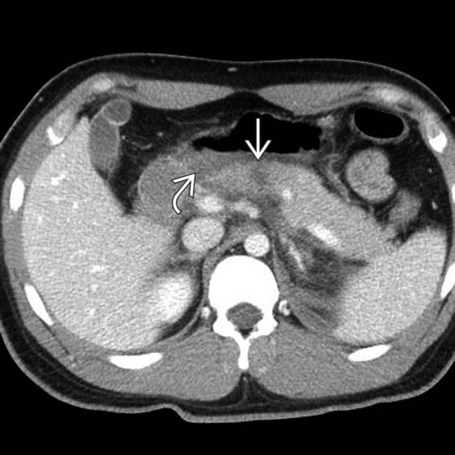
through the body of the pancreas. There is a small fluid collection in the lesser sac image.

SELECTED REFERENCES

1. Dreizin, D, et al. Evaluating blunt pancreatic trauma at whole body CT: current practices and future directions. Emerg Radiol. 2013; 20(6):517–527.

Panda, A, et al. Evaluation of diagnostic utility of multidetector computed tomography and magnetic resonance imaging in blunt pancreatic trauma: a prospective study. Acta Radiol. 2014. [ePub].

Debi, U, et al. Pancreatic trauma: a concise review. World J Gastroenterol. 2013; 19(47):9003–9011.

Gordon, RW, et al. Blunt pancreatic trauma: evaluation with MDCT technology. Emerg Radiol. 2013; 20(4):259–266.

Lahiri, R, et al. Pancreatic trauma. Ann R Coll Surg Engl. 2013; 95(4):241–245.

Maeda, K, et al. Management of blunt pancreatic trauma in children. Pediatr Surg Int. 2013; 29(10):1019–1022.

Kaman, L, et al. Current management of pancreatic trauma. Trop Gastroenterol. 2012; 33(3):200–206.

Lee, KJ, et al. Management of blunt pancreatic injury by applying the principles of damage control surgery: Experience at a single institution. Hepatogastroenterology. 59(118–119), 2011.

Duchesne, JC, et al. Selective nonoperative management of low-grade blunt pancreatic injury: are we there yet? J Trauma. 2008; 65(1):49–53.

Buccimazza, I, et al. Isolated main pancreatic duct injuries spectrum and management. Am J Surg. 2006; 191(4):448–452.

Fang, JF, et al. Usefulness of multidetector computed tomography for the initial assessment of blunt abdominal trauma patients. World J Surg. 2006; 30(2):176–182.

Cay, A, et al. Nonoperative treatment of traumatic pancreatic duct disruption in children with an endoscopically placed stent. J Pediatr Surg. 2005; 40(12):e9–12.

Krige, JE, et al. The management of complex pancreatic injuries. S Afr J Surg. 2005; 43(3):92–102.

Stringer, MD. Pancreatitis and pancreatic trauma. Semin Pediatr Surg. 2005; 14(4):239–246.

Lin, BC, et al. Management of blunt major pancreatic injury. J Trauma. 2004; 56(4):774–778.

Kao, LS, et al. Predictors of morbidity after traumatic pancreatic injury. J Trauma. 2003; 55(5):898–905.

Mayer, JM, et al. Pancreatic injury in severe trauma: early diagnosis and therapy improve the outcome. Dig Surg. 2002; 19(4):291–297. [discussion 297-9].