Paediatric Anaesthesia

Published on 27/02/2015 by admin

Filed under Anesthesiology

Last modified 22/04/2025

Print this page

rate 1 star rate 2 star rate 3 star rate 4 star rate 5 star
Your rating: none, Average: 0 (0 votes)

This article have been viewed 1843 times

Paediatric Anaesthesia

The differences in anatomy and physiology between children, especially infants, and adults have important consequences in many aspects of anaesthesia. The differences also account for the different patterns of disease seen in intensive care units (ICUs). Although major psychological differences persist throughout adolescence, a 10- to 12-year-old child may be thought of, anatomically and physiologically, as a small adult.

PHYSIOLOGY IN THE NEONATE

Respiration

Control of respiration in newborn infants, especially premature neonates, is poorly developed. The incidence of central apnoea (defined as a cessation of respiration for 15 s or longer) is not uncommon in this group. The likelihood of this increases if the patient is given a drug with a sedative effect. Potentially life-threatening apnoea may occur. The incidence is reduced by postoperative administration of xanthine derivatives such as caffeine and theophylline which act as central respiratory stimulants. Because of this problem, it is wise to admit for overnight oximetry and apnoea monitoring all children under 60 weeks’ postconceptual age who have had surgical procedures, no matter how minor. Hypoxaemia in the neonate and small child appears to inhibit rather than stimulate respiration and this is contrary to what one might expect.

The newborn has between 20 and 50 million terminal air spaces. At 18 months of age, the adult level of 300 million is reached by a process of alveolar multiplication. This explains why infants who suffer with respiratory distress of the newborn improve as they grow older. Subsequent lung growth occurs by an increase in alveolar size. The lung volume in infants is disproportionately small in relation to body size. The metabolic rate is nearly twice that of the adult, and therefore ventilatory requirement per unit lung volume is increased. Thus, they have far less reserve for gas exchange.

Before the age of 8 years, the calibre of the airways is relatively narrow. Airway resistance is therefore relatively high. Small decreases in the diameter of the airways as a result of oedema or respiratory secretions significantly increase the work of breathing. Elastic tissue in the lungs of small children is poorly developed. As a result of this, compliance is decreased. This has important consequences in that airway closure may occur during normal tidal ventilation, thereby bringing about an increase in alveolar–arterial oxygen tension difference (PA − aO2). This explains why PaO2 is lower in the infant than in the child. The decreased compliance results in ventilatory units with short time constants. Consequently, the infant is able to achieve adequate alveolar ventilation whilst maintaining a high respiratory rate. However, because of the increased resistance and decreased compliance, the work of breathing may represent up to 15% of total oxygen consumption (Table 36.1). The high respiratory rate is necessary because the metabolic rate of the infant is nearly twice that of the adult. The high alveolar minute ventilation explains why induction and emergence from inhalational anaesthesia are relatively rapid in small children. The high metabolic rate also explains why desaturation occurs very rapidly in children.

TABLE 36.1

Lung Mechanics of the Neonate Compared with the Adult

Neonate Adult
Compliance (mL cmH2O−1) 5 100
Resistance (cmH2O L−1 s−1) 30 2
Time constant (s) 0.5 1.3
Respiratory rate (breath min−1) 32 15

The ratio of physiological dead space to tidal volume (VD/VT) is similar to that of the adult at about 0.3. However, because the volumes are smaller, modest increases in VD produced by equipment such as humidification filters may have a disproportionately greater effect (Table 36.2).

TABLE 36.2

Respiratory Variables in the Neonate

Tidal volume (V) 7 mL kg−1
Dead space (VD) (VT)  ×  0.3 mL
Respiratory rate 32 breath min−1

Ventilation in small children is almost entirely diaphragmatic. Because the ribs are horizontal, there is no ‘bucket handle’ movement of the ribs as occurs in the adult. It is therefore important to appreciate that normal minute ventilation is respiratory rate-dependent. The infant’s diaphragm is made of fast twitch fibres. This type of muscle fibre exhausts easily if it has to work against a load. This implies that in infancy, when lung compliance is low, the work of breathing is reduced by breathing rapidly. Consequently if the work of breathing is increased by an increase in airway resistance, respiratory failure may easily ensue.

It is important to appreciate that the infant’s response to hypoxaemia may be bradypnoea and not tachypnoea as occurs in the adult.

Cardiovascular System

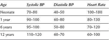
The process of growth demands a high metabolic rate. It is, therefore, not surprising that infants and children have a higher cardiac index compared with the adult, so that oxygen and nutrients may be delivered to actively growing tissues. The ventricles of neonates and infants are poorly compliant, so even though the ventricles of infants demonstrate the Frank-Starling mechanism, the main determinant of cardiac output is heart rate. Infants tolerate heart rates of 200 beat min−1 with ease (Table 36.3). Bradycardia may occur readily in response to hypoxaemia and vagal stimulation and it results in a decrease in cardiac output. Immediate cessation of the stimulus, and treatment with oxygen and atropine, are absolutely crucial. A heart rate of 60 beat min−1 in an infant is considered a cardiac arrest and requires cardiac massage. Arrhythmias are rare in the absence of cardiac disease. The usual cardiac arrest scenarios are electromechanical dissociation and asystole, not ventricular fibrillation.

Even though infants and children have a higher cardiac index, arterial pressure tends to be lower than in adults because of a reduced systemic vascular resistance associated with an abundance of vessel-rich tissues in the infant. The arterial pressure increases from approximately 80/50 mmHg at birth to the normal adult value of 120/70 mmHg by the age of 16 years. Children under the age of 8 years who are normovolaemic at the start of anaesthesia tend not to exhibit a decrease in arterial pressure when central neural blockade such as spinal anaesthesia is administered. They do not require fluid preloading as an adult would to avoid hypotension, because venous pooling tends not to occur as venous capacitance cannot increase. The reasons for this are, first, that the sympathetic nervous system is less well developed and so infants tend to be venodilated at rest. Second, they have a lower extremity:body surface ratio and as a consequence have a smaller venous capacitance.

As in all patients, the cardiovascular system must be monitored carefully. Pulse oximeter probes placed on the extremities provide a good index of peripheral perfusion. Auscultation of heart sounds, especially by an oesophageal stethoscope, is useful as the volume of heart sounds tends to be diminished as cardiac output decreases. Non-invasive measurement of arterial pressure is undertaken easily using an appropriately sized cuff. Complications preclude the use of invasive monitoring of arterial and central venous pressures for all but major cases.

Blood Volume

The stage at which the umbilical cord is clamped determines the circulating blood volume of the neonate. Variations of up to ± 20% may occur. The average blood volume at birth is 90 mL kg−1, and this decreases in the infant and young child to 80 mL kg−1, attaining the adult level of 75 mL kg−1 at the age of 6–8 years. Blood losses of greater than 10% of the red cell mass should be replaced by blood, especially if additional losses are expected. However, most children who have a normal haemoglobin concentration at the start of surgery can tolerate losses of up to 20% of their red cell mass. Children may tolerate a haematocrit of 25% and the decision to transfuse blood must be balanced against the risks, which include transmitted infection and antibody formation. The latter may cause problems in later life, especially in female children during child-bearing years.

Haemoglobin

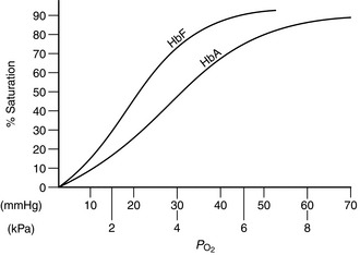
At birth, 75–80% of the neonate’s haemoglobin is fetal haemoglobin (HbF). By the age of 6 months, adult haemoglobin (HbA) haematopoiesis is fully established. HbF has a higher affinity for oxygen than HbA. This is demonstrated by the leftward shift of the oxygen haemoglobin dissociation curve (Fig. 36.1). Low tissue PO2 and metabolic acidosis in the tissues result in the avidity of HbF for oxygen being reduced, thereby aiding delivery of oxygen. Alkalosis produced by hyperventilation results in less oxygen being available and it is therefore sensible to maintain normocapnia.

If blood transfusion is required, it is crucial that blood is filtered and warmed – the smaller the child, the more important is this precaution. A syringe used via a tap in the intravenous giving set is probably the safest way of avoiding inadvertent overtransfusion. The circulating volume of a 1 kg neonate is of the order of 80 mL. Common sense dictates that blood loss should be monitored carefully, so swabs should be weighed and, if possible, all suction losses collected in a graduated container.

Renal Function and Fluid Balance

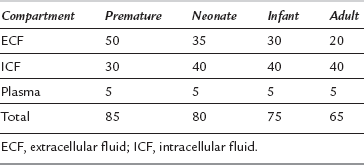
Body fluids constitute a greater proportion of body weight in the infant, particularly the premature infant, compared with the adult (Table 36.4). In an adult, most of the total body water is in the intracellular compartment. In a newborn infant, most of the total body water is in the extracellular compartment. With increasing age, the ratio reverses. Plasma volume remains constant throughout life at about 5% of body weight.

The kidneys are immature at birth. Both glomerular filtration rate (GFR) and subsequent reabsorption by the renal tubules are reduced. The GFR at birth is of the order of 45 mL min−1 1.7 m−2. This increases rapidly to about 65 mL min−1 1.7 m−2 and then gradually approaches the adult value of 125 mL min−1 1.7 m−2 by the age of 8 years. Thus, there is inability to handle excessive water and sodium loads. Overtransfusion may lead to pulmonary oedema and cardiac failure. The maturation in renal function is produced by hyperplasia in the first 6 months of life and then by a process of hypertrophy in the first year. Care must also be exercised when drugs eliminated by the renal route are used in infants; either reduced doses or an increased dosage interval should be employed. Renal maturation is not just an increase in size but also of function. The ability to modify the ultrafiltrate produced at the glomerulus increases with age. It follows that sodium bicarbonate and glucose homeostasis mechanisms are not fully developed. Medical intervention may be required to ensure that biochemical values are kept within normal ranges.

Poorly developed mechanisms exist for conserving water in the kidneys and gastrointestinal tract. Increased cutaneous water loss because of a large surface area:volume ratio through poorly keratinized skin may lead to a turnover of fluid in the infant of about 15% of total body water per day. Dehydration ensues very rapidly in an infant who is kept fasted.

Fluid Therapy

An intravenous infusion delivering maintenance fluids should be in place for all neonates requiring surgery. Maintenance fluid requirements increase over the first few days of life (Tables 36.5, 36.6). The normal infant requires of the order of 3–5 mmol kg−1 of sodium and an equivalent amount of potassium per day to maintain normal serum electrolyte concentrations. The ability of the infant’s kidneys to eliminate excess sodium is limited. Exceeding this amount in the absence of loss results in hypernatraemia and its sequelae. Infants undergoing any procedure more than the briefest should also have their calorific needs addressed. This may be achieved by including glucose-containing fluids in the regimen; failure to do so results in hypoglycaemia and ketosis. This may occur rapidly because of the limited glycogen stores and high metabolic rate of the infant.

TABLE 36.5

Fluid Requirements in the First Week of Life

Day Rate (mL kg−1 Day−1)
1 0
2, 3 50
4, 5 75
6 100
7 120

TABLE 36.6

Maintenance Fluid Requirements

Weight (kg) Rate (mL kg−1 Day−1)
Up to 10 kg 100
10–20 kg 1000 + 50  ×  [weight (kg) – 10] mL
20–30 kg 1500 + 25  ×  [weight (kg) – 20] mL

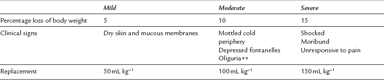
It is imperative that the anaesthetist recognizes and resuscitates the dehydrated infant appropriately before surgery. Clinical examination of skin turgor, capillary refill, tension of fontanelles, arterial pressure and venous filling may aid estimation of hydration, but electrolyte and haemoglobin concentrations and haematocrit, urine volumes and plasma and urine osmolalities should be monitored if problems of fluid balance exist (Table 36.7).

Intravenous fluids should be administered using a system that allows small volumes to be given accurately. This may vary from the anaesthetist injecting fluid using a syringe to microprocessor-controlled syringe driver pumps. The latter are preferable, as fluid is given at a steady rate and the anaesthetist’s hands are free to attend to other tasks. During surgery, fluid administration should be increased to account for increased losses occurring through evaporation from exposed viscera and third-space losses.

The intraosseous route may be used to carry out fluid resuscitation and drug therapy in shocked children. The needle should be inserted in an aseptic fashion to minimize the risk of osteomyelitis. Although various sites have been described for needle insertion, the proximal end of the tibia below the tuberosity is probably the easiest to perform. The intraosseous route is safer than attempting central venous cannulation in the shocked child in whom veins are difficult to discern. The usual fluid administered in this situation is a colloid solution. This is given as a 10 mL kg−1 bolus and repeated until clinical improvement occurs.

Temperature Regulation and Maintenance

Homeothermic animals possess the ability to produce and dissipate heat. Heat loss occurs by one of four processes: radiation, convection, evaporation and conduction. The environment in which the patient is situated governs the relative contribution of each. The neutral thermal environment is defined as the range of ambient temperatures at which temperature regulation is achieved by non-evaporative physical processes alone.

The metabolic rate at this temperature is minimal. The temperature of such an environment is 34°C for the premature neonate, 32°C for the neonate at term and 28°C for the adult.

Heat may be produced by one of three processes: voluntary muscle activity, involuntary muscle activity and non-shivering thermogenesis. Infants under the age of 3 months do not shiver. The only method available to increase their temperature in the perioperative period is non-shivering thermogenesis. The process is mediated by specialized tissue termed brown fat. It differentiates in the human fetus between 26 and 30 weeks of gestation. It comprises between 2% and 6% of total body weight in the human fetus and is located mainly between the scapulae and in the axillae. It is also found around blood vessels in the neck, in the mediastinum and in the loins. Brown fat is made of multinucleated cells with numerous mitochondria and has an abundant blood and nerve supply. Its metabolism is mediated by catecholamines. The substrate used for heat production is mainly fatty acids.

Radiation accounts for about 60% of the heat loss from a neonate in a 34°C incubator placed in a room at 21°C. If the infant was in a thermoneutral environment of 34°C, the percentage loss by radiation would decrease to about 40% of the total heat loss, and, in addition, the total heat loss in this environment would be lower. The reason for this is that heat loss by radiation is a function of skin surface area and the difference in temperature between the skin and the room. The second major source of heat loss in the neonate is convection. This is a function of skin temperature and ambient temperature. The neonate possesses minimal subcutaneous fat that may act as thermal insulation and as a barrier to evaporative loss. A neonate has a body surface area:volume ratio about 2.5 times greater than the adult; thus, a neonate may become hypothermic very rapidly.

If neonates are allowed to become hypothermic during anaesthesia, unlike adults they attempt to correct this by non-shivering thermogenesis. Metabolic rate increases and oxygen consumption may double. The increase in metabolic rate puts an additional burden on the cardiorespiratory system and this may be critical in neonates with limited reserves. The release of noradrenaline in response to hypothermia causes vasoconstriction, which in turn causes a lactic acidosis. The acidosis favours an increase in right-to-left shunt, which causes hypoxaemia. As a result, a vicious positive feedback loop of hypoxaemia and acidosis is set up. The protective airway reflexes of a hypothermic neonate are obtunded, thereby increasing the risks of regurgitation and aspiration of gastric contents. The action of most anaesthetic drugs is potentiated by hypothermia. This effect is particularly important with regard to neuromuscular blocking drugs. The combination of hypothermia and prolonged action of these drugs increases the chances of the neonate hypoventilating after surgery.

Many precautions should be taken to ensure that the neonate’s body temperature is maintained. First, the child must be transported to theatre wrapped up and in an incubator set at the thermoneutral temperature. The theatre should be warmed up to the thermoneutral temperature, ideally a few hours before the planned start of surgery. This interval allows the walls of the theatre to warm up and this reduces the net heat loss by radiation. Heat loss by radiation is a two-way process. The child loses heat by radiation to the walls and also gains heat from the walls. All body parts which are not needed for insertion of cannulae and for monitoring should remain covered until the child has been draped with surgical towels. If the child has to be exposed, overhead radiant heaters may be used. During surgery, the child should lie on a thermostatically controlled heated blanket. Forced air warming systems are effective in maintaining the child’s temperature during surgery; these work on the principle of blowing filtered, warmed air into quilted blankets with perforations. This allows warmed air to come into direct contact with the patient. Simple measures such as using a bonnet to reduce heat loss from the head are very effective. Intravenous fluids and fluids used to perform lavage of body cavities must be warmed. Anaesthetic gases should be humidified and warmed in order to preserve ciliary function and to reduce heat loss from the respiratory tract.

Monitoring

It is important in all procedures to measure temperature. For short procedures, an axillary temperature probe may be sufficient. In longer operations, core temperature should be measured at one of a variety of sites, such as rectal, bladder, nasopharyngeal or oesophageal. The oesophageal probe is often the preferred method, as most modern oesophageal probes may be connected to a stethoscope. The anaesthetist is therefore able to listen to heart sounds in addition to recording the patient’s temperature. When active heating methods such as cascade humidifiers and heated blankets are used, it is important that temperature gradients between the patient and the warming device are kept to less than 10°C. Failure to observe this may result in burns to the skin and the respiratory tract. In the ICU, simultaneous measurement of core and peripheral temperatures, though not often used in theatre, may serve as a useful guide to adequacy of the cardiac output. Decreases in cardiac output result in a reduction of blood flow to the peripheries and this is reflected in a core-peripheral temperature gradient greater than 3–4°C.

PHARMACOLOGY IN THE NEONATE

Developmental Pharmacology

Drugs given via the oral or rectal route are absorbed by a process of passive absorption. This process is dependent on the physicochemical properties of the drug and the surface area available for absorption. Most drugs are either weak bases or weak acids. The un-ionized portion of the drug therefore depends on the pH of the fluid in the gut. The gastric pH of the neonate is higher than that of the older child and adult. The consequence is that drugs inactivated by a low pH undergo greater absorption. Examples of these include antibiotics such as penicillin G.

Factors which determine the distribution of intravenously administered drugs include protein and red cell binding, tissue volumes, tissue solubility coefficients and blood flow to tissues. Neonates, in particular preterm infants, have lower plasma concentrations of albumin. In addition, the albumin is qualitatively different in that its ability to bind drugs is lower than that of adult albumin. The concentration of α1-acid glycoprotein is also lower in this group of patients; this protein is the major binding protein for alkaline drugs, which include opioid analgesics and local anaesthetics.

The blood–brain barrier is immature at birth; thus, it is more permeable to drugs. In addition, the neonate’s brain receives a larger proportion of the cardiac output than does the adult brain. Consequently, brain concentrations of drugs are higher in neonates than in adults. For example, administration of morphine, which has low lipid solubility, results in high concentrations in the neonate’s brain and therefore it should be used with caution and in reduced amounts.

In a neonate, total body water, extracellular fluid and blood volume are proportionally larger in comparison with an adult. This results in a larger apparent volume of distribution for a parenterally administered drug. This explains in part why neonates appear to require larger amounts of some drugs on a weight basis to produce a given effect. However, plasma concentrations tend to remain high for longer because they have smaller muscle mass and fat stores to which drugs redistribute.

The action of most drugs is terminated by metabolism or excretion through the liver and kidneys. In the liver, phase I reactions convert the original drug to a more polar metabolite by the addition or unmasking of a functional group such as -OH, -NH2 or -SH. These reduction/oxidation reactions are a function of liver size and the metabolizing ability of the appropriate microsomal enzyme system. The volume of the liver relative to body weight is largest in the first year of life. The enzyme systems in the liver responsible for the metabolism of drugs are incompletely developed in the neonate. Their activity appears to be a function of postnatal rather than post-conceptual age, because premature and full-term infants develop the ability to metabolize drugs to the same degree in the same period after birth. Adult levels of activity are achieved within a few days of birth. Phase II reactions which involve conjugation with moieties such as sulphate, acetate, glucuronic acid, etc., are severely limited at birth. Most of these conjugation reactions are in place by the age of 3 months. The kidneys ultimately eliminate most drugs. As mentioned above, GFR is lower in young children than in adults. However, by the age of 3 months, the clearance of most drugs approaches adult values.

Specific Drugs in Paediatric Anaesthesia

Inhalational Agents

Alveolar and brain concentrations of inhalational anaesthetic agents increase rapidly in children, because they have a greater alveolar ventilation rate in relation to functional residual capacity (FRC) and because of the preponderance of vessel-rich tissues. Induction and excretion of the agent at the termination of anaesthesia are more rapid.

The minimum alveolar concentration (MAC) of anaesthetic agents changes with age, because of age-related differences in blood/gas solubility coefficients. From birth, MAC increases to a peak at the age of 6 months and then declines gradually until the adult value is reached. It is worth stating at this juncture that malignant hyperthermia has been reported or is possible with all the presently available volatile anaesthetic agents and that all potentiate the duration of neuromuscular blocking drugs.

Nitrous Oxide: Nitrous oxide is used as a carrier gas for most inhalational anaesthetic agents. It is also used for its MAC-sparing effect. The effect is most marked with halothane, with which a 60% reduction can be achieved. However, with the newer agents such as sevoflurane, only a 25% reduction may be produced. Thus, there would appear to be little to be gained by adding nitrous oxide to a sevoflurane anaesthetic. A major problem with nitrous oxide is its greater solubility compared with nitrogen. It diffuses into closed nitrogen-containing spaces at a greater rate than nitrogen leaves, thereby causing expansion. This effect is particularly important in lung lesions such as pneumothorax and congenital lobar emphysema. Expansion of the bowel in exomphalos or gastroschisis may make surgical reduction into the peritoneal cavity difficult.

Halothane: This agent has been the gold standard for induction of anaesthesia in children, because until recently its odour was one of the least pungent. It shares with all anaesthetic agents the ability to depress the myocardium. However, it also slows heart rate, causing a decrease in cardiac output. It is therefore prudent to give an anticholinergic before its administration. Induction with halothane is smooth, and because most vaporizers allow 5  ×  MAC to be administered, it may be given in almost 100% oxygen. This is a useful feature when anaesthetizing a child with an airway problem. Another property which makes halothane useful in this situation is its prolonged action compared with the newer volatile agents, as it is undesirable that anaesthesia ‘lightens’ during instrumentation of the airway. In adult anaesthetic practice, repeat administrations of halothane within a period of less than 3 months may be associated with hepatic dysfunction and occasionally with fulminant hepatic failure. The exact mechanism of this toxic effect is not clear, but some have speculated that a reductive metabolite of halothane is responsible. Reductive hepatic metabolism of drugs is developed poorly in children and this may explain why this problem is extremely rare in children. However, if a child needs a second anaesthetic within 3 months of a first halothane anaesthetic, a risk–benefit assessment has to be undertaken. Economic considerations dictate that this is the most widely used inhalational agent for induction and maintenance of anaesthesia in children worldwide. As one might predict from its physical characteristics, emergence from halothane anaesthesia tends to take longer compared with the newer agents. It is acceptable to induce anaesthesia with halothane and then to use a less lipid-soluble agent for maintenance.

Sevoflurane: The blood/gas partition coefficient of 0.68 results in rapid induction of anaesthesia with this agent and also a quick recovery. It is the least pungent of the currently available agents. It is possible to turn the vaporizer to its maximum output of 8% without experiencing significant problems of coughing, breath-holding or laryngeal spasm. There is little to be gained by including nitrous oxide during induction, as the MAC-sparing effect on sevoflurane is not as great as with other agents. It is not unusual to observe slowing of the heart rate during induction, but it is not usually necessary to give an anticholinergic. Cardiac arrhythmias do not commonly occur during induction or maintenance with sevoflurane. Economic considerations dictate that the agent is used mainly for induction, followed by a cheaper agent such as isoflurane for maintenance. Sevoflurane is an excellent choice for induction in children with upper airway obstruction. The agent is partly degraded by soda lime to compound A, which is nephrotoxic in rats because they possess the enzyme β-lyase. This hazard would appear to be theoretical in humans.

Intravenous Agents

The availability of topical local anaesthetic creams has resulted in venepuncture and cannulation being relatively atraumatic for children. As a consequence, intravenous induction has become more common.

Ketamine: The current formulation of this drug is a racemic mixture of the S(+) and R(–) enantiomers. It is possible to separate the two enantiomers, although at present this does not appear to be a commercially viable proposition. The reason for doing this is that the S(+) enantiomer is twice as potent, recovery is quicker and the incidence of emergence reactions is lower. One of the major advantages of ketamine is the intense analgesia it provides. The analgesia has both spinal and supraspinal components. Epidural administration of the drug in combination with local anaesthetic significantly prolongs the duration of analgesia compared with local anaesthetic alone. The lack of cardiovascular depression in vivo is a feature which allows the drug to be used for inducing anaesthesia in children with congenital heart disease. Occasionally, pulmonary vascular resistance may increase, and as a consequence pulmonary pressures also increase. Even though upper airway reflexes are relatively well preserved, aspiration of gastric contents may still occur. The drug has bronchodilator properties and may be used for sedating the child with status asthmaticus to allow artificial ventilation in the ICU. It is prudent to administer an anti-cholinergic when the drug is used for maintenance of anaesthesia, because increased salivation and bronchial secretions are potential problems. Emergence from ketamine anaesthesia is slower than with other agents. It may be accompanied by emergence phenomena such as hallucinations and unpleasant dreams. The incidence of these can be reduced by concurrent administration of a benzodiazepine.

Opioids: These may be used in large doses as the sole agent to provide stable haemodynamic conditions for children with cardiac disease. The major disadvantage of using this technique is that drug effects persist into the postoperative period, causing respiratory depression. As a result, postoperative mechanical ventilation is mandatory. Morphine is the drug used most commonly for the management of severe pain in children. Hepatic glucuronidation is the process by which it is eliminated. Morphine is converted to morphine-3- and morphine-6-glucuronide. These metabolites are active. Morphine-3-glucuronide antagonizes the analgesic effects of morphine-6-glucuronide. The very young suffer from respiratory depression at lower morphine infusion rates. This is probably because of sensitivity of the brainstem and also because the ratio of morphine-3- to morphine-6-glucuronide is lower. Remifentanil is the newest of the synthetic opioids. This drug is unique in that its metabolism to a virtually inactive metabolite is by non-specific esterases in blood and tissue. The half-life of the drug is independent of the duration of infusion. There are few data on the use of this drug in infants and children, and at present it is unlicensed for use in children under the age of 2 years. However, it is likely to be valuable in infants because of its lack of accumulation and short half-life. Opioid-induced respiratory depression may be reversed by naloxone. The reversal is short-lived and it is probably wise to mechanically ventilate the lungs of children in this state.

Neuromuscular Blocking Drugs and their Antagonists

The neuromuscular junction in infants is not mature. Electrophysiological studies demonstrate that the response of the junction is similar to what one might observe in a patient with myasthenia gravis. In other words, the junction is very sensitive to the effects of neuromuscular blockers. These drugs are polar and as a result distribute mainly to the extracellular space. Because this space is larger in the infant, the dose of drug required to depress twitch tension is similar or slightly larger than that required for adults on a dose/unit weight basis. The larger volume of distribution explains why drugs that depend on the kidneys or liver for elimination have a longer duration of action. Conversely, drugs such as atracurium, which is degraded by a combination of ester hydrolysis and Hoffman elimination, act for a shorter time because of the larger extracellular space.

Anticholinesterases are used to antagonize residual neuromuscular blockade. The appropriate anticholinergic to match the duration and onset should be combined with the anticholinesterase in order to minimize muscarinic side-effects. Atropine with edrophonium and glycopyrronium with neostigmine are the recommended combinations. The dose requirements are similar to those of adults and reversal should not be attempted in the presence of profound blockade. The edrophonium/atropine combination has a quicker onset of action and a shorter duration of action. This combination, although not readily available, might be more appropriate for reversing the currently widely used intermediate-duration drugs.

Succinylcholine: This depolarizing neuromuscular blocking drug has the most rapid onset of action of any currently readily available agent. It is therefore the drug of choice for the patient with a full stomach and also for the treatment of laryngeal spasm. In the infant, it has the ability to cause bradycardia after only a single dose. It is wise to administer an anticholinergic before administration. A hyperkalaemic response is not seen after administration to children with myelomeningocoele or cerebral palsy. It is one of the most potent triggers for malignant hyperthermia. The incidence of this increases if succinylcholine is preceded by induction of anaesthesia with halothane. Fatal cardiac arrest has occurred in a small number of patients. It is presumed that these patients had unsuspected muscular dystrophies and that the drug caused massive muscle breakdown. As it is not possible to predict which patients might exhibit this response, it is wise to limit the use of the drug to patients with a full stomach and for the relief of laryngospasm.

Non-Depolarizing Agents: Onset and duration of action and the response in patients with renal and hepatic disease are probably the most important considerations when choosing one of these agents. Rocuronium has the most rapid onset of all the currently available agents. The onset of all these drugs may be increased by giving larger doses, but this is counterbalanced by a correspondingly longer duration of action. Recently, a new amino-steroid, rapacuronium, was evaluated in paediatric practice. Data suggested that its onset of action was similar to that of succinylcholine and that its duration of action was comparable to that of mivacurium. However, there were several reports of bronchospasm and hypoxaemia, especially in small children, and the drug was withdrawn. Mivacurium has the shortest duration of action of the currently available drugs. Mivacurium is best suited for the short surgical procedure, which usually matches its duration of action. Very occasionally, a patient may be cholinesterase-deficient, in which case its duration is long. Atracurium, rocuronium and vecuronium have an intermediate duration of action, making them the most commonly used, as the duration of most paediatric operations falls into this category. Cisatracurium and pancuronium are best reserved for longer procedures. Pancuronium is excreted renally and should be used with caution in the patient with renal failure. In spite of the fact that vecuronium and rocuronium are excreted by the liver, their duration of action is minimally affected by hepatic disease. Atracurium or cisatracurium are the most obvious choices for patients with renal or hepatic disease because elimination is altered minimally by organ failure.

ANAESTHETIC MANAGEMENT

Preoperative Preparation

For all elective surgery, it should be possible to prepare the child and family for what is to be expected in the perioperative period. This may be done in a wide variety of ways, including hospital tours, educational videotapes and pamphlets. The optimum choice depends on the age and intellectual ability of the child. Children possess great insight, and to attempt to keep forthcoming events secret is only likely to lead to mistrust and fear. All children should be visited preoperatively by the anaesthetist responsible for caring for them in the perioperative period. This is the opportunity not only to assess fitness for anaesthesia and surgery but also, when appropriate, to allay anxiety, answer questions and to find out what the child’s preferences are for mode of induction, pain relief, etc.

Children who are systemically unwell should not have elective surgery. It is not unusual for a child to present with coryzal symptoms alone. There is an increased incidence of airway problems during anaesthesia; these children are more at risk of laryngeal spasm, breath-holding and bronchospasm, and in the postoperative period the chance of post-intubation croup is increased. The decision to proceed should be made only by a senior anaesthetist. Occasionally, these symptoms precede a more serious upper or lower respiratory tract infection. In very rare cases, the viraemic phase of the illness may be associated with a myocarditis. Each case should be dealt with on its merits. Children who have active viral illnesses such as chickenpox should not have elective surgery, nor should children who have recently been immunized using live vaccines, for two reasons: first, there is an associated myocarditis or pneumonitis; and, secondly, to protect others on the ward who may be immunocompromised.

It is extremely important that the child is weighed before arrival in theatre, because body weight is the simplest and most reliable guide to drug dosage. Veins suitable for insertion of a cannula should be identified and, if possible, local anaesthetic cream applied and covered with an occlusive dressing. If it has not been possible to weigh the child, the weight may be estimated from the child’s age (Table 36.8).

TABLE 36.8

Estimates of Children’s Weight

Age Approximate Body Weight (kg)
Neonate 3
4 months (Age in months × 0.5) + 4 = 6
1–8 years (age in years × 2) + 8
9–13 years (age in years × 3) + 7

Preoperative Fasting

Morbidity and mortality caused by aspiration of gastric contents are extremely rare in children undergoing elective surgery. What is becoming increasingly clear is that prolonged periods of starvation in children, especially the very young infant, are harmful. These children, who have a rapid turnover of fluids and a high metabolic rate, are at risk of developing hypoglycaemia and hypovolaemia. Research has shown that children allowed unrestricted clear fluids up to 2 h before elective surgery have a gastric residual volume equal to or less than that of children who have been fasted overnight. The essential message is that children should, rather than could, be given clear fluids up to 2 h before induction. Solids (including breast and formula milk) should not be given for at least 6 h before the anticipated start of induction. In the emergency setting, e.g. the child who has sustained trauma shortly after ingesting food, it is probably best (if possible) to wait 4 h before inducing anaesthesia. Clearly, in this situation risk–benefit judgements have to be made. If it is surgically possible to wait 4 h, an i.v. infusion of a glucose-containing solution such as 5% dextrose with 0.9% NaCl, must be commenced and, if necessary, appropriate fluid resuscitation undertaken.

Premedication

The advent of local anaesthetic creams has reduced the necessity for sedative premedication. Currently, two formulations are available:

image EMLA (eutectic mixture of local anaesthetics) has been available for nearly two decades. Venepuncuture is usually painless if it has been applied and an occlusive dressing placed over the site at least 1 h before the planned procedure. It is wise to apply it over at least two locations marked by the anaesthetist in case the first attempt fails. It should not be used in the very small child or on mucous membranes because of the danger of systemic absorption of prilocaine that results in methaemoglobinaemia. It should not be left on the skin for more than 5 h. A major disadvantage of EMLA is that it causes some venoconstriction and this may obscure the vein.

image Tetracaine gel is the other agent available for this purpose. It has the advantage of a quicker onset of action and also provides analgesia for a considerable period of time after the occlusive dressing has been removed (4 h). This is an advantage in the day-care unit, because it may be applied as part of the admission procedure for all the children, left on for about 45 min and then removed, as small children often object to the presence of the occlusive dressing.

Occasionally, a sedative premedicant drug is required. This is particularly useful for the child who, in spite of good preoperative preparation, remains apprehensive. Currently, the injectable form of midazolam given orally is gaining widespread popularity. The dose used is 0.5 mg kg−1. An effect occurs within 10 min, with the peak at 20–30 min after administration. It may be used for day-case patients without a significant effect on discharge time. The bitter taste is a disadvantage. This should be eliminated when an oral formulation becomes available. One should err on reducing the dose if the patient is concurrently taking drugs which inhibit hepatic enzymes, because the duration of action of midazolam may be significantly prolonged.

An alternative to midazolam is oral ketamine in a dose 3–10 mg kg−1. An antisialagogue (e.g. atropine 0.02 mg kg−1) should be added to prevent excess salivation. The larger the dose, the more likely it is that the child may experience postoperative nausea and vomiting. If profound degrees of sedation are required, it is possible to combine midazolam and ketamine. The incidences of nausea and vomiting and of excess sedation in the postoperative period are increased.

Intramuscular premedication is generally not tolerated well by children. Often, it is the event in their hospital stay which they dislike the most. Rectal administration of induction agents has been used, such as thiopental in doses of 25–30 mg kg−1. This form of premedication should be used only under the direct supervision of the anaesthetist, as respiratory depression is a distinct possibility. A relatively new route for premedication administration is the intranasal route. This is particularly useful for the child who refuses to swallow an oral premedication. Drugs which have been used by this route include ketamine and midazolam in the doses mentioned above. At this stage, it is not known how much of the drug goes through the cribriform plate directly into the central nervous system. Until this issue is clarified, it is best not to use this route routinely, because midazolam or its preservative and the preservative used with ketamine are neurotoxic when applied directly to neural tissue.

Induction

It is important that children are accompanied into the anaesthetic room by someone with whom they are familiar. This person is usually a parent but may be a ward play specialist with whom the child feels comfortable. It is equally important that whoever accompanies the child is not coerced into doing so. Children usually detect anxiety in their parents and this tends to have an adverse effect on their behaviour.

The person accompanying the child should be informed on the ward of what to expect in the anaesthetic room. For example, if an inhalational induction is planned, he or she should be made aware of some of the signs of the excitation phase that the child might exhibit. If an i.v. induction is planned, the person should be made aware of how to assist the anaesthetist by distracting the child.

Unlike adult practice, it is not possible to have all the necessary monitoring devices placed on the child before induction. In most cases, it should be possible to place an appropriately sized pulse oximeter probe on a digit. Most children also allow the placement of a precordial stethoscope. The appropriate monitoring should be applied as soon as possible after the start of anaesthesia. The anaesthetist must always have present an assistant who is used to paediatric anaesthesia.

When inhalational induction is planned, clear, scented plastic masks are much more acceptable to small children than the traditional Rendell–Baker rubber masks. Clear masks allow respiration and the presence of vomitus to be observed. An alternative to using a mask is cupping the hands over the face of the child while holding the T-piece. It is important to ensure that the flow of fresh gas is directed away from the child’s eyes because anaesthetic gases may be irritant.

Airway Management

The ratio of dead space to tidal volume tends to remain constant at about 0.3 throughout life in the healthy person. Anaesthetic apparatus such as connectors and humidification devices significantly increase dead space and should be kept to the minimum. This is especially important if the child breathes spontaneously during anaesthesia.

The Rendell–Baker masks were developed to fit around the facial anatomy of the child in an attempt to minimize equipment dead space. However, the flow of gas in a clear mask is such that the advantage of using Rendell-Baker masks is minimal. These masks are much more difficult to use than the clear ones with a pneumatic cushion. When using a face mask, it is important that the soft tissue behind the chin is not pushed backwards by the fingers, thereby obstructing the airway. The anaesthetist’s fingers should rest only on the mandible.

The Jackson–Rees modification of the Ayre’s T-piece is the breathing system used traditionally for children under 20 kg in weight. It has been designed to be lightweight with a minimal apparatus dead space. The apparatus may be used for both spontaneous and controlled ventilation. The open-ended reservoir bag is used for manually controlled ventilation. This mode of ventilation is especially useful in the neonate and infant, as the anaesthetist is able to detect changes in compliance produced by tube displacement. The reservoir bag also allows the application of continuous positive airway pressure for both the spontaneously breathing child and one undergoing artificial ventilation. This may be helpful in improving oxygenation. The bag may be removed and an appropriate ventilator such as the Penlon 200 attached to the expiratory limb. A minimum gas flow rate of 3 L min−1 is required to operate this apparatus satisfactorily. Fresh gas flows of 300 mL kg−1 for spontaneous respiration and flows of 1000 mL plus 100 mL kg−1 for controlled ventilation usually result in normocapnia. It is difficult to scavenge the T-piece system. For the older child, it is satisfactory to use a Bain, Humphrey ADE or circle absorber system. It is easy to scavenge the waste gases from these systems with the resultant benefit of reducing pollution of the theatre environment. In addition, the circle system offers economic advantages because of the low fresh gas flows required.

The Guedel airway is a useful adjunct in maintaining the airway of a child undergoing anaesthesia. It is important that the appropriate size of airway is used. If an airway is too small or too large, it may obstruct the child’s airway completely. A reliable way of selecting the correct size is to place the flange of the airway at the angle of the mouth. The correctly sized airway should reach the angle of the mandible. The tongue should be depressed using a depressor or even the blade of the laryngoscope and the airway inserted. The method used in adults of rotating the airway through 180° during insertion is not recommended for small children because of the possibility of damaging the pharynx and subsequently compromising the airway. It is important that all procedures involving the airway of a child, including suction of the pharynx, are performed under direct vision.

The laryngeal mask airway is a major advance in anaesthetic airway management. It does not protect the airway against aspiration of refluxed gastric contents. It should be used only when it is planned that the child is to breathe spontaneously during surgery. It follows that it is unwise to use the device when neuromuscular blocking drugs are used. The mask may be displaced easily, which may result in airway obstruction and gastric insufflation. With these provisos, it may be used for a variety of operations where in the past tracheal intubation would have been mandatory, such as squint correction and tonsillectomy. Because of the large cross-sectional area of the mask tube, airway resistance increases only a small amount, if at all. Masks are available to fit all children, including neonates. The neonatal (size 1) mask is not popular for several reasons: it is relatively difficult to insert; it may be displaced very easily; and it increases apparatus dead space, resulting in rebreathing and hypercapnia.

It is mandatory to intubate the trachea during artificial ventilation. Intubation of the trachea confers many advantages. The lungs are protected against aspiration of gastric contents, ventilation is controlled and bronchoalveolar toilet may be performed. Operations in the oral cavity of a small child are not possible without tracheal intubation. It is very difficult to maintain the airway of a neonate using an airway and a face mask for any but the shortest surgical procedure requiring general anaesthesia. It is usually wise to intubate the trachea electively in most situations. Insertion of a tracheal tube results in a reduction of the cross-sectional area of the airway. A 3.5 mm tube in a neonate causes an increase in resistance by a factor of 16. Neonates with a tracheal tube must undergo artificial ventilation in order to reduce the work of breathing.

The vocal cords should be visible in order to be certain of intubating the trachea. In order to be able to do this, the anaesthetist has to align three imaginary axes: one through the trachea, one through the pharynx and one through the mouth. In the older child and adult, this is usually achieved by placing a pillow under the head – the familiar ‘sniffing the morning air’ position. A laryngoscope blade is then put into the vallecula in front of the epiglottis and the laryngeal structures lifted. Because of anatomical differences, the technique needs to be modified for the infant. Infants have a head which is large and a neck which is short relative to the size of the body. Instead of placing a pillow under the head, it is usually necessary to place a small pad or pillow under the torso. An alternative is to ask the assistant to gently raise the torso off the surface on which the child is lying. The larynx of a child under the age of 2 years tends to sit higher in the neck opposite the vertebral bodies of C3–4, whereas in the older child it is opposite C5–6. This results in the larynx being more anterior during laryngoscopy. The epiglottis of the infant is relatively large and, because the cartilaginous support is not fully developed, tends to be floppy. The anaesthetist cannot usually elevate the epiglottis sufficiently in order to be able to see the vocal cords if a curved blade such as the Macintosh is used. Instead, the anaesthetist has to use a straight blade and place it on the posterior surface of the epiglottis whilst lifting. In addition to the above, gentle cricoid pressure helps to bring the three axes into alignment. This may be performed with the little finger of the left hand.

In the adult, the narrowest part of the airway is the glottic opening. In the child, the narrowest part is the cricoid ring, which cannot be seen during laryngoscopy. It is very important that the correct size of tube is selected. If too large a tube is selected, the tracheal mucosa is damaged and the child may develop post-intubation croup; if it is too small, excessive leak makes effective positive pressure ventilation impossible. The ring forms a natural cuff around the tube, thereby eliminating the need for a pneumatic cuff. Generally, cuffed tubes are used only in children above the age of 8 years. The reason for this is that the pressure may render the underlying trachea ischaemic and subsequently lead to post-intubation croup. If possible, tracheal intubation should not be performed in children having day-case procedures. The laryngeal mask has eliminated the need for this.

The following formulae are used to calculate the internal diameter of the appropriate size of tube for children greater than one year:

(age/4) + 4.5 mm (uncuffed)

(age/4) = 3.5 mm (cuffed)

An alternative is to use a tube with an external diameter similar to that of the child’s little finger. It is important that tubes with internal diameters 0.5 mm larger and smaller than the predicted size are readily available.

The tip of the tracheal tube should lie at the mid-trachea. For oral intubation, the measurement from the alveolar ridge to the mid-trachea is about (age/2) + 12 cm. An alternative is three times the internal diameter of the tube. For nasal intubation, the measurement is (age/2) + 15 cm.

For neonates, the best guide is related to weight (Table 36.9).

TABLE 36.9

Estimates of Tracheal Tube Size in Neonates

Weight (kg) Internal Diameter (mm) Length from Alveolar Ridge (cm)
1 2.5 7
2 3 8
3 3.5 9

After tracheal intubation has been performed, the lung fields and epigastrium should be auscultated to confirm correct placement. Additional confirmation of correct placement using a capnograph is essential. If intubation has been preceded by a period of difficult mask ventilation, it is not unusual for the stomach to become inflated. The inflated stomach decreases excursion of the lungs and results in arterial desaturation. If this has occurred, the stomach should be deflated by passing an orogastric tube, which is removed as soon as the task is complete.

Because children have a relatively short trachea, it is easy for the tube to become displaced and enter a main bronchus or for the trachea to become extubated. It is vital that the tube is well secured. It is best to use adhesive tape and secure the tube to the immobile maxilla rather than to the mandible. Preformed tubes such as the RAE are not recommended for the infant because inadvertent bronchial intubation easily occurs.

Day Surgery

Day-case surgery confers many advantages in children. Children who are admitted to hospital often develop behavioural problems, perhaps as a result of separation from parents and disruption of family life. These problems may manifest as an alteration of sleep pattern, bedwetting and regression of developmental milestones.

Most children make excellent candidates for day-case surgery. They are usually healthy and the procedures performed are usually of short to intermediate duration. Only experienced surgeons and anaesthetists should undertake day-case surgery. Because this form of surgery is performed by experienced personnel, even ASA III patients may be considered. Children who are under 60 weeks’ postconceptual age, those who have diseases that are not well controlled (e.g. poorly controlled epilepsy) and those with metabolic disease (e.g. insulin-dependent diabetes) which may result in hypoglycaemia should always be admitted electively for overnight stay.

Parents must be given clear written instructions well before the planned date of surgery. They should be told how long their child should be fasted before surgery. They should also be asked to make arrangements so that two responsible adults with their own transport accompany the child home.

Sedative premedication is rarely required for a child who has been well prepared. Children accompanied to the anaesthetic room by their parents usually remain calm. It makes sense to use agents with the shortest half-life. Regional anaesthesia performed after induction is useful in reducing the amount of anaesthetic needed intraoperatively and also provides excellent postoperative analgesia, especially when long-acting agents such as bupivacaine or ropivacaine are used. Paracetamol or diclofenac given as suppositories at the end of surgery ensure that the child remains comfortable when the local anaesthetic has regressed. It is essential to seek the parent’s informed consent for regional anaesthesia and rectal analgesics.

After surgery, the child should be allowed to recover in a fully equipped and staffed recovery ward. The child is returned to the day ward only when protective reflexes have returned. The child is discharged home when oral fluids are tolerated, but if the child has received intraoperative hydration, it is possible to ignore this criterion. Another yardstick used is whether the child has passed urine or not; this is particularly important if the child has been given a caudal block. Occasionally, caudal blocks and inguinal blocks result in weakness of the leg. In this case, it is advisable to wait for the block to regress before discharging the child; clearly, this applies only to children who are walking. Ondansetron is useful in the treatment of postoperative nausea and vomiting, as the lack of any sedative effect is conducive to an early discharge. Children who have undergone tracheal intubation should remain on the day ward for at least 2 h to ensure that post-intubation croup does not occur.

Parents should be given an adequate supply of postoperative analgesics. It is crucial to emphasize the importance of giving analgesics pre-emptively ‘by the clock’ instead of waiting for the child to complain of pain.

Paediatric Regional Anaesthesia

Most children admitted to hospital experience regional anaesthesia. The commonest use is probably the application of topical preparations of lidocaine/prilocaine or tetracaine before venesection or intravenous cannulation. Topical anaesthesia is not limited to these uses. It may be used as the sole anaesthetic, in suitable children, for procedures such as division of preputial adhesions and removal of simple skin lesions. Procedures performed in this way often do not involve an anaesthetist.

Whenever possible, regional anaesthesia is combined with general anaesthesia. Children who are anaesthetized in this way wake up quickly and appear to be less troubled by nausea and vomiting than when general anaesthesia is used without a regional block. In the day-case surgery setting, this leads to earlier discharge and a more pleasant experience for the child and the family.

Wound infiltration is a technique which is not usually performed by, but which is supervised by, the anaesthetist. Examples include wound infiltration after pyloromyotomy or herniotomy. The anaesthetist advises the surgeon as to the volume of anaesthetic that may be used safely. This technique is mostly used at the end of surgery because of the distortion of anatomy that the infiltration of local anaesthetic might cause. Occasionally, the surgeon may be persuaded to inject at the beginning, especially if local anaesthetic with a vasoconstrictor is used, as this provides the surgeon with a ‘dry’ field. A good example of this would be for removal of accessory auricles or encapsulated subcutaneous lesions.

Central neuraxial and peripheral nerve blocks are almost always performed on anaesthetized children. The anaesthetist must therefore be experienced in identifying fascial planes with needles and must also be confident with the use of the nerve stimulator. A peripheral block is usually preferred to a neuraxial block because of a lower complication rate. Complications associated with neuraxial blocks include inadvertent intravascular or intrathecal injection and permanent nerve injury. The risk:benefit ratio and surgical/ patient considerations have to be taken into account before selecting a block. For example, a penile ring block, while safe and simple, might make it difficult for the surgeon to perform a circumcision.

The ilioinguinal/iliohypogastric block is used for surgery in the groin area. It provides excellent analgesia for inguinal herniotomy. This block may be combined with infiltration anaesthesia if the child needs orchidopexy. Local anaesthetic is deposited under the aponeurosis of the external oblique muscle at a point a finger’s breadth medial to the anterior superior iliac spine. Very occasionally, the local anaesthetic may block the femoral nerve. The parents should be warned about this beforehand so that they can assist their child when taking the first steps after anaesthesia.

The dorsal nerves of the penis are blocked by injection of local anaesthetic, each side of the midline, into the subpubic space. If more than 0.1 mL kg−1 is injected, the anatomy may be distorted. The block is appropriate for circumcision but for hypospadias surgery a caudal epidural is probably a better choice. The dorsal penile arteries, which run alongside the nerves, are end arteries. The local anaesthetic solution should not contain vasoconstrictor.

The paediatric equivalent of the 3-in-1 femoral nerve block is the fascia iliaca block. The femoral, obturator and lateral cutaneous nerves of the thigh are blocked reliably using this approach. The block has the advantage of being distant from neurovascular structures, thereby reducing the opportunity for complications. The injection point is as follows. A line is drawn from the anterior superior iliac spine to the pubic tubercle. The needle is inserted 0.5 cm below this line at the junction of the medial two-thirds and lateral one-third. The regional block needle is advanced until two ‘pops’ have been felt and the local anaesthetic is then deposited. The two ‘pops’ represent the needle traversing the fascia lata and fascia iliaca, respectively. The block may be used to reduce femoral fractures in the Accident and Emergency department. An elegant technique is to apply topical local anaesthetic to the injection site before performing the block. The block may also be used to obtain muscle biopsies. Metatarsal and metacarpal blocks are usually the only blocks required for surgery on the extremities. It is unusual to require a brachial plexus block.

The combination of auriculotemporal and great auricular nerve blocks is useful for pinnaplasty. Operation on the ears is associated with nausea and vomiting and it is probable that these blocks help to reduce the incidence of these side-effects. The great auricular nerve is a branch of the superficial cervical plexus. It is blocked by infiltrating about 3 mL of local anaesthetic anterior to the tip of the mastoid process. This anaesthetizes the posterior aspect and the lower third of the anterior surface of the ear. The auriculotemporal nerve is a branch of the mandibular division of the trigeminal nerve and it supplies the superior two-thirds of the anterior surface of the ear. An additional 1–2 mL of local anaesthetic deposited subcutaneously immediately anterior to the external auditory meatus blocks this nerve.

Caudal epidural blockade is probably the most commonly used block in children. The sacrococcygeal membrane (otherwise known as the sacral hiatus) represents the unfused laminae of the S5 vertebral body. The sacral hiatus is at the apex of an equilateral triangle, the base of which is a line joining the posterior iliac spines. In children, this point is higher than might be expected and it is always above the natal cleft. The epidural space in children is devoid of fat. This implies that by increasing the volume of solution injected, a predictably higher dermatomal level may be reached. The Armitage formula is the most commonly used to calculate the volume of local anaesthetic. Analgesia over the sacral dermatomes is achieved by injecting 0.5 mL kg−1 volume of solution. It is necessary to inject 1 mL kg−1 to reach the lower thoracic dermatomes and 1.25 mL kg−1 to reach the mid-thoracic dermatomes. Another benefit derived from the absence of epidural fat is that it is possible to advance a catheter from the sacral region to the mid-thoracic region. This is used to provide analgesia after laparotomy in neonates. In small babies, the epidural space may be found at a depth of approximately 1 mm kg−1 from skin. In older children, the thoracic dermatomes may be blocked by threading the catheter from the lumbar epidural space to the thoracic. It is safer to avoid a direct thoracic approach because the tributaries to the anterior spinal artery may be damaged easily. Sometimes, preservative-free clonidine or ketamine is added to the local anaesthetic mixture to prolong the duration of a single-shot caudal. The dural sac in babies usually ends opposite the S3–S4 intervertebral space and, because of differential growth, rises to end opposite the S1–S2 intervertebral space. Therefore, there is a potential for a total spinal block after caudal injection. The chances of this complication occurring may be reduced significantly by careful attention to how far the needle or cannula is inserted into the space. After insertion of the needle or cannula, the anaesthetist should wait a short while with the hub open to air so that blood or CSF may drip out; if this happens, the technique has to be abandoned.

In babies, the spinal cord ends opposite the body of L3. Because of differential growth, the spinal cord ‘ascends’ in the spinal canal so that by the time the child is 1 year old it is at the adult level opposite the L1–2 intervertebral space. In the UK, spinal anaesthesia may be used to provide anaesthesia for infraumbilical surgery in the ex-premature infant who is less than 60 weeks’ postconceptual age. At any age, damage to the spinal cord is avoided by injecting below an imaginary line joining the iliac crests.

The introduction of portable ultrasound to the theatre environment has greatly facilitated the provision of regional anaesthesia for children. The fact that neural structures and the anatomical planes through which they traverse are quite superficial means that high resolution ultrasound can be used. Consequently, local anaesthetic can be deposited very precisely in patients who are under general anaesthetic. Smaller volumes of local anaesthetic can be used and the quality of analgesia is generally excellent. When ultrasound is used, peripheral blocks can be a reliable alternative to central neuraxial blockade. In the very young, ultrasound can aid the performance of central neuraxial blocks both for the definition of anatomy and visualization of the catheter.

SPECIFIC OPERATIONS IN THE NEONATE

Inguinal Hernia Repair

This is one of the commonest operations in the neonatal period. The incidence in this age group is highest amongst preterm infants. There is debate about the most appropriate method of anaesthesia; the choice is based on individual patient factors and the particular skills and experience of the surgeon and anaesthetist. Spinal anaesthesia offers the advantage of a quick onset with profound muscle relaxation in less than 2 min. Unfortunately, the duration of action is very short – of the order of about 40 min; the reason for this is probably a higher cardiac index than in adults. Addition of an α1-agonist such as phenylephrine or adrenaline may prolong the block to about 1 h. Caudal anaesthesia is also possible in this situation, the main disadvantages being a slower onset time and the potential complications of injecting large quantities of local anaesthetic. However, the block lasts a long time so that bilateral hernia repair is easily possible. The child should have an intravenous cannula in situ, but unlike adult practice, there is no need for volume preloading, nor is there a need to administer vasoactive drugs such as ephedrine.

Pyloromyotomy

Pyloric stenosis usually presents in weeks 4–8 of life. A previously well male child develops projectile vomiting. Untreated, the child becomes severely dehydrated with a hypokalaemic, hypochloraemic metabolic alkalosis. Because the obstruction is at the level of the pylorus, the body loses hydrogen and chloride ions but none of the alkaline small bowel secretions. The kidney is thus presented with a large bicarbonate load, which exceeds its absorptive threshold and this results initially in alkaline urine. As further fluid depletion occurs, the renin-angiotensin-aldosterone axis is activated in an attempt to preserve circulating volume. This results in an exchange of sodium ions for hydrogen and potassium ions, which leads to a paradoxical aciduria with a worsening hypokalaemia and metabolic alkalosis.

The initial management is insertion of a nasogastric tube and an intravenous cannula. A solution of 5% glucose in 0.45% saline to which 40 mmol L−1 of potassium chloride has been added is given at a rate of 6 mL kg−1 h−1. The nasogastric tube is aspirated and the aspirate replaced with 0.9% saline. The child is ready for surgery between 24 and 48 h after this regimen is started. A normal serum potassium concentration and a bicarbonate concentration of 25 mmol L−1 are used to indicate that sufficient volume replacement has taken place.

In theatre, the child’s stomach should be washed with warm saline until the aspirated fluid is clear. Induction should be smooth, by either the inhalational or intravenous route according to the experience of the anaesthetist. Postoperative analgesia is provided by wound infiltration followed by either rectal or oral paracetamol, depending on when the surgeon decides that the child may be fed.

Tracheo-Oesophageal Fistula and Oesophageal Atresia

Six types of this condition (A–F) have been described. The commonest is C in which the proximal oesophagus ends as a diverticulum and the lower part exists as a fistula off the trachea just above the carina. Cardiovascular anomalies such as a septal defect or coarctation of the aorta often coexist with this condition. An echocardiogram should always be performed before surgery. The corrective surgery should be performed as a matter of urgency as a one-stage repair because delay results in soiling of the lungs and pneumonitis. Preoperatively, the child should be nursed in an upright position to prevent soiling of the lungs by gastric fluid. It is important that a tube is placed in the diverticulum and continuous suction applied to aspirate the saliva that the child cannot swallow. If the lungs become soiled, then antibiotics and physiotherapy are required and the operation should be performed as soon as the child’s condition has been optimized.

An inhalational induction is the preferred method for induction. Positive-pressure ventilation results in distension of the stomach and subsequent impairment of oxygenation. The tracheal tube should be inserted with the bevel facing up so that the posterior wall of the tube occludes the fistula. Initially, the tube should be inserted further than predicted and then withdrawn gently until both lungs are being ventilated. Manual ventilation is recommended because surgical traction may easily occlude the neonate’s soft trachea.

Postoperatively, the lungs should be ventilated artificially so that adequate amounts of analgesia may be given and also to prevent traction on the oesophageal anastomosis by movement of the head.

Diaphragmatic Hernia

In this condition, the abdominal contents herniate through a defect in the diaphragm, usually on the left side. The abdominal contents exert pressure on the developing lung and, if the defect is large enough, the mediastinum is shifted to the right and the growth of the contralateral lung is also impaired. Repair of the hernia is not an emergency and the child should be managed medically. Problems which have to be managed include ventilation, acidosis and pulmonary hypertension. Surgery is considered when the child’s condition has been optimized medically. Positive-pressure ventilation by bag and mask may expand the abdominal viscera and should be avoided. Nitrous oxide should also be avoided for the same reason. The defect is usually repaired through an abdominal incision. It is not always possible to fit the viscera in the peritoneal cavity, in which case a silastic silo may be used and the contents introduced gradually. It is wise to avoid cannulation of veins in the lower extremity because the return of abdominal viscera increases the pressure in the inferior vena cava. Infants who present soon after birth with severe symptoms do not usually survive because they have inadequate amounts of lung tissue to sustain life.

Exomphalos and Gastroschisis

Embryologically, these are two separate conditions. However, both present similar challenges to the anaesthetist. The abdominal contents, which have herniated through the abdominal wall, offer a large surface area from which heat and fluid may be lost. It is imperative that the abdominal contents are placed into a clear sterile polythene bag as soon as possible after birth. The defects should be corrected as a matter of urgency. Nitrous oxide should be avoided to facilitate surgery and a nasogastric tube must be in place to decompress the stomach. If it is not possible to return all the viscera into the peritoneal cavity a silastic silo may be used. It is usual to ventilate the child’s lungs postoperatively because of the reduction in compliance caused by return of the viscera to the peritoneum. As with all congenital anomalies, associated abnormalities are described with these conditions, particularly with exomphalos.

Postoperative Care

Unless they are to be admitted to an ICU, all children should be nursed in a properly equipped and staffed recovery unit. Oxygen should be administered until the child has a good oxygen saturation breathing room air. The cardiovascular and respiratory systems should be monitored and interventions carried out appropriately. It is becoming increasingly popular to have a step-down area attached to the recovery unit. This is an area where the child may be accompanied by the parents but may still be monitored closely. The child is returned to the ward when warm, pain-free and haemodynamically stable. It is the anaesthetist’s responsibility to ensure that appropriate analgesia and intravenous fluids have been prescribed.

Useful information for dealing with paediatric emergencies is shown in Table 36.10.

FURTHER READING

Anon. Postgraduate educational issue: the paediatric patient. Br. J. Anaesth. 1999;83:16–129.

Arthurs, G., Nicholls, B. Ultrasound in anaesthetic practice. Cambridge: Cambridge University Press; 2009.

Baum, V., O’Flaherty, J. Anesthesia for genetic, metabolic and dysmorphic syndromes of childhood. Baltimore: Lippincott Williams & Wilkins; 2006.

Dalens, B. Regional anesthesia for infants, children and adolescents, second ed. Baltimore: Williams & Wilkins; 2001.

McKenzie I., Gaukroger P.B., Ragg P., Brown T.C.K., eds. Manual of acute pain management in children. Edinburgh: Churchill Livingstone, 1997.

Motoyama, E.K., Davis, P.J. Smith’s anesthesia for infants and children. St Louis: Mosby; 2006.

Peutrell, J.M., Mather, S.J. Regional anaesthesia for babies and children. Oxford: Oxford University Press; 1997.

Rowney, D.A., Doyle, E. Epidural and subarachnoid blockade in children. Anaesthesia. 1998;53:980–1001.