Other Protozoa

Published on 08/02/2015 by admin

Filed under Basic Science

Last modified 22/04/2025

Print this page

rate 1 star rate 2 star rate 3 star rate 4 star rate 5 star
Your rating: none, Average: 0 (0 votes)

This article have been viewed 2526 times

Other Protozoa

Free-Living Amebae

Infections caused by small, free-living amebae belonging to the genera Naegleria, Acanthamoeba, and Balamuthia are generally not very well-known or recognized clinically. Also, methods for laboratory diagnosis are unfamiliar and not routinely offered by most laboratories. However, approximately 310 cases of primary amebic meningoencephalitis (PAM) caused by Naegleria fowleri and more than 150 cases of granulomatous amebic encephalitis (GAE) caused by Acanthamoeba spp. and Balamuthia mandrillaris (including several cases in patients with acquired immunodeficiency syndrome [AIDS]) have been documented. Other infections caused by these organisms result in Acanthamoeba keratitis, now numbering more than 750 cases and related primarily to poor lens care in contact lens wearers. Additionally, both Acanthamoeba spp. and B. mandrillaris can cause cutaneous infections in humans. Sappinia pedata, a free-living ameba normally found in soil contaminated with the feces of elk and buffalo, was identified in an excised brain lesion from a 38-year-old immunocompetent man who developed a frontal headache, blurry vision, and loss of consciousness following a sinus infection. Additionally, Paravahlkampfia francinae, a new species of the free-living ameba genus Paravahlkampfia, was recently isolated from the cerebrospinal fluid (CSF) of a patient with a headache, sore throat, and vomiting, symptoms typical of primary amebic meningoencephalitis (PAM) caused by Naegleria fowleri from the environment.

Naegleria Fowleri

General Characteristics

There are both trophozoite and cyst stages in the life cycle, with the stage present primarily dependent on environmental conditions. Trophozoites can be found in water or moist soil and can be maintained in tissue culture or other artificial media. The amebae may enter the nasal cavity by inhalation or aspiration of water, dust, or aerosols containing the trophozoites or cysts. N. fowleri is incapable of survival in clean, chlorinated water. Following inhalation or aspiration, the organisms then penetrate the nasal mucosa, probably through phagocytosis of the olfactory epithelium cells, and migrate via the olfactory nerves to the brain.

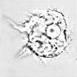
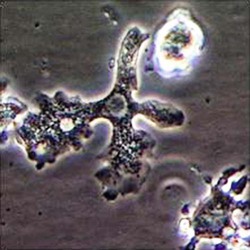
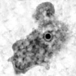
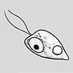
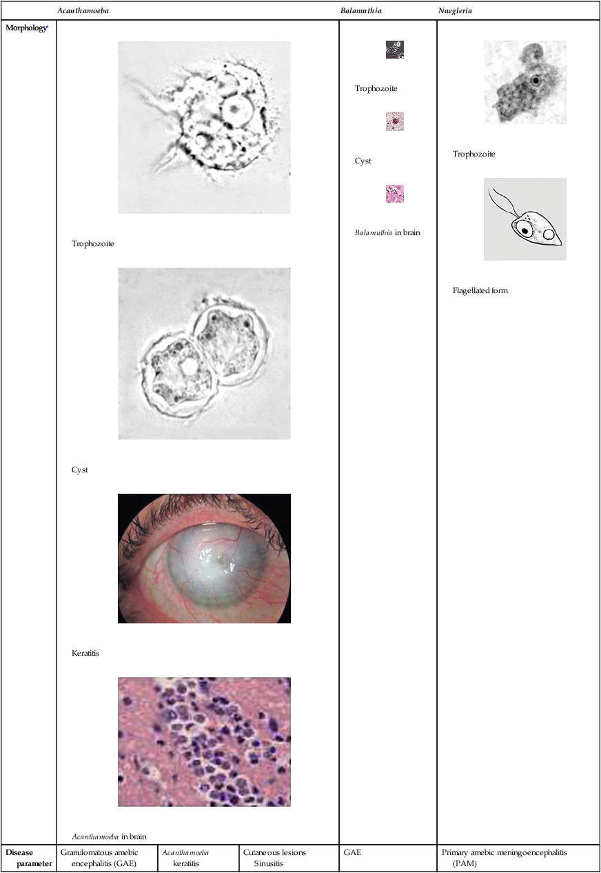
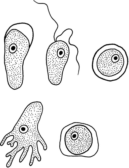
The trophozoites can occur in two forms: ameboid and flagellate (Table 50-1, Figure 50-1). The size ranges from 7 to 35 µm. The diameter of the rounded forms is usually 15 µm. There is a large, central karyosome and no peripheral nuclear chromatin. The cytoplasm is somewhat granular and contains vacuoles. The ameboid form organisms change to the transient, pear-shaped flagellate form when they are transferred from culture or teased from tissue into water and maintained at a temperature of 27° to 37° C. These flagellate forms do not divide, but when the flagella are lost, the ameboid forms resume reproduction. Cysts are generally round, measuring from 7 to 15 µm with a thick double wall.

TABLE 50-1

Free-Living Amebae Causing Disease in Humans

  Acanthamoeba Balamuthia Naegleria
Morphologya
image

Trophozoite

image

Cyst

image

Keratitis

image

Acanthamoeba in brain

image

Trophozoite

image

Cyst

image

Balamuthia in brain

image

Trophozoite

image

Flagellated form

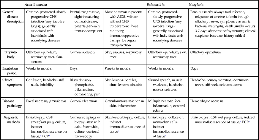
Disease parameter Granulomatous amebic encephalitis (GAE) Acanthamoeba keratitis Cutaneous lesions
Sinusitis
GAE Primary amebic meningoencephalitis
(PAM)
General disease description Chronic, protracted, slowly progressive CNS infection (may involve lungs); generally associated with individuals with underlying diseases Painful, progressive, sight-threatening corneal disease; patients generally immunocompetent Most common in patients with AIDS, with or without CNS involvement; those receiving immunosuppressive therapy for organ transplantation Chronic, protracted, slowly progressive CNS infection (may involve lungs); generally associated with individuals with underlying diseases Rare, but nearly always fatal infection; migration of amebae to brain through olfactory nerve; symptoms can mimic bacterial meningitis; death usually occurs 3-7 days after onset of symptoms; clinical suspicion based on history critical
Entry into body Olfactory epithelium, respiratory tract, skin, sinuses Corneal abrasion Skin, sinuses, respiratory tract Olfactory epithelium, skin, respiratory tract Olfactory epithelium
Incubation period Weeks to months Days Weeks to months Weeks to months Days
Clinical symptoms Confusion, headache, stiff neck, irritability Blurred vision, photophobia, inflammation, corneal ring, pain Skin lesions, nodules, sinus lesions, sinusitis Slurred speech, muscle weakness, headache, nausea, seizures Headache, nausea, vomiting, confusion, fever, stiff neck, seizures, coma
Disease pathology Focal necrosis, granulomas Corneal ulceration Granulomatous reaction in skin, inflammation Multiple necrotic foci, inflammation, cerebral edema Hemorrhagic necrosis
Diagnostic methods Brain biopsy, CSF smear/wet prep, culture, indirect immunofluorescence on tissue,b PCRb Corneal scrapings or biopsy, stain with calcofluor white, culture, confocal microscopy Skin lesion biopsy, culture, indirect immunofluorescence of tissuea Brain biopsy, culture on mammalian cells, indirect immunofluorescence of tissuea Brain biopsy, CSF wet prep, culture, indirect immunofluorescence of tissue,a PCRa

image

image

aAcanthamoeba in brain and Balamuthia in brain courtesy Dr. Govinda Visvesvara, Centers for Disease Control and Prevention.

bIndirect immunofluorescence on tissue and PCR methods available from Centers for Disease Control and Prevention.

Pathogenesis and Spectrum of Disease

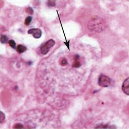
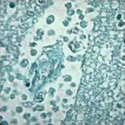
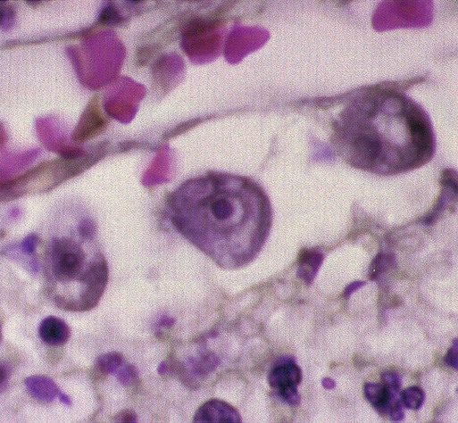
Primary amebic meningoencephalitis (PAM) caused by N. fowleri is an acute, suppurative infection of the brain and meninges (Figure 50-2). With extremely rare exceptions, the disease is rapidly fatal in humans. The period between organism contact and onset of symptoms such as fever, headache, and rhinitis varies from a few days to 2 weeks. Early symptoms include vague upper respiratory tract distress, headache, lethargy, and occasionally olfactory problems. The acute phase includes sore throat; a stuffy, blocked, or discharging nose; and severe headache. Progressive symptoms include pyrexia, vomiting, and stiffness of the neck. Mental confusion and coma usually occur approximately 3 to 5 days before death, which is usually caused by cardiorespiratory arrest and pulmonary edema.

PAM resembles acute bacterial meningitis, and these conditions may be difficult to differentiate. Unfortunately, if the CSF Gram stain is interpreted incorrectly as a false positive, the resulting antibacterial therapy has no impact on the amebae and the patient will usually die within a few days.

Laboratory Diagnosis

Routine Methods

Clinical and laboratory data usually cannot be used to differentiate pyogenic meningitis from PAM. A high index of suspicion is often critical for early diagnosis. Most cases are associated with exposure to contaminated water through swimming or bathing. There is normally an incubation period of 1 day to 2 weeks, and then a course of 3 to 6 days, most often ending in death.

Analysis of the CSF will show decreased glucose and increased protein concentrations. The leukocyte count will range from several hundred to >20,000 cells per mm3. Although Gram stains and bacterial cultures of CSF will be negative, serious patient complications can occur as the result of incorrect therapy if false-positive Gram stains are reported.

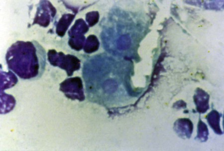
A confirmed diagnosis is made by the identification of amebae in the CSF or in biopsy specimens. CSF should be placed on a slide, under a cover slip, and observed for motile trophozoites; smears can be stained with any of the blood stains. It is important not to mistake leukocytes for actual organisms or vice versa. This type of misidentification often occurs when using a counting chamber and the amebae sink to the bottom and round up, hence the recommendation to use just a regular slide and cover slip. Depending on the temperature and lag time between specimen collection and examination, motility may vary. Slides may be warmed slightly to improve motility. The most important differential characteristic is the spherical nucleus with a large karyosome.

Specimens should never be refrigerated before examination, and CSF should be centrifuged at a slow speed (250× g). If N. fowleri is the causative agent, only trophozoites are normally seen, whereas cysts and trophozoites can be seen with Acanthamoeba spp.

Acanthamoeba Spp.

General Characteristics

Unlike N. fowleri, Acanthamoeba spp. do not have a flagellate stage in the life cycle, only the trophozoite and cyst. Several species of Acanthamoeba cause granulomatous amebic encephalitis (GAE), primarily in immunosuppressed, chronically ill, or otherwise debilitated individuals. These patients usually have no relevant history involving freshwater exposure. Acanthamoeba spp. also cause amebic keratitis, and it is estimated that the incidence in the United States may be one to two cases per million contact lens users. Apparently, the incidence of Acanthamoeba keratitis in the United Kingdom is 15 times higher than that in the United States and 7 times higher than that in Holland.

Motile organisms have spine-like pseudopods; there is a wide organism size range (25 to 40 µm), with the average diameter of the trophozoites being 30 µm. The nucleus has the typical large karyosome, similar to that found in N. fowleri. This morphologic characteristic can be seen on a wet preparation.

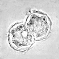
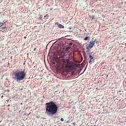
The cysts are usually round with a single nucleus, also having the large karyosome as in the trophozoite nucleus. The double wall is usually visible, with the slightly wrinkled outer cyst wall and what has been described as a polyhedral inner cyst wall. This cyst morphology is identifiable in organisms cultured on agar plates.

Pathogenesis and Spectrum of Disease

GAE

Meningoencephalitis caused by Acanthamoeba spp. may present as an acute suppurative inflammation of the brain and meninges similar to N. fowleri infection. The incubation period of GAE is unknown; several weeks or months are probably necessary to establish disease. The clinical course tends to be subacute or chronic and is usually associated with trauma or underlying disease, not as a result of swimming. GAE may present with symptoms of confusion, dizziness, drowsiness, nausea, vomiting, headache, lethargy, stiff neck, seizures, and sometimes hemiparesis. Unlike PAM caused by N. fowleri, both trophozoites and cysts are found throughout the tissue. Also, dissemination to other tissues such as the liver, kidneys, trachea, and adrenals can occur in immunocompromised individuals; or additional unusual sites also include the ear and necrotic bone from a bone graft of the mandible. Some patients, especially those with AIDS, can develop erythematous nodules, chronic ulcerative skin lesions, or abscesses.

Keratitis

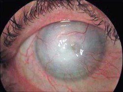
Acanthamoeba spp. also cause keratitis and corneal ulceration. Clinicians need to consider acanthamoebic infection in the differential diagnosis of eye infections that are not responding to bacterial, fungal, or viral therapy. These infections are often due to direct exposure of the eyes to contaminated materials or solutions. Use of contact lenses is the leading risk factor for keratitis. Conditions that are linked with disease include the use of home-made saline solutions, poor contact lens hygiene, and corneal abrasions. A contact lens can act as a mechanical vector for transport of amebae present in the storage case onto the cornea. Subsequent multiplication and invasion of the tissue may occur. Decreased corneal sensation has contributed to the misdiagnosis of Acanthamoeba keratitis as herpes simplex keratitis. Acanthamoeba keratitis may be present as a secondary or opportunistic infection in patients with herpes simplex keratitis. Unfortunately, as a result, treatment can be delayed for 2 weeks to 3 months.

Laboratory Diagnosis

Routine Methods

The most effective culture approach uses non-nutrient agar plates with Page’s saline and an overlay growth of Escherichia coli on which the amebae feed. Tissue stains are also effective, and cysts isolated from cultures can be stained with Gomori’s silver methenamine, periodic acid-Schiff, and calcofluor white. Identification of Acanthamoebae in ocular samples and other tissues can be difficult, even for trained laboratory professionals; in histologic preparations, the organisms appear similar to keratoplasts, as well as neutrophils and monocytes. It has been estimated that up to 70% of clinical Acanthamoeba keratitis cases are misdiagnosed as viral keratitis. Also, the average time to diagnosis of keratitis attributable to Acanthamoeba infection can average 2.5 weeks longer for non–contact lens wearers than for contact lens users.

Other Methods

CSF or bronchoalveolar lavage fluid cytospin preparations can be used to look for amebae in patients with GAE or respiratory symptoms. The characteristic morphology of the Acanthamoeba trophozoites, such as the prominent nucleolus, contractile vacuole, and cytoplasmic vacuoles, can be seen more easily using trichrome or hematoxylin and eosin stains on fixed preparations after cytocentrifugation.

In the differential diagnosis of GAE, other space-occupying lesions of the central nervous system (CNS) (e.g., tumor, abscess, fungal infection) must also be considered. Predisposing conditions include Hodgkin’s disease, diabetes, alcoholism, pregnancy, and corticosteroid therapy. Organisms have also been found in the adrenal gland, brain, eyes, kidneys, liver, pancreas, skin, spleen, thyroid gland, and uterus.

In infections caused by Acanthamoeba spp., periodic acid-Schiff stains the cyst wall red and methenamine silver stains the cyst black. Normally, Naegleria and Acanthamoeba isolates are identified to the species level by a reference laboratory, such as the Centers for Disease Control and Prevention, using indirect fluorescent antibody procedures with a monoclonal or polyclonal antibody.

Balamuthia Mandrillaris

General Characteristics

Balamuthia mandrillaris is uncommon and was thought to be a harmless soil organism, with no relevance for infecting mammals. However, since the appearance of B. mandrillaris was first seen at the San Diego Wild Animal Park in a gibbon that died of meningoencephalitis, a number of primates, as well as dogs, sheep, and horses, have died of CNS infection caused by this organism. Approximately 100 cases of human amebic encephalitis worldwide have been identified with about half of the cases diagnosed within the United States. Death can occur from a week to several months after the onset of symptoms. Patients eventually die with a massive CNS infection. Genotyping studies indicate that lethal infections caused by B. mandrillaris are due to a single species with a global distribution.

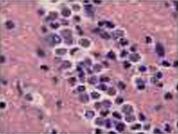
The life cycle is similar to that of Acanthamoeba spp.; like Acanthamoeba spp., Balamuthia does not have a flagellated stage in the life cycle. Both trophozoites and cysts are found in CNS tissue, and their sizes are similar to those of Acanthamoeba trophozoites and cysts. It is difficult to differentiate Balamuthia from Acanthamoeba spp. in tissue sections under a light microscope. Using electron microscopy, the cysts are characterized by having three layers in the cyst wall: an outer wrinkled ectocyst, a middle structure–less mesocyst, and an inner thin endocyst. Under light microscopy, they appear to have two walls: an outer irregular wall and an inner round wall. In some cases, Balamuthia trophozoites in tissue sections appear to have more than one nucleolus in the nucleus. In such cases, it may be possible to distinguish Balamuthia amebae from Acanthamoeba organisms on the basis of nuclear morphology, because Acanthamoeba trophozoites have only one nucleolus.

Trichomonas Vaginalis

General Characteristics

Infection is acquired primarily through sexual intercourse, hence the need to diagnose and treat asymptomatic males. The organism is capable of survival for extended periods in a moist environment such as damp towels and underclothes; however, this mode of transmission is thought to be very rare. Infection with Trichomonas vaginalis occurs worldwide. It is estimated that 5 million women and 1 million men in the United States have trichomoniasis, with an estimated 7.4 million new cases occurring annually. The prevalence of trichomoniasis worldwide is estimated to be more than 170 million cases, which does not include the number of asymptomatic cases that remain untreated. In North America, more than 8 million new cases are reported yearly, with an estimated rate of asymptomatic cases being as high as 50%. Trichomoniasis is the primary non-viral sexually transmitted disease worldwide. Infection with T. vaginalis has major health consequences for women, including complications in pregnancy, association with cervical cancer, and predisposition to HIV infection.

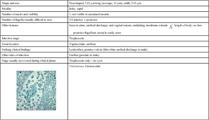
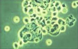
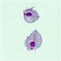
The life cycle of T. vaginalis has a single trophozoite stage, and is very similar in morphology to other trichomonads (Table 50-2). The trophozoite is 7 to 23 µm long and 5 to 15 µm wide. The axostyle is usually obvious and protrudes through the bottom of the organism, whereas the undulating membrane ends halfway down the side of the trophozoite. There are a large number of granules evident along the axostyle.

TABLE 50-2

Characteristics of Trichomonas vaginalis

Shape and size Pear-shaped, 7-23 µm long (average, 13 µm); width, 5-15 µm
Motility Jerky, rapid
Number of nuclei and visibility 1; not visible in unstained mounts
Number of flagella (usually difficult to see) 3-5 anterior, 1 posterior
Other features Seen in urine, urethral discharge, and vaginal smears; undulating membrane extends image length of body; no free posterior flagellum; axostyle easily seen
Infective stage Trophozoite
Usual location Vagina (male, urethra)
Striking clinical findings Leukorrhea, pruritus vulvae (thin white urethral discharge in male)
Other sites of infection Urethra (prostate in male)
Stage usually recovered during clinical phase Trophozoite only—no cyst
image

Trichomonas, Giemsa stain

image

Pathogenesis and Spectrum of Disease

Growth of the organism results in inflammation and large numbers of trophozoites in the tissues and the secretions. As the acute infection becomes more chronic, the purulent discharge diminishes, with a decrease in the number of organisms. Symptoms such as vaginal or vulval pruritus and discharge are often sudden and occur during or after menstruation as a result of the increased vaginal acidity. Symptoms include vaginal discharge (42%), odor (50%), and edema or erythema (22% to 37%). Complaints also include dysuria and lower abdominal pain.

From 25% to 50% of infected women may be asymptomatic and have a normal vaginal pH of 3.8 to 4.2 and normal vaginal flora. Even in the carrier form, about 50% of women will become symptomatic during the following 6 months.

Although vaginitis is the most common finding in women with trichomoniasis, other complications include distention of a fallopian tube with pus, endometritis, infertility, low birth weight, and cervical erosion. There is also an increased association with HIV transmission and cervical dysplasia.

Dysuria, often the earliest symptom, occurs in about 20% of women with vaginal trichomoniasis. Infected males may be asymptomatic, or the infection may be self-limited, persistent, or result in recurring urethritis. In nonspecific urethritis, T. vaginalis has been detected in 10% to 20% of subjects and in 20% to 30% of those whose sexual partners had vaginitis. Once established, the infection persists for an extended period in females but only for about 10 days or less in males. T. vaginalis is the cause of 11% of all cases of non–gonococcal urethritis in males.

Respiratory distress has been reported in a full-term, normal male infant with T. vaginalis with severe respiratory problems following delivery. A wet preparation of thick, white sputum demonstrated few leukocytes and motile flagellates, which were identified as T. vaginalis. This study supports previous data confirming that the organism may cause neonatal pneumonia.

Laboratory Diagnosis

Humans are the only natural host for T. vaginalis, and organisms reside in the vagina and prostate; they usually do not survive outside the urogenital tract. The parasites feed on the mucosal surface of the vagina, where bacteria and leukocytes are abundant. The preferred pH for good parasitic growth in females is slightly alkaline or acidic (6.0 to 6.3 optimal), not the normal pH (3.8 to 4.2) of the healthy vagina. The organisms can also be recovered in urine, in urethral discharge, or after prostatic massage. Often, the organisms are recovered in centrifuged urine sediment from both male and female patients.

Pentatrichomonas Hominis

Pentatrichomonas hominis derives its name on the basis of the morphologic structure of the trophozoite. The organism has five anterior flagella and a parabasal body. The organism is recovered worldwide and is considered to be nonpathogenic although it has been isolated from patients with diarrhea.

Toxoplasma Gondii

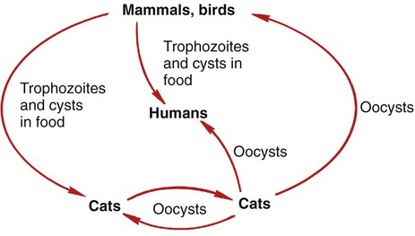
Toxoplasma gondii is a protozoan parasite that infects most species of warm-blooded animals, including humans. Members of the cat family, Felidae, are the only known definitive hosts for the sexual stages of T. gondii and serve as the main reservoirs of infection. Cats become infected with T. gondii through carnivorism or by ingestion of oocysts. Outdoor cats are much more likely to become infected than domestic cats that are confined indoors. After tissue cysts or oocysts are ingested by the cat, organisms are released and invade epithelial cells of the cat small intestine, where they undergo an asexual cycle followed by a sexual cycle with the formation of oocysts, which are excreted in the feces. The uninfective oocyst takes 1 to 5 days after excretion to become infective. Cats shed oocysts for 1 to 2 weeks and large numbers may be shed, often more than 100,000 per gram of feces. Oocysts survive in the environment for several months to more than 1 year and are resistant to disinfectants, freezing, and drying. However, they are killed by heating to 70° C for 10 minutes. The life cycle in the cat takes approximately 19 to 48 days after infection with the oocysts but only 3 to 10 days after the ingestion of meat infected with cysts (e.g., a mouse) (Figure 50-3).
General Characteristics

There are three infectious stages of T. gondii: the tachyzoites (in groups or clones), the bradyzoites (in tissue cysts), and the sporozoites (in oocysts from cat feces). Tachyzoites rapidly multiply in any cell of the intermediate host and in epithelial cells of the definitive host (cats). Bradyzoites are found within the tissue cysts and usually multiply very slowly; the cyst may contain few to hundreds of organisms, and intramuscular cysts may reach 100 µm in size. The tissue cysts can be found in visceral organs such as the lungs, liver, and kidneys; however, they are more prevalent in the brain, eyes, and skeletal and cardiac muscle. Intact tissue cysts can persist for the life of the host and do not cause an inflammatory response.

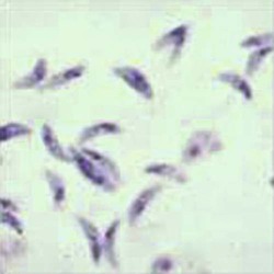
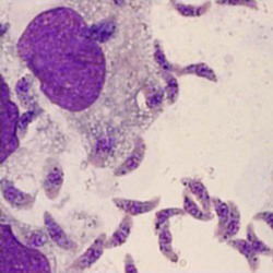
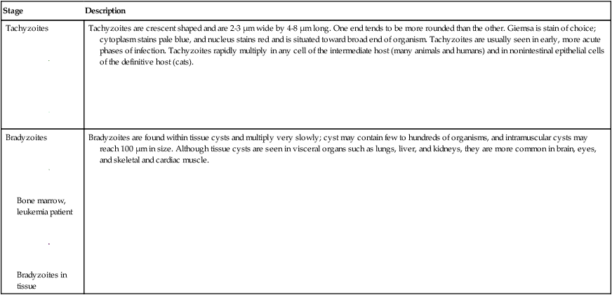
Tachyzoites are crescent-shaped and are 2 to 3 µm wide by 4 to 8 µm long (Table 50-3). One end tends to be more rounded than the other. Giemsa is the stain of choice; the cytoplasm stains pale blue, and the nucleus stains red and is situated toward the broad end of the organism.

TABLE 50-3

Morphology of Toxoplasma gondii Stages Found in Humans

Stage Description
Tachyzoites

image

image

Tachyzoites are crescent shaped and are 2-3 µm wide by 4-8 µm long. One end tends to be more rounded than the other. Giemsa is stain of choice; cytoplasm stains pale blue, and nucleus stains red and is situated toward broad end of organism. Tachyzoites are usually seen in early, more acute phases of infection. Tachyzoites rapidly multiply in any cell of the intermediate host (many animals and humans) and in nonintestinal epithelial cells of the definitive host (cats).
Bradyzoites

image

Bone marrow, leukemia patient

image

Bradyzoites in tissue

Bradyzoites are found within tissue cysts and multiply very slowly; cyst may contain few to hundreds of organisms, and intramuscular cysts may reach 100 µm in size. Although tissue cysts are seen in visceral organs such as lungs, liver, and kidneys, they are more common in brain, eyes, and skeletal and cardiac muscle.

image

Cysts are formed in chronic infections, and the bradyzoites within the cyst wall are strongly periodic acid-Schiff positive. During the acute phase, there may be groups of tachyzoites that appear to be cysts; however, they are not strongly periodic acid-Schiff positive and have been termed pseudocysts.

Pathogenesis and Spectrum of Disease

As the tachyzoites actively grow, increase in number, and eventually rupture from the cell, they invade adjacent cells. This process creates additional lesions. Once the cysts are formed, the process becomes quiescent, with little or no multiplication and spread. In the immunocompromised or immunodeficient patient, a cyst rupture or primary exposure to the organisms often leads to lesions. The organisms can be disseminated via the lymphatic system and the bloodstream to other tissues.

Toxoplasmosis can be categorized into four groups: (1) acquired in the immunocompetent patient; (2) acquired or reactivated in the immunodeficient patient; (3) congenital; and (4) ocular.

Diagnosis and their interpretations may differ for each clinical category.

Immunocompromised Individuals

Infections in the compromised patient can lead to severe complications (Table 50-4). Underlying conditions that may impact the disease outcome include Hodgkin’s disease, non-Hodgkin’s lymphomas, leukemia, solid tumors, collagen vascular disease, organ transplantation, and AIDS. In the immunocompromised patient, the CNS is primarily involved, but these patients may also have myocarditis or pneumonitis. More than 50% of these patients will show altered mental state, motor impairment, seizures, abnormal reflexes, and other neurologic sequelae. Toxoplasmosis in patients being treated with immunosuppressive drugs may be due to either newly acquired or reactivated latent infection.

TABLE 50-4

People at Risk for Severe Toxoplasmosis

Category Comments
Infants born to mothers who are first exposed to Toxoplasma infection several months before or during pregnancy Mothers who are first exposed to Toxoplasma more than 6 months before becoming pregnant are not likely to pass infection to their children
Persons with severely weakened immune systems Infection that occurred at any time during life can reactivate in an immunocompromised individual

In transplant recipients, the disease presentation depends on prior exposure to T. gondii by the donor and recipient, the type of organ involved, and the patient’s level of immunosuppression. Reactivation of a latent infection or an acute primary infection acquired directly from the transplanted organ can lead to severe disease. Stem cell transplant (SCT) recipients are particularly susceptible to severe toxoplasmosis, primarily attributable to reactivation of a previously acquired latent infection.

Before the use of highly active antiretroviral therapies (HAARTs), Toxoplasma encephalitis (TE) was a life-threatening opportunistic infection among patients with AIDS and was usually fatal if not treated. In AIDS patients with reactivated latent infections, psychiatric manifestations of T. gondii are seen, including altered mental status (60%) with delusions, auditory hallucinations, and thought disorders. T. gondii enhances HIV-1 replication within reservoir host cells and, at the same time, HIV-1 undermines acquired immunity to the parasite, promoting reactivation of chronic toxoplasmosis.

Congenital Infections

Congenital infection results when the mother acquires a primary infection during pregnancy. The majority of patients remain asymptomatic during the acute infection. However, congenital infections may be severe if the mother becomes infected during the first or second trimester. At birth or soon thereafter, symptoms in these infants may include retinochoroiditis, cerebral calcification, and occasionally hydrocephalus or microcephaly. Because treatment of the mother may reduce the severity of disease in the infant, prompt and accurate diagnosis is mandatory. Many infants who are asymptomatic at birth will subsequently develop symptoms of congenital toxoplasmosis; however, treatment may help prevent subsequent sequelae. Central nervous system involvement may not appear until several years later.

Laboratory Diagnosis

The most common method of diagnosis for toxoplasmosis is serologic testing for T. gondii– specific antibodies. Other procedures include PCR; examination of biopsy specimens, buffy coat cells, or cerebrospinal fluid; or isolation of the organism in tissue culture or in laboratory animals. It is important to remember that many individuals have been exposed to T. gondii and may have cysts within the tissues. Recovery of organisms from tissue culture or animal inoculation may be misleading, because the organisms may be isolated but may not be the etiologic agent of disease. However, two situations in which organism detection may be very significant are (1) tachyzoites in smears and/or tissue cultures inoculated from cerebrospinal fluid and (2) tachyzoites in patients with acute pulmonary disease and the demonstration of intracellular and extracellular tachyzoites in Giemsa-stained smears of bronchoalveolar lavage (BAL) fluid.

When laboratory personnel decide to initiate Toxoplasma-specific antibody testing or switch to a different antibody detection kit, the user must carefully review the manufacturer’s package insert and published literature for information on the sensitivity and specificity rates.

An in-laboratory comparison of kits should be performed, using positive and negative samples confirmed by a toxoplasmosis reference laboratory.

The serologic diagnosis of toxoplasmosis is very complex and has been discussed extensively in the literature (Wilson & McAuley, 2003); a number of additional procedures include enzyme immunoassays, enzyme-linked immunosorbent assays (ELISAs), direct agglutination, an immunosorbent agglutination assay, an indirect immunofluorescence assay (IFA), immunocapture, and immunoblot tests.

Therapy

Treatment is recommended for the following conditions: clinically active disease, diagnosed congenital toxoplasmosis in newborns, pregnant women with infection during gestation, patients with chorioretinitis, and disease in symptomatic compromised patients. Therapy is also recommended for preventive or suppressive treatment in HIV-infected persons. The currently recommended drugs work primarily against the actively dividing tachyzoite form of T. gondii and do not eradicate encysted organisms (bradyzoites).

The most common drug combination used to treat congenital toxoplasmosis consists of pyrimethamine and a sulfonamide (sulfadiazine is recommended in the United States), plus folinic acid in the form of leucovorin calcium to protect the bone marrow from the toxic effects of pyrimethamine. Spiramycin is recommended for pregnant women with acute toxoplasmosis when fetal infection has not been confirmed in an attempt to prevent transmission of T. gondii from the mother to the fetus.

In immunosuppressed persons with toxoplasmosis, the combination of pyrimethamine and sulfadiazine plus leucovorin is the preferred treatment. Clindamycin is a second alternative for use in combination with pyrimethamine and leucovorin in those who cannot tolerate sulfonamides. Because relapse often occurs after toxoplasmosis in HIV-infected patients, maintenance therapy with pyrimethamine plus sulfadiazine or pyrimethamine plus clindamycin is recommended. For prophylaxis to prevent an initial episode of toxoplasmosis in Toxoplasma-seropositive persons with CD4+ T-lymphocyte counts of less than 100 cells/mL, trimethoprim-sulfamethoxazole is recommended as the first choice, with alternatives consisting of dapsone plus pyrimethamine or atovaquone with or without pyrimethamine. Leucovorin is given with all regimens, including pyrimethamine. HIV-infected persons who are serologically negative for Toxoplasma IgG should be advised to protect themselves from primary infection by eating well-cooked meats and washing their hands after possible soil contact. Cats kept as pets should be fed commercial or well-cooked food, should be kept indoors, and should have their litter box changed each day.

Pyrimethamine and sulfadiazine are often used for persons with ocular disease. Clindamycin, in combination with other antiparasitic medications, is frequently used for the treatment of ocular disease. In addition to antiparasitic drugs, physicians may add corticosteroids to reduce ocular inflammation.