Orthopedics and podiatry
A Arthroscopy
2. Preoperative assessment and patient preparation
a) Arthroscopic procedures may be anesthetically managed by almost any of the available anesthesia techniques (general anesthesia, regional anesthesia, combined regional and general anesthesia, local blockade, and sometimes monitored anesthesia care).
b) Patient selection for a given anesthetic technique is crucial with arthroscopic procedures, as with all operative procedures. Critical factors in the selection of the available anesthesia techniques appropriate for arthroscopic procedures are the patient positioning necessary to facilitate the proposed arthroscopic procedure and the overall state of health of the patient.
c) The choice of position is determined by the surgeon’s operating requirements. Reviewing the patient’s chart and, most important, personally interviewing the patient, along with understanding the physiologic changes associated with various positions, will assist the anesthesia provider in offering the best suggestion for anesthesia care for each patient.
d) The factors in the decision are listed in the following box.
a) Complications: Complications from arthroscopic procedures represent a small percentage of the total number of procedures performed.
(1) The full range of potential anesthetic complications associated with patient positioning applies (e.g., inadvertent extubation, eye or corneal injury, and nerve injury from improper patient positioning).
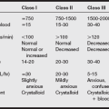
(2) Because of the less invasive nature of arthroscopic procedures, concerns over blood loss are typically minimal. However, sudden sustained hypotension is a cause for immediate investigation.
(3) Perforation of a major blood vessel may occur during trocar insertion and may not be detected until the tourniquet is deflated. Such vascular injury may result from pressure exerted by excessive extravasated irrigation fluid during the procedure.
(1) To provide optimal visualization of joint structures during arthroscopic procedures, the irrigating fluid used to distend the operative joint is instilled under pressure.
(2) Take note of any deficits of inflow versus outflow of irrigating solution throughout the procedure.
(a) Depending on the complexity of the arthroscopic procedure, a large number of irrigation fluid bags may be required.
(b) Small individual inflow/outflow deficits may result in significant fluid absorption by the patient over the course of an extended procedure.
(c) Fluid absorption is of particular concern for shoulder or hip arthroscopic procedures in which fluid absorption is not relatively limited by the use of the pneumatic tourniquet.
(d) Absorption of excessive extravasated fluid may lead to the development of signs and symptoms of congestive heart failure, pulmonary edema, volume overload, or hyponatremia.
(e) If the patient experiences these symptoms, treatment with fluid restriction, supplemental oxygen, and diuresis should be instituted.
(3) Although the mechanism of occurrence has not been delineated, subcutaneous emphysema, tension pneumothorax, and pneumomediastinum have been reported during shoulder arthroscopy, specifically subacromial decompression.
a) History and physical examination: Individualized
(1) Radiographs of the affected extremity
(2) Chest radiography, electrocardiography (ECG), and laboratory tests as indicated
a) Standard monitoring equipment
b) Standard drugs for general or regional anesthesia
e) Circuit extension if table positioned away from anesthesia practitioner
a) Induction: Standard induction with routine medications are used.
(1) Most often, the supine position is used for arthroscopic procedures of both the upper and lower extremities.
(2) Arthroscopy on the knee requires the supine position with the foot of the operating room bed lowered. The nonoperative leg should either have a sequential compression device or some form of antiembolic stocking in place to reduce pooling of blood and reduce the potential for thrombus formation.
(3) Patients undergoing elbow arthroscopy may be placed in the supine, lateral decubitus, or prone position; the position is dictated by operative necessity and surgeon preference. The prone position is more advantageous primarily because of the better limb stability during the procedure.
(4) Shoulder arthroscopy is usually accomplished by either the modified Fowler position (beach chair position) or the lateral decubitus position, based on optimal access to the injury and surgeon preference. Because this procedure does not use a tourniquet, deliberate hypotension may be requested by surgeons. Blood pressure cuff measurements taken on the arm are not representative (underestimate) perfusion pressure in the brain when patients are in a sitting position. Therefore, it is recommended to maintain preoperative mean arterial pressures to avoid hypoxic brain injury.
(5) Hip arthroscopy is also typically accomplished by the lateral decubitus position or the supine position, with the patient on a fracture table. The fracture table is used to provide greater stability while traction is applied using either weights and counterweights (lateral decubitus position) or mechanical traction attached to the leg-holding device of the fracture table (supine position).
c) Tourniquet use: See the discussion of knee arthroscopy later in this section.
d) Emergence: The patient is usually extubated in the operating room unless there was preoperative respiratory compromise.
a) Pain is usually minimal to moderate, unless reconstruction was performed.
(1) Intraarticular injections of local anesthetics or opioids are now widely used in an attempt to provide postoperative analgesia.
(2) Inadequate pain control can lead to decreased mobility and an increased incidence of postoperative complications.
b) Swelling or edema: Assess capillary refill in the affected extremity and avoid overhydration intraoperatively.
B Foot and ankle surgery
Plantar fasciotomy is indicated for severe foot pain during or after ambulating or on arising after sleep, resulting from chronic plantar fasciitis that has not responded to conservative therapy. Open fasciotomy is accomplished through a small incision along the posterior surface of the calcaneus. The plantar fascia is incised to relieve the tension across the plantar arch. Endoscopic plantar fasciotomy is accomplished via two “miniature” incisions, one medial and one lateral, at the beginning of the plantar arch. A small trocar is inserted through these incisions. The sheath of the trocar is slotted to allow visualization of the plantar fascia with the endoscope. The full thickness of the plantar fascia is incised, and the skin incisions are closed.
a) Patients scheduled for foot or ankle surgery are excellent candidates for regional anesthesia.
b) Most surgical procedures on the foot or ankle can be accomplished within a 2-hour time frame, often on an outpatient basis.
c) Spinal anesthesia provides sufficient surgical anesthesia to allow completion of most procedures. However, the postanesthesia recovery phase may be unacceptably long and may require the patient to spend a night in the hospital or outpatient facility, which may be unacceptable to the patient.
d) Nerve blocks are especially effective for surgical procedures on the foot or ankle. Posterior tibial nerve block, Mayo blockade, and Bier block are examples of blocks that are effective for foot and ankle procedures.
e) One may provide IV sedation by either continuous infusion or intermittent bolus to provide amnesia and to minimize or eliminate any anxiety the patient may have. The surgeon can inject the surgical site with long-acting local anesthetic (e.g., bupivacaine) to maintain the patient’s comfort immediately and for several hours postoperatively.
C Forearm and hand surgery
a) Patients scheduled for surgical procedures on the forearm or hand are excellent candidates for regional anesthesia.
b) Axillary block and Bier block provide excellent surgical anesthesia for most surgical procedures of the forearm and hand that are anticipated to require 1 hour or less to accomplish.
c) For procedures precipitated by traumatic injury, such as complex, comminuted fractures or reconstruction of the vascular and nerve structures of the hand or forearm (procedures that may require considerable amounts of time to accomplish), the better anesthetic choice may be general anesthesia.
d) Tourniquet pain becomes an issue with such longer procedures if regional anesthesia is chosen.
e) In addition, for a patient requiring surgery as the result of traumatic injury, the issue of the patient’s nothing by mouth (NPO) status becomes important. Frequently, trauma patients have eaten or ingested liquids close to the time of the traumatic injury. Alcohol may be a precipitating factor in the traumatic injury as well. For these reasons, rapid-sequence induction of general anesthesia may be a more appropriate anesthetic course.
D Hip arthroplasty
2. Preoperative assessment and patient preparation
a) History and physical examination
(1) With this elderly population, assess for coexisting medical diseases.
(2) Carefully assess blood volume, central venous pressure, and orthostatic hypotension because dehydration may mask hemoglobin changes resulting from hematoma formation.
(1) Radiographs: Hip and chest
(2) Laboratory tests: Complete blood count, electrolytes, glucose, blood urea nitrogen, creatinine, urinalysis, prothrombin time, partial thromboplastin time, bleeding time of the patient on aspirin, and type and crossmatch
c) Preoperative medications and IV therapy
(1) Anticoagulants: Heparin, low-molecular-weight heparin, oral anticoagulants
(2) Antirheumatic or anti-inflammatory medications
(4) Sedatives and narcotics: Used with caution in the elderly population.
(5) Two peripheral, large-bore (16- to 18-gauge) IV lines with moderate fluid replacement
(6) Epidural catheter placement: Test dose performed on an awake patient
(2) Indwelling urinary catheter: Controversial
(3) Central venous pressure: Trend of volume status.
(4) Warming modalities are used.
(5) ECG leads V5 and II detect myocardial ischemia and diagnose tachyarrhythmias in the elderly population.
(6) Transesophageal echocardiography: Assess fat and bone deposits when the acetabulum is reamed and curetted.
(7) Use x-ray shields for self-protection.
(8) Arterial line monitoring is indicated if hypotensive techniques are used.
b) Pharmacologic agents: Vasopressors
c) Position: Lateral; a special orthopedic table may be used.
a) Considerations: Regional blockade, general anesthesia, or a combination of both
(1) An endotracheal tube must be inserted.
(2) Combination with regional anesthetic allows reduced dosages of agents and control of airway.
(a) Thorough airway assessment is done in an arthritic population.
(b) Induction is performed with the patient on the stretcher.
(c) Succinylcholine may be contraindicated with crush injuries if large amounts of muscle tissue are devitalized.
(a) Monitor fluid and blood replacement therapy to minimize blood loss and transfusion that are applicable during hip arthroplasty (e.g., autologous donation and deliberate hypotension).
(b) Laminar flow is used to minimize infections and can increase evaporative fluid and heat losses from the operative site.
(c) Controlled hypotensive techniques facilitate surgical exposure and decrease blood loss.
(3) The anesthesia provider must be particularly cognizant of the possible occurrence of hypotension, hypoxia, and potential cardiovascular collapse. These complications are observed most often during insertion of the femoral prosthesis during total hip arthroplasty.
(a) Possible causes of these complications include the MMA cement, fat embolism, air embolism, thromboembolism, and bone marrow embolism.
(b) MMA has been demonstrated to produce significant increases in both pulmonary vascular resistance and pulmonary wedge pressure while decreasing systemic vascular resistance, cardiac output, and arterial pressure.
(c) MMA cement is used to distribute the forces of the femoral and acetabular prosthetic components.
(i) Mixing the cement causes the monomer portion to polymerize (an exothermic reaction).
(ii) Problems with MMA relate to cementing the femoral prosthesis; unpolymerized monomer can be absorbed into the circulation.
(iii) It causes direct vasodilation, usually within the first minute; it can last as long as 10 minutes.
(iv) Venous embolism can occur when the femoral prosthesis is inserted into the femoral canal.
(v) Hypotension, hypoxia, and cardiovascular collapse after prosthesis insertion have been reported.
(vi) Prevent complications: Communicate with the surgeon regarding application.
(vii) Use 100% oxygen, decrease the vasodilating agent, maximize fluid status, and have vasopressor support available.
a) Obtain laboratory results: Hemoglobin and hematocrit; watch for hidden bleeding.
b) Fat embolism typically appears 12 to 48 hours after a long bone fracture.
(b) Adult respiratory distress syndrome
(c) Central nervous system dysfunction (confusion, coma, and seizures)
(d) Immobilization of long bone fractures
(e) Other potential complications include DVT and PE. DVT is a precursor to PE development. DVT has been demonstrated in 40% to 60% of patients undergoing total hip arthroplasty and in approximately 80% of patients having total knee arthroplasty. From within the two patient populations, PE is believed to develop 1% to 5% of the time.
E Hip pinning (open reduction and internal fixation)
2. Preoperative assessment and patient preparation
a) History and physical examination: Obtain a verbal history from the patient or family member. Note any preexisting disease processes, social history, current medications, surgical history, and allergies.
b) Laboratory tests: Hemoglobin, hematocrit, complete blood count, prothrombin time, partial thromboplastin time, and others are obtained as indicated by the history and physical examination.
c) Diagnostic tests: 12-lead ECG, chest radiography, and others are obtained as indicated by the history and physical examination.
a) Monitoring equipment: Standard, arterial line, central line as needed
(1) Positioning devices and operating table: The patient is usually placed in a lateral position. Aging skin atrophies and is prone to trauma from adhesive tape, electrocautery pads, and ECG electrodes. Arthritic joints may interfere with positioning; when possible, the elderly patient should be positioned for comfort. Meticulous padding of the axilla and all bony prominences decreases the risk of nerve injury and ischemia. Prevent pressure to the ears and eyes. Maintain the neck in neutral alignment.
(2) Because of the length of the procedure and surgical exposure, warming modalities should be implemented (fluid warmer, warming blankets).
(1) Continuous infusion: Consider the use of a continuous epidural infusion intraoperatively or for postoperative pain control.
(2) IV fluids: Estimated blood loss may be greater than 1000 mL. Type and crossmatch for 2 units of packed red blood cells. Blood loss is replaced 1:1 with blood products or colloid solutions or 3:1 if crystalloid solutions are used. Maintain urine output at 0.5 mL/kg/hr. Keep in mind any preexisting disease processes that may easily place the elderly patient in a state of fluid overload. Consider the use of a cell saver intraoperatively. The insertion of 2 large bore IV’s is warranted.
(3) Tabletop: Standard. All equipment needed to implement the anesthetic plan should be available.
4. Perioperative management and anesthetic technique
a) Regional and general anesthesia are both options for elderly patients.
b) Hip pinning may be performed using subarachnoid block or continuous epidural infusion extending to the T8 sensory level. Keep in mind the expected length of the procedure, the patient’s history and physical examination findings, and the patient’s level of cooperation and ability to lie still. Major advantages of regional anesthesia is a decreased incidence of postoperative thromboembolism and another is reduced blood loss. This is thought to be the result of peripheral vasodilation and maintenance of venous blood flow in the lower extremities. Local anesthetics also inhibit platelet aggregation and stabilize endothelial cells. Difficult patient positioning and altered landmarks related to degenerative changes of the spine may increase the technical difficulty of performing a regional block. Postdural puncture headaches are not as prevalent in the elderly population.
c) If a general anesthetic is the best choice for the patient, drugs for induction and maintenance should reflect findings from the patient’s history and physical examination. One advantage of general anesthesia is that the anesthetic can be induced with the patient on the bed or stretcher before moving to the operating table, thus avoiding painful positioning. A disadvantage of general anesthesia is that the elderly patient cannot be positioned for maximal comfort. Consider the use of nondepolarizing muscle relaxants during induction if there are no airway concerns. Continued muscle relaxation is optional and is left to the discretion of the anesthesia provider or the request of the surgeon. The effects of nondepolarizing muscle relaxants that are renally excreted may be slightly prolonged in elderly persons because of reduced drug clearance.
d) Emergence: An epidural catheter may be placed for supplemental use with general anesthesia or for postoperative pain control.
e) Fat embolization: See the discussion of pelvic reconstruction later in this section.
a) Consider the use of a continuous epidural infusion for patient-controlled analgesia for postoperative pain control.
b) A marked decrease in blood pressure postoperatively may be related to hematoma formation.
c) DVT prophylaxis should be instituted postoperatively (support hose and deep venous thrombosis prophylaxis).
F Knee (total knee replacement) arthroplasty
a) Respiratory: These patients may have rheumatoid arthritis and associated pulmonary conditions. Pulmonary effusions may be present. Rheumatoid arthritis involving the cricoarytenoid joints may exhibit itself by hoarseness. A narrow glottic opening may lead to a difficult intubation. Arthritic involvement of the cervical spine and temporomandibular joint may also complicate airway management.
b) Cardiovascular: Depending on the severity of the arthritis, the patient may have a lowered exercise tolerance. Rheumatoid arthritis is associated with pericardial effusion. Cardiac valve fibrosis and cardiac conduction abnormalities can occur with possible aortic regurgitation. Test with an ECG and, if possible, an echocardiogram and cardiac nuclear imaging.
c) Neurologic: A thorough preoperative neurologic examination may yield evidence of cervical nerve root compression. If indicated, obtain lateral neck radiographs for the determination of stability of the atlanto-occipital joint.
d) Musculoskeletal: Positioning may be difficult because of pain and the decreased mobility of the joints.
e) Hematologic and laboratory: Obtain hemoglobin and hematocrit and other tests related to the history and physical examination.
f) Premedication is individualized based on the patient’s need.
a) Standard monitoring equipment. A tourniquet may or may not be used.
b) The patient should have one large-bore IV line.
c) Fluid requirements include normal saline or lactated Ringer’s solution at 4 to 6 mL/kg/hr.
d) Standard drugs for general or regional anesthesia are used.
a) This procedure can be done using general or regional anesthesia.
b) Regional anesthesia could be with a subarachnoid block or placement of an epidural catheter.
a) Induction: Standard induction with routine medications. Muscle relaxation is needed for the placement of the prosthesis.
b) Monitor fluid and blood therapy.
c) Requires the supine position with the foot of the operating room bed lowered. The nonoperative leg should either be wrapped with an elastic bandage or have some form of antiembolic stocking in place to reduce pooling of blood and reduce the potential for thrombus formation.
d) Monitor for physiologic changes that are caused by tourniquets.
e) Safety measures for preventing tourniquet complications are listed in the box below. Use of MMA can increase both pulmonary vascular resistance and pulmonary wedge pressure while decreasing systemic vascular resistance, cardiac output, and arterial pressure. MMA causes direct vasodilation, and these effects can last up to 10 minutes.
(1) The tourniquet should be applied where the nerves are best protected in the underlying musculature. The pressure is frequently set at two times the systolic systolic blood pressure.
(2) Check the proper availability and functioning of the equipment before it is operated.
(3) The tourniquet should be used for no longer than 2 hours.
(4) Only the minimally effective pressure should be used for occluding blood flow to the extremity. For the lower extremity, twice the patient’s systolic pressure should be used.
(5) The pressure display must accurately reflect the pressure in the tourniquet bladder.
(6) The cuff must properly fit the extremity.
(7) The limb must be padded, and the cuff must be properly applied to the limb with care and attention.
f) Emergence: These patients are usually extubated in the operating room unless there was preoperative respiratory compromise.
G Pelvic reconstruction
a) History and physical examination: Obtain a verbal history from the patient or family member. Note any preexisting disease processes, social history, current medications, surgical history, and allergies.
(1) Cardiac: Assess for cardiac contusion or aortic tear. Tests include 12-lead ECG, creatine phosphokinase isoenzymes, and chest radiography (wide mediastinal silhouette suggests aortic tear). Transesophageal echocardiography or angiography is indicated if an aortic tear is suspected. Consult with a cardiologist if indicated.
(2) Respiratory: Assess for possible hemothorax, pneumothorax, pulmonary contusion, fat embolism, and aspiration. The patient may require supplemental oxygen or mechanical ventilation to correct hypoxemia. Coexisting trauma to the head or cervical spine may require fiberoptic intubation. Tests include chest radiography and arterial blood gases.
(3) Neurologic: A thorough neurologic evaluation including mental status and peripheral sensory examination. Note any preexisting deficits. Consult with a neurologist if necessary. For tests, computed tomography of the head is indicated before anesthesia for patients who experience a loss of consciousness.
(4) Renal: Renal injury is possible with high-impact trauma. Rule out a urethral tear before placing the Foley catheter. A suprapubic catheter may be necessary. Intraoperative monitoring of urine output is mandatory to assess adequate renal perfusion. Consult a urologist if necessary. Tests include urinalysis, blood urea nitrogen, serum creatinine, hematuria, and myoglobinuria.
(5) Musculoskeletal: Cervical spine clearance may be required before neck manipulation (i.e., laryngoscopy). Consider evaluating thoracic and lumbar radiographs to rule out any deformity or instability before anesthesia. Tests include cervical spine radiography and others as indicated from the history and physical examination.
(6) Hematologic: Large blood loss associated with traumatic injury may occur. The patient’s hematocrit should be restored to greater than 25% before induction of anesthesia. Type and crossmatch for 6 units of packed red blood cells. Consider the use of a cell saver intraoperatively.
(7) Gastrointestinal: Patients should be assessed for abdominal injury associated with trauma. The test used is diagnostic peritoneal lavage.
(1) Laboratory tests: Hemoglobin, hematocrit, electrolytes, prothrombin time, partial thromboplastin time, and others are obtained as indicated from the history and physical examination.
(2) Medications: Anxiolytics, narcotics, antibiotics, and others as indicated from the history and physical examination. The patient may also be receiving anticoagulant therapy for the prevention of DVT. A broad-spectrum antibiotic should be administered preoperatively.
a) Monitoring equipment: Standard, arterial line, central venous pressure, two large peripheral IV catheters
(1) Positioning devices and operating table: The patient may be placed in the supine, lateral, or prone position. A fracture table may be used. Meticulously pad the chest, axilla, pelvis, and extremities to prevent potential nerve injury and ischemia. Prevent pressure to the downward ear and eye if the patient is in the lateral position. Maintain the patient’s neck in neutral alignment and support as necessary.
(2) Because of the length of the procedure and surgical exposure, warming devices should be implemented (i.e., fluid warmer, warming blanket).
(3) A nasogastric tube should be used to decompress the stomach if rapid-sequence induction is performed.
(1) Continuous infusions: Consider the use of an IV narcotic infusion. If an epidural catheter is placed for postoperative pain control, consider an intraoperative continuous infusion to decrease anesthetic requirements.
(2) IV fluids and blood: Estimated blood loss is greater than 1000 mL. Type and crossmatch the patient for 6 units of packed red blood cells. Blood loss is replaced 1:1 with blood products or colloid solutions or 3:1 if crystalloid solutions are used. Consider the intraoperative use of a cell saver. Maintain urine output at 0.5 to 1 mL/kg/hr. Consider the use of deliberate hypotension to control blood loss in patients without cardiovascular disease or carotid stenosis. Isoflurane, esmolol, sodium nitroprusside, or a combination thereof, titrated to decrease mean arterial pressure by 30% (but not less than 60 mmHg), is commonly used. Any fluid deficits must be replaced before the institution of deliberate hypotension.
4. Perioperative management and anesthetic technique
(1) Rapid-sequence induction must be used for trauma patients to decrease the risk of aspiration.
(2) A standard induction may be used if the procedure is performed electively. Severe hypotension can occur because of hypovolemia.
(3) Because of the painful nature of the injury, induction is best performed on the patient’s bed or stretcher before moving to the operating table.
(4) Drugs for induction and maintenance should reflect the patient’s history and physical examination and should account for any significant medical history, current physiologic states, and drug allergies.
(5) Consider the use of a nondepolarizing muscle relaxant during induction if there are no airway concerns.
(6) Anesthetic gases should be warmed and humidified. Continued muscle relaxation is optional and is left to the discretion of the anesthesia provider or the request of the surgeon.
(1) An epidural catheter may be placed for supplemental use intraoperatively and for postoperative pain control.
(2) Trauma patients undergoing rapid-sequence induction should be fully awake and reflexive before extubation.
(3) Patients who have pulmonary complications related to trauma or the surgical procedure (i.e., fat embolism, pulmonary contusion, or aspiration) should not be extubated.
(1) About 30% to 90% of patients with fractures are reported to experience fat embolization. Most patients remain asymptomatic. The incidence of fat embolization is higher in patients with long bone or pelvic fractures.
(2) Signs and symptoms include hypoxemia; tachycardia; tachypnea; respiratory alkalosis; mental status changes; petechiae on the chest, upper extremities, axillae, and conjunctivae; fat bodies in the urine; and diffuse pulmonary infiltrates.
(3) Treatment is supportive and prophylactic: Oxygen therapy and continuous positive airway pressure by mask or endotracheal tube, judicious fluid management to help decrease the severity of pulmonary capillary leaks, heparin, and high-dose corticosteroids may all be considered.
a) Consider the use of continuous epidural infusion or patient-controlled analgesia for postoperative pain control.
b) The L4 to S5 nerve roots may be damaged from the primary traumatic event or the operation. The result is hemiplegia with bladder and bowel dysfunction. Intraoperative pressure on the ilioinguinal ligament may cause neuropathy of the femoral genitofemoral or femoral cutaneous nerve.
c) A marked postoperative decrease in blood pressure may be related to retroperitoneal hematoma formation.
d) DVT prophylaxis should be instituted postoperatively (i.e., support hose and DVT prophylaxis).
H Upper extremity arthroplasty
(1) Patients with rheumatoid arthritis may show signs of pleural effusion or pulmonary fibrosis. Hoarseness may result from cricoarytenoid joint involvement. Inflammation and destruction of laryngeal structures may make intubation difficult in these patients.
(2) Tests: Chest radiography and pulmonary function tests (if indicated; arterial blood gases in compromised patients) are obtained.
b) Cardiovascular: Patients with rheumatoid arthritis may have chronic pericardial tamponade, valvular disease, and cardiac conduction defects. If indicated, a consult with cardiologist may be warranted.
(1) Because of the modified Fowler position, blood pressure measurements taken on the arm underestimated cerebral artery autoregulation. Cerebral hypoxia has occurred. Maintenance of the preoperative mean arterial pressure may help to decrease the incidence of brain injury.
(2) Patients with arthritis may have cervical or lumbar radiculopathies. Preoperative documentation of these conditions is essential. Head flexion may cause cervical spinal cord compression.
(3) Patients with neck or upper extremity radiculopathy require cervical spine radiographs to rule out subluxation.
d) Musculoskeletal: With the possibility of limited neck and jaw mobility, special intubation may be indicated. Special attention must be paid to positioning in patients with bony deformities or contractures to prevent pressure ulcers and neuropathies.
e) Hematologic: Almost all nontrauma patients will be receiving some type of nonsteroidal anti-inflammatory drug, which should be stopped approximately 5 days before the procedure.
f) Endocrine: Patients with rheumatoid arthritis will most likely be receiving some type of corticosteroid; therefore, supplemental steroids should be given to treat adrenal suppression (e.g. IV hydrocortisone 100 mg) in patients on chronic corticosteroid therapy.
g) Laboratory tests: Hemoglobin and hematocrit are obtained from healthy patients; other tests are as indicated from the history and physical examination.
h) Premedication: If a peripheral nerve block is performed, moderate to heavy premedication is indicated.
a) Standard monitoring equipment is required.
b) The patient will be in a modified Fowler (beach chair) position. A precordial Doppler device may be used to detect venous air embolism.
c) One large-bore IV line will be needed on the nonoperative side.
d) If the patient is hemodynamically compromised, continuous hemodynamic monitoring may be indicated.
e) Fluid replacement using normal saline or lactated Ringer’s solution at 6 mL/kg/hr is indicated.
4. Anesthetic technique: Shoulder
a) Patients undergoing total shoulder arthroplasty can be managed by general anesthesia, interscalene block, or supraclavicular block.
b) If general anesthesia is chosen, caution is advised to observe for the ongoing potential for inadvertent extubation as a result of the surgical manipulations necessary while in close proximity to the patient’s head and neck. The patient’s neck may be subjected to excessive stretch during the surgical manipulations, and if the patient’s head becomes dislodged from the supportive device used, there is the potential for cervical spine injury.
c) For the patient undergoing shoulder arthroscopy, pulmonary function will more closely resemble “normal” function as a result of being in the modified Fowler position. This position puts the patients at increased risk for a venous air embolism.
d) Recognize the risk of fat or bone marrow embolism and thromboembolism incumbent with the required reaming of the shaft of one of the body’s long bones. The potential cardiovascular effects of MMA cement must also be considered if the humeral component is cemented in place.
e) Blood loss during shoulder arthroplasty can be significant, just as it can with hip arthroplasty. Minimize blood loss and transfusion (e.g., autologous donation). Deliberate hypotension can be used if the patient is positioned supine and the patient’s cardiovascular and neurologic status are not compromised.
5. Anesthetic technique: Elbow
a) Elbow arthroplasty can be performed in any of three positions: supine, lateral decubitus, or prone.
b) The deciding factors on which position will be used for this procedure are based on surgeon preference and the health of the patient.
c) Elbow arthroplasty can be managed by general anesthesia or supraclavicular, infraclavicular, interscalene, or axillary block.
d) With the patient under general anesthesia, be mindful of the potential complications and physiologic changes associated with each position.
e) During elbow arthroplasty, a pneumatic tourniquet may be used to minimize blood loss and to provide a clear surgical field. Tourniquet: For the upper extremity, the tourniquet is set 50 to 100 mmHg greater than the patient’s systolic blood pressure.
f) Be prepared to treat the patient’s tourniquet pain whenever regional anesthesia is used.
g) Be aware of the increased risk of thromboembolism development when the pneumatic tourniquet is used, particularly in patients with a history of DVT.
a) Induction: Standard. If the patient has a limited range of motion, awake fiberoptic intubation may be indicated.
b) Maintenance: Standard maintenance with muscle relaxation
c) Position: The patient is in the beach chair position or the lateral decubitus position.
d) Considerations: If using nitrous oxide during placement of the humeral component, discontinue it because of the increased risk of a venous air embolism with moderate muscle relaxation at that time.
e) After surgical completion the patient may be extubated in the operating room. The patient should be back to a supine position before reversing the muscle relaxant.
Consider a patient-controlled analgesia pump or a regional block technique for postoperative pain.