Chapter 626 Orbital Infections
Orbital infections are common in children. It is important to be able to distinguish the different forms of infection that occur in the orbital region to allow rapid diagnosis and treatment to prevent loss of vision or spread of the infection to the nearby intracranial structures (see Table 618-1).
Preseptal Cellulitis
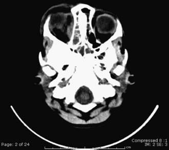
Inflammation of the lids and periorbital tissues without signs of true orbital involvement (such as proptosis or limitation of eye movement) is generally referred to as periorbital or preseptal cellulitis and is a form of facial cellulitis. This is common in young children and may be caused by bacteremia, trauma, an infected wound, or an abscess of the lid or periorbital region (pyoderma, hordeolum, conjunctivitis, dacryocystitis, insect bite). Patients present with eyelid swelling; the edema may be so intense as to make it difficult to evaluate the globe. Prior to the Haemophilus influenzae type B vaccine, the most common cause of pediatric preseptal (facial) cellulitis was bacteremia due to H. influenzae type B. Group A streptococcal infection a common is cause. Clinical examination shows lack of proptosis, normal ocular movement, and normal pupil function. CT examination demonstrates edema of the lids and subcutaneous tissues anterior to the orbital septum (Fig. 626-1). Antibiotic therapy and careful monitoring for signs of sepsis and local progression are essential.
Orbital Cellulitis
Orbital cellulitis can follow direct infection of the orbit from a wound, metastatic deposition of organisms during bacteremia, or more often direct extension or venous spread of infection from contiguous sites such as the lids, conjunctiva, globe, lacrimal gland, nasolacrimal sac, or more commonly the paranasal (ethmoid) sinuses (Table 626-1). In some cases, primary or metastatic tumor in the orbit can produce the clinical picture of orbital cellulitis. The most common cause of orbital cellulitis in children is paranasal sinusitis. The spread of infection to the orbit from the sinuses is more prevalent in children because of their thinner bony septa and sinus wall, greater porosity of bones, open suture lines, and larger vascular foramina. The spread of infection is also facilitated by the venous and lymphatic communication between the sinuses and surrounding structures, which allow flow in either direction, facilitating retrograde thrombophlebitis. Common pathogenic organisms include Staphylococcus species, including methicillin-resistant S. aureus (MRSA), Streptococcus species, and Haemophilus species.
Table 626-1 CHANDLER CLASSIFICATION OF ORBITAL COMPLICATIONS OF SINUSITIS, A CLINICAL DESCRIPTION
| CHANDLER CLASS | STAGE | CLINICAL DESCRIPTION AND DEFINITION |
|---|---|---|
| I | Inflammatory edema | Eyelid edema and erythema |
| Normal extraocular movement | ||
| Normal visual acuity | ||
| II | Orbital cellulitis | Diffuse edema of orbital contents without discrete abscess formation |
| III | Subperiosteal abscess | Collection of purulent exudate* beneath periosteum of lamina papyracea |
| Displacement of globe downward/laterally | ||
| IV | Orbital abscess | Purulent collection within orbit* |
| Proptosis | ||
| Chemosis | ||
| Ophthalmoplegia | ||
| Decreased vision | ||
| V | Cavernous sinus thrombosis | Bilateral eye findings |
| Prostration | ||
| Meningismus |
* The radiographic correlation of a subperiosteal or orbital abscess seen with CT is a contrast-enhancing mass in the extraconal or intraconal space, possibly with areas of cavitation, because purulence cannot be determined with CT scanning.
From Rudloe TF, Harper M, Prabhu SP, et al: Acute periorbital infections: who needs emergent imaging? Pediatrics 125:e719–e726, 2010.
The potential for complications is great. Vision loss can occur secondary to an increase in orbital pressure that causes retinal artery occlusion or optic neuritis. This is more likely to occur in the presence of an orbital abscess. Extension of infection from the orbit into the cranial cavity can lead to cavernous sinus thrombosis or meningitis, epidural or subdural empyema, or brain abscesses (see Table 626-1).
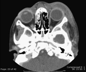
Orbital cellulitis must be recognized promptly and treated aggressively. Hospitalization and systemic antibiotic therapy are usually indicated. All patients require CT imaging of the orbit (including the surrounding CNS), preferably with intravenous contrast to detect a subperiosteal abscess, orbital abscess, or intracranial extension. Parenteral antibiotics must be started immediately. Antimicrobial agents that generally provide coverage for methicillin-sensitive S. aureus as well as aerobic and anaerobic sinus-derived bacteria include vancomycin and a third-generation cephalosporin (cefotaxime, ceftriaxone), and amoxicillin or ticarcillin combined with a β-lactamase inhibitor (clavulanic acid). Metronidazole may be administered in combination with an agent effective against aerobic or facultative streptococci and S. aureus if anaerobes are suspected or in the presence of intracranial extension. If there is no evidence of improvement or if there are signs of progression, sinus drainage may be required. An orbital or subperiosteal abscess (Fig. 626-2) can require urgent drainage of the orbit. The clinical presentation and course of each individual patient should dictate the need and timing of abscess drainage. The use of adjunctive corticosteroids and anticoagulation for cavernous venous thrombosis and or superior ophthalmic vein thrombosis is controversial.
Ambati BK, Ambati J, Azar N, et al. Periorbital and orbital cellulitis before and after the advent of Haemophilus influenzae type B vaccination. Ophthalmology. 2000;107:1450-1453.
Brook I. Microbiology and antimicrobial treatment of orbital and intracranial complications of sinusitis in children and their management. Int J Pediatr Otorhinolaryngol. 2009;73:1183-1186.
Campolattaro BN, Lueder GT, Tychsen L. Spectrum of pediatric dacryocystitis: medical and surgical management of 54 cases. J Pediatr Ophthalmol Strabismus. 1997;34:143-153.
Garcia GH, Harris GJ. Criteria for nonsurgical management of subperiosteal abscess of the orbit: analysis of outcomes 1988–1998. Ophthalmology. 2000;107:1454-1456.
Greenberg MF, Pollard ZF. Medical treatment of pediatric subperiosteal orbital abscess secondary to sinusitis. J AAPOS. 1998;2:351-355.
Nageswaran S, Woods CR, Benjamin DKJr, et al. Orbital cellulitis in children. Pediatr Infect Dis J. 2006;25:695-699.
Rudloe TF, Harper M, Prabhu SP, et al. Acute periorbital infections: who needs emergent imaging? Pediatrics. 2010;125:e719-e726.


