Omental Infarct

Published on 19/07/2015 by admin

Filed under Radiology

Last modified 22/04/2025

Print this page

rate 1 star rate 2 star rate 3 star rate 4 star rate 5 star
Your rating: none, Average: 3 (1 votes)

This article have been viewed 2627 times

 Heterogeneous, encapsulated mass located within omentum (usually in right lower quadrant)

– Surrounding inflammatory change and fat stranding ± small free fluid
image Variable attenuation, but usually foci of fat attenuation
image Usually well circumscribed with peripheral hyperdense rim 

– May appear ill defined as poorly marginated fat stranding without discrete mass in earliest stages
image No central dot sign (seen with epiploic appendagitis)
image Whorled pattern of vessels leading to infarct may reflect torsion of vessels feeding omentum
• Ultrasound appearance

image Hyperechoic, nonmobile, noncompressible fixed mass arising in omentum

– Decreased or absent flow within echogenic mass
image Focal tenderness with graded compression

PATHOLOGY

• Most cases idiopathic (obesity is predisposing factor)
• Other causes include abdominal surgery, trauma, incarcerated hernia, etc.

CLINICAL ISSUES

• Clinical presentation may mimic acute appendicitis

image Usually normal WBC and lack of nausea, vomiting, diarrhea, or constitutional symptoms
• Benign disorder almost never associated with complications
• Usually self-limiting process that resolves spontaneously and should be treated only with pain management
• If diagnosed prospectively on CT, surgery should not be performed
image
(Left) Axial CECT in a patient with abdominal pain demonstrates an ill-defined, fat-containing mass image in the omentum with a subtle peripheral rim, in keeping with an omental infarct. The patient’s pain resolved in a few days with conservative therapy.

image
(Right) Axial CECT demonstrates a well-circumscribed fatty mass image 
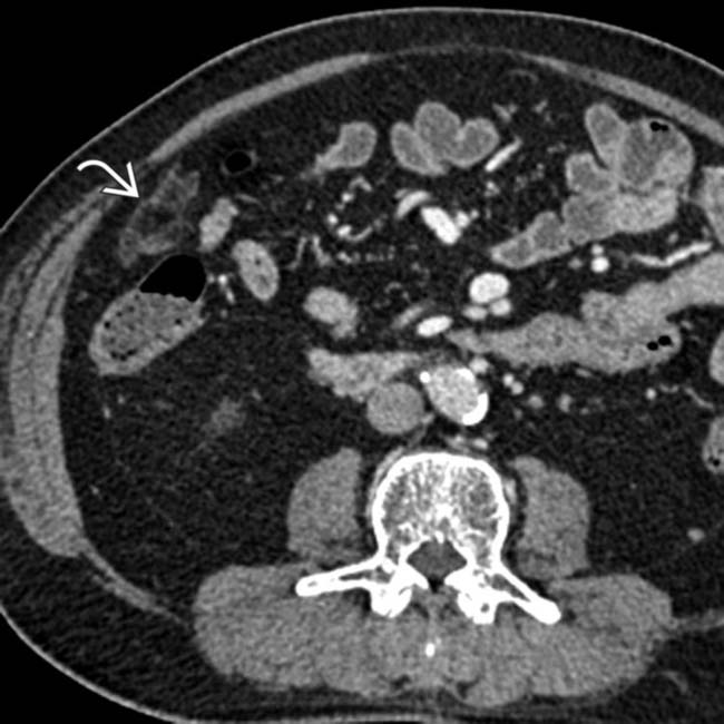
with a peripheral hyperdense rim in the right omentum. The patient had experienced RLQ pain about 1 week earlier, and this was thought to be a subacute omental infarct.
image
(Left) Axial CECT in a patient with abdominal pain shows a classic omental infarct as a fatty mass image 
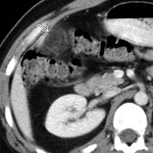
with a hyperdense rim adjacent to the ascending colon.

image
(Right) Axial CECT in the same patient demonstrates a “swirled” appearance of an omental vessel image 
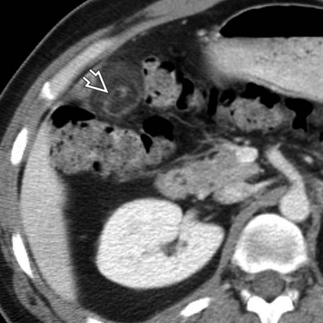
within the infarcted omentum, indicating twisting of the omental pedicle, which may be the etiology of the infarct in some cases.

TERMINOLOGY

Definitions

• Fat necrosis caused by interruption of arterial blood supply to omentum

IMAGING

General Features

• Best diagnostic clue

image Focal mass of heterogeneous density within omental fat with surrounding soft tissue inflammation
• Size

image Varies from 3.5-15 cm
image Larger infarcts often associated with surgery
• Morphology

image Focal heterogeneous mass composed of inflamed omental fat ± hemorrhage
image Usually well circumscribed, and appear triangular, ovoid, or cake-like in shape

CT Findings

• Heterogeneous, encapsulated mass located within omentum between anterior abdominal wall and colon

image Can have variable internal attenuation, but usually some internal foci of fat attenuation (-20 to -50 HU)
image Usually well-circumscribed margins with peripheral hyperdense rim, particularly when occurring after surgery

– Infarcts in earliest stages may appear ill defined: Sites of poorly marginated fat stranding without discrete mass
image Does not have central dot sign seen with epiploic appendagitis
image Whorled pattern of vessels leading to infarct may reflect torsion of vessels feeding omentum
• Often associated with surrounding inflammatory change and fat stranding ± small free fluid
• May be adherent to either colon or parietal peritoneum

image Only rarely causes reactive colonic wall thickening
image Rarely causes reactive thickening of overlying abdominal wall
• Usually located in right inferior omentum, especially when idiopathic

image Usually located adjacent to site of surgery when occurring in postoperative setting

Ultrasonographic Findings

• Grayscale ultrasound

image Echogenic, nonmobile, noncompressible fixed mass arising in omentum

– Associated with focal tenderness when graded compression applied to site
– Echogenic fat surrounding mass (reflects inflammation) ± small free fluid
• Color Doppler

image Decreased or absent flow within echogenic mass

Nuclear Medicine Findings

• PET/CT

image Omental infarcts may demonstrate FDG avidity suggesting tumor (if not correlated with CT appearance)

Imaging Recommendations

• Best imaging tool

image CECT
• Protocol advice

image 

DIFFERENTIAL DIAGNOSIS

Acute Appendicitis

• Can mimic omental infarction clinically, but distinction readily made with cross-sectional imaging
• Dilated appendix with wall thickening, mucosal hyperemia, and periappendiceal fat stranding, inflammation, and free fluid

image May demonstrate reactive wall thickening of cecum or terminal ileum
image Fluid collection, abscess, or ectopic gas in setting of perforation
image Appendicolith may be seen in 10-15% of cases
• Noncompressible appendix ≥ 7 mm on graded compression US often with increased color flow vascularity

Epiploic Appendagitis

• Benign and self-limited disorder: 1% of patients with acute right lower quadrant pain

image Most commonly caused by primary thrombosis or torsion of epiploic appendage, but rarely secondary to adjacent inflammation (diverticulitis, appendicitis, etc.)
• May appear similar to omental infarct, but distinction is irrelevant, as both entities treated conservatively
• CT: Small fatty mass with hyperattenuating ring abutting colon with mild adjacent fat stranding

image Central dot sign due to thrombosed vessel commonly present, not seen with omental infarcts
image May rarely cause reactive wall thickening of colon and thickening of parietal peritoneum
image Most common in left lower quadrant (rectosigmoid), unlike omental infarcts which occur in right lower quadrant

Liposarcoma

• Large omental infarcts can mimic fatty tumor (i.e., liposarcoma)
• Distinction based on clinical presentation (omental infarcts present with acute pain) and change over time (tumor grows over time while infarct should get smaller)
• Biopsy may be required in some rare cases

Pancreatitis With Extrapancreatic Fat Necrosis

• Focal or diffuse enlargement of pancreas with peripancreatic fluid and stranding ± fluid collections
• Release of pancreatic enzymes into surrounding tissues can cause extrapancreatic fat necrosis

image May appear focal and mimic omental infarct or tumor

Fibrosing/Sclerosing Mesenteritis

• Spiculated soft tissue mass usually found in root of small bowel mesentery

image May be associated with calcification, mimicking mesenteric carcinoid metastasis
• Often results in thickening, infiltration, displacement, and narrowing of adjacent bowel loops
• Usually located in left upper quadrant mesentery (not omentum)

Omental Torsion

• Due to omental cysts, hernias, tumors, adhesions
• Fibrous and fatty folds converging toward torsion
• Primary omental torsion due to anomaly

image Bifid
image Accessory omental tissue
• Secondary omental torsion

image More common than primary
image Postoperative adhesions &/or hernias
image Cysts, tumors

Peritoneal Carcinomatosis

• Peritoneal tumor implants usually appear more nodular or solid in appearance

image Early carcinomatosis may appear as ill-defined nodularity and stranding in omentum, potentially mimicking infarct
• Usually multifocal, whereas infarct is solitary
• Known history of primary malignancy (ovarian, gastrointestinal, etc.)

PATHOLOGY

General Features

• Etiology

image Rare entity due to abundant collateral vessels supplying omentum

– Omental infarcts tend to occur in right inferior omentum due to its more tenuous blood supply
– Right epiploic vessels involved in 90% of cases
– Hemorrhagic infarction with fat necrosis → inflammatory infiltrate → fibrosis and retraction → healing or autoamputation
image Most cases are idiopathic

– Obesity is known predisposing factor
image Multiple other secondary causes reported

– Abdominal surgery accounts for vast majority of cases with known cause
– Blunt trauma
– Incarcerated hernia
– Omental torsion (“twisting” of omentum with compromise of omental blood supply)

image Sometimes idiopathic with predisposition for children
image More commonly secondary to omental cysts, tumors, post-surgical scarring, or hernias
– Vigorous activity (reported in marathon runners)

image May result in “low flow” to omental blood vessels
– Vascular congestion, kinking of vessels (usually caused by sudden positional changes), and sudden increases in intraabdominal pressure
– Heart failure and digitalis
– Rarely superior mesenteric artery occlusion

Gross Pathologic & Surgical Features

• Infarcted omentum often adherent to parietal peritoneum or colon
• Serosanguineous fluid in peritoneal cavity

Microscopic Features

• Fat necrosis with inflammatory infiltrate

image Predominantly plasmocytic, lymphocytic, and histiocytic cells
• Collagenous scarring in chronic cases

CLINICAL ISSUES

Presentation

• Most common signs/symptoms

image Subacute right lower quadrant abdominal pain

– Right-sided in 90% of cases
image Mimics acute appendicitis or rarely acute cholecystitis
image Fever, nausea, vomiting, diarrhea, and other constitutional symptoms are uncommon (unlike appendicitis) but do rarely occur

– Children are more likely to exhibit constitutional symptoms than adults
• Other signs/symptoms

image Lab data

– WBC and ESR are normal or mildly elevated
image Physical examination

– Rebound tenderness in right lower quadrant ± palpable mass

Demographics

• Age

image Elderly obese people (85% of cases)
image Less common in children (15% of cases)
• Gender

image M:F = 2:1
• Epidemiology

image Very rare (1/250 as common as appendicitis)
image Adults (85%)
image Children (15%)

– Primary infarction more prevalent in young patients

Natural History & Prognosis

• Benign, self-limited disorder
• Complications (very rare)

image Abscess
image Adhesions with eventual development of bowel obstruction
• Prognosis

image Usually self-limiting process that resolves spontaneously in 1-4 months
• Surgery can be avoided if omental infarct prospectively differentiated from other entities (such as appendicitis) on imaging

Treatment

• Conservative management

image Pain management with NSAIDs
image If diagnosed prospectively on CT, surgery should not be performed
• Laparoscopic excision sometimes considered if patient mistakenly underwent laparoscopy for suspected appendicitis

image May be missed at laparoscopy unless there is careful evaluation of omentum
image Theoretically might reduce risk of developing adhesions and prevent future bowel obstruction

DIAGNOSTIC CHECKLIST

Consider

• Look carefully for omental infarction in patients with suspected appendicitis with normal appendix on CT

Image Interpretation Pearls

• Heterogeneous, encapsulated, fat-containing mass in right lower quadrant omentum with surrounding fat stranding, inflammation, and small free fluid

image
(Left) Axial CECT demonstrates a small, fat-containing mass image 
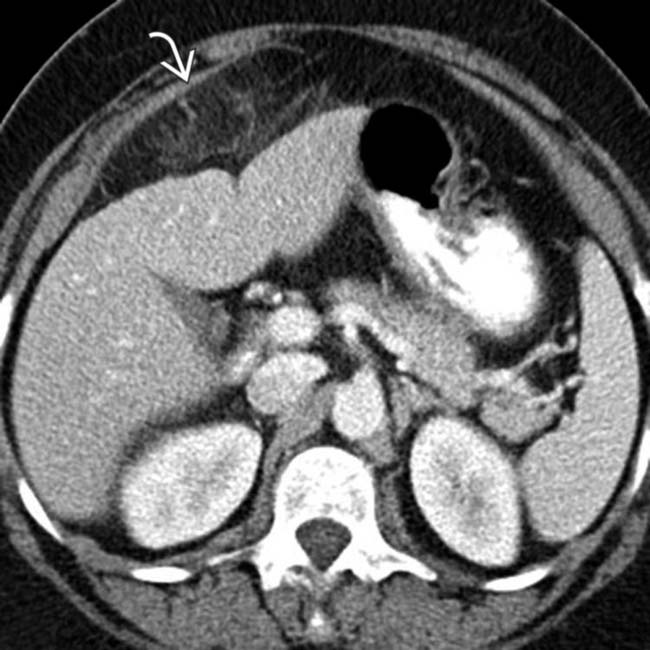
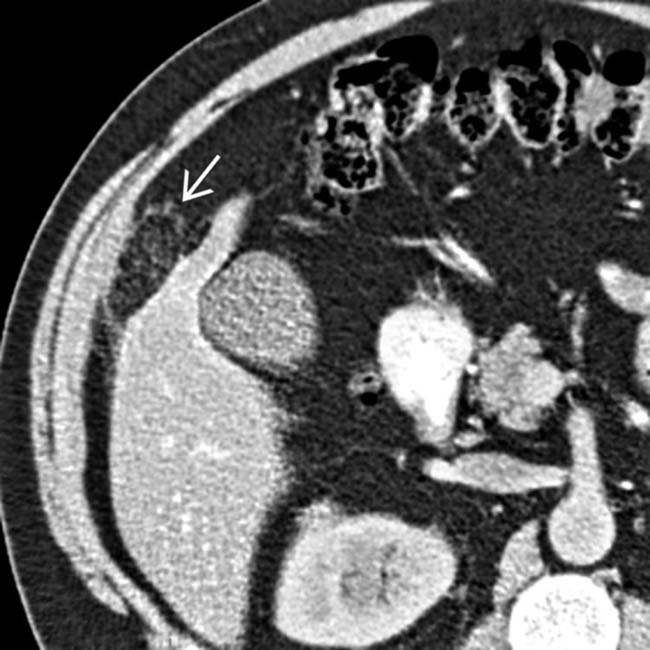
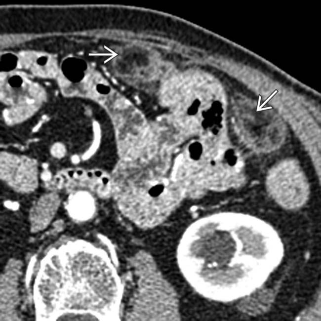
with a hyperdense rim and adjacent inflammation in the right lower quadrant (RLQ) omentum, a classic appearance and location for an omental infarct.
image
(Right) Coronal CECT in a patient after laparoscopic distal pancreatectomy demonstrates a large fat-containing mass image 
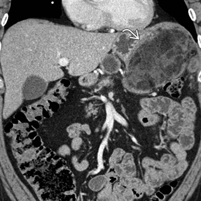
in the left upper quadrant omentum, representing a large omental infarct. Omental infarcts after surgery can be quite large and in proximity to the surgical bed.
image
(Left) Coronal CECT in a patient with pain shows an encapsulated fatty mass image 
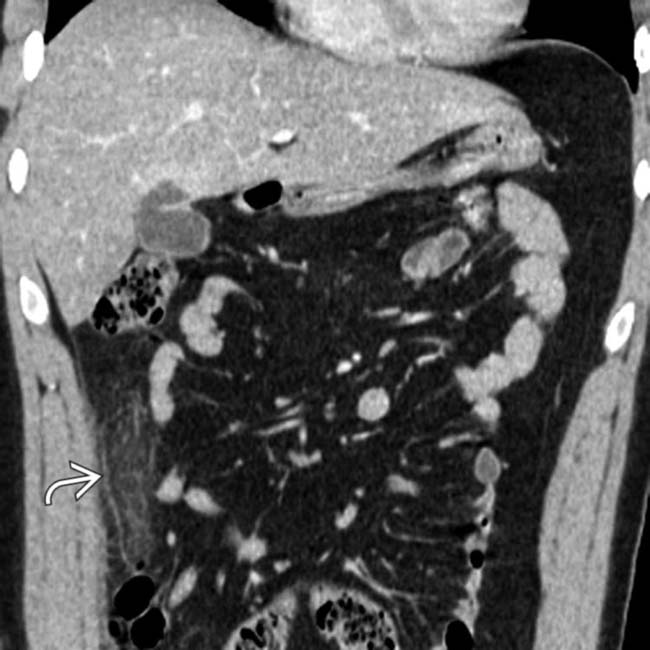
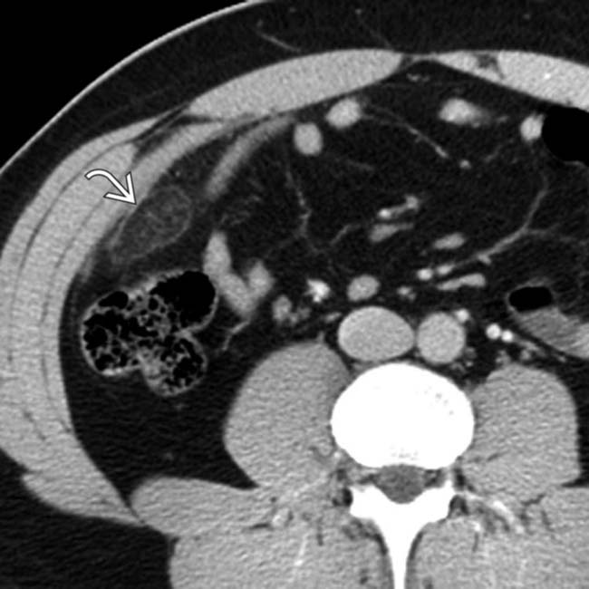
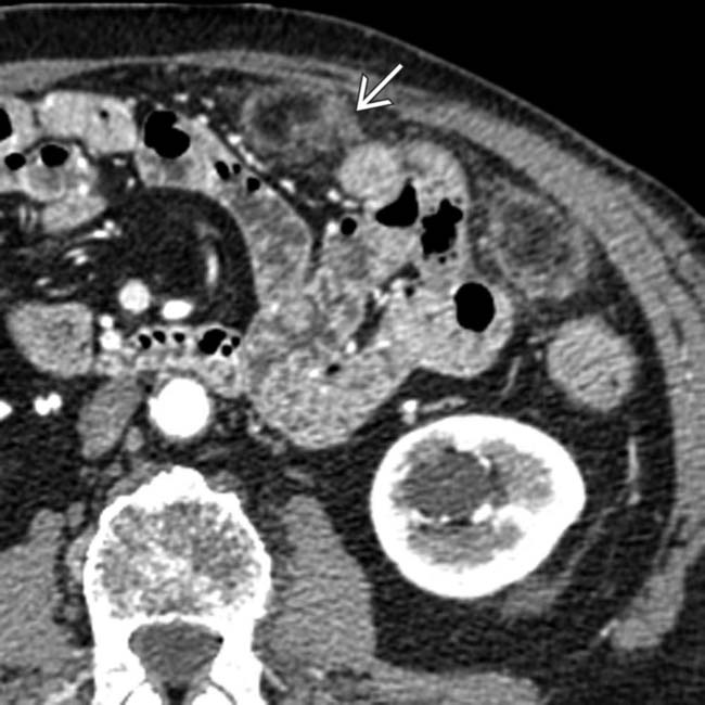
in the RLQ omentum with surrounding fat stranding, characteristic of an omental infarct.

image
(Right) Axial CECT in a patient who had recently undergone distal pancreatectomy shows a large circumscribed mass image 
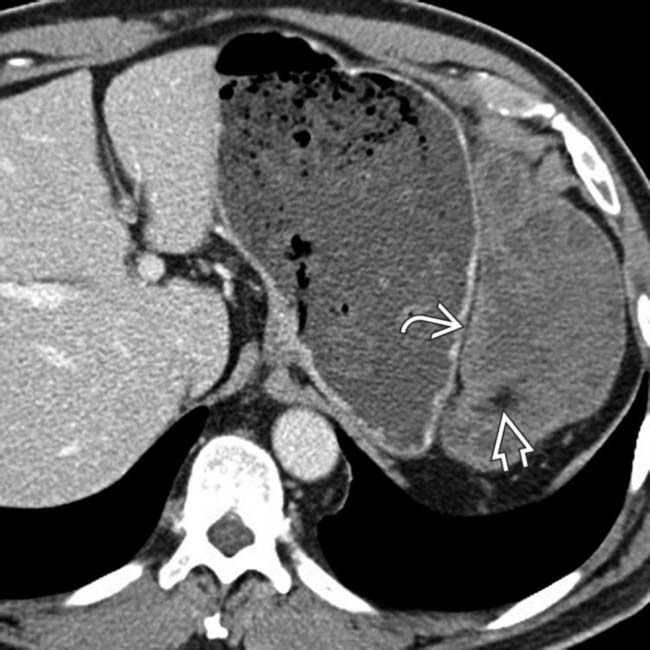
in the LUQ omentum. Note the presence of internal fat density image within the mass. Omental infarcts, as in this case, can be quite large and mimic a tumor (such as a liposarcoma) or carcinomatosis.
image
(Left) Axial CECT in a patient with polyarteritis nodosa and abdominal pain shows focal fat stranding image 
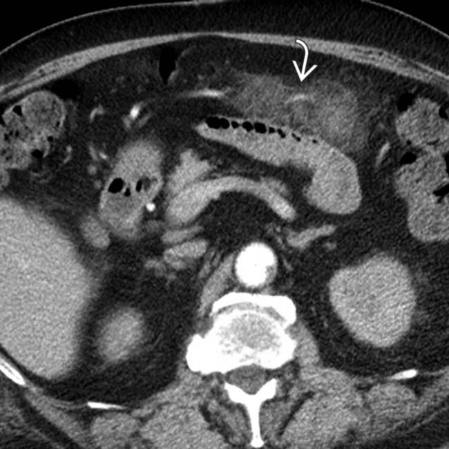
in the anterior omentum, corresponding to the site of the patient’s pain. This represents an omental infarct likely related to the vasculitis.

image
(Right) Axial CECT in an 11-year-old boy with RLQ pain shows a well-circumscribed, oval, fat-density mass image 
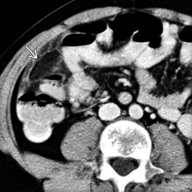
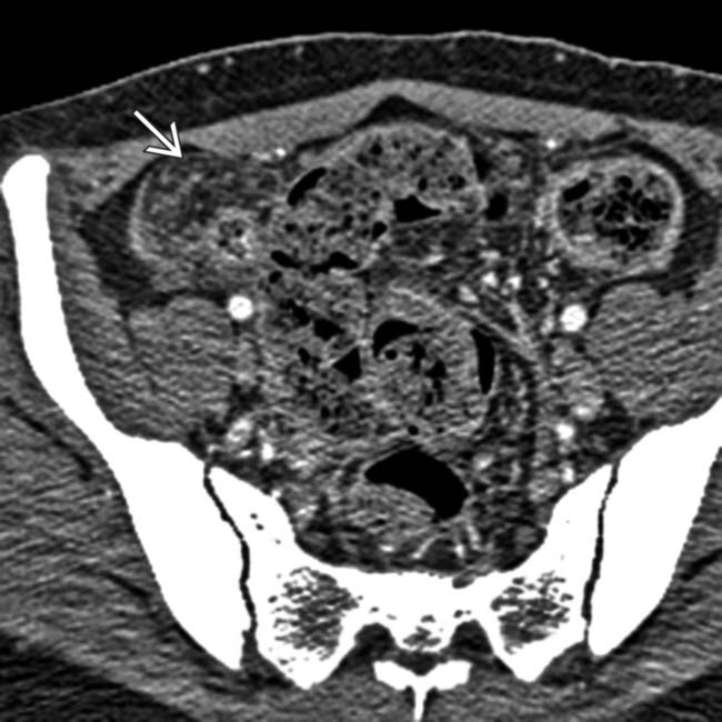
in the RLQ with adjacent fat stranding. The referring physician favored a diagnosis of appendicitis and opted for surgery, where an omental infarct was confirmed.
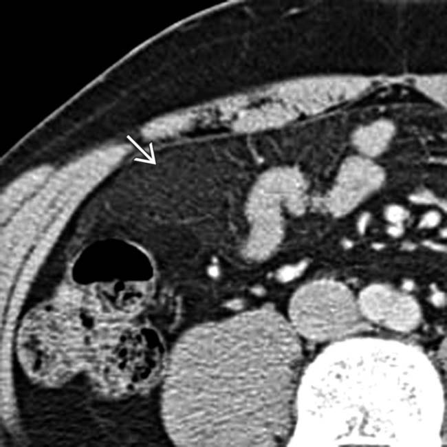
image
Axial CECT shows a very subtle omental infarct. Note the subtle edematous changes within the omental fat anterior to the right colon image.

image
Axial CECT shows omental infarct. Note the rounded mass-like area of edematous fat in the right lower quadrant image.
image
Axial CECT shows a classic right-side omental infarct. Note the oval, encapsulated, fat-containing lesion image 
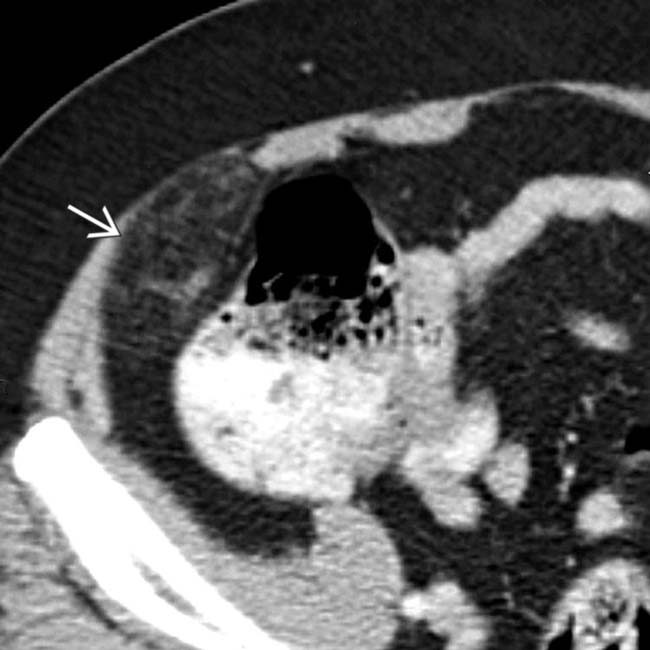
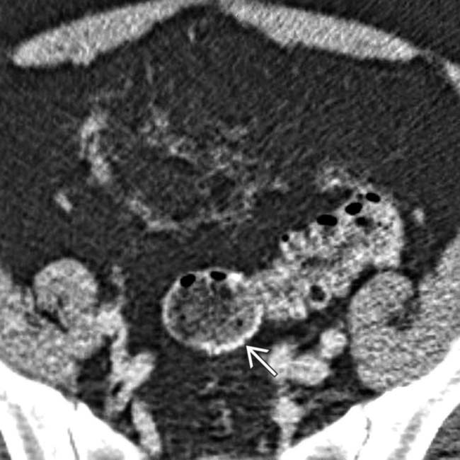
within the omentum overlying the hepatic flexure. This is the typical site and appearance for spontaneous omental infarction.
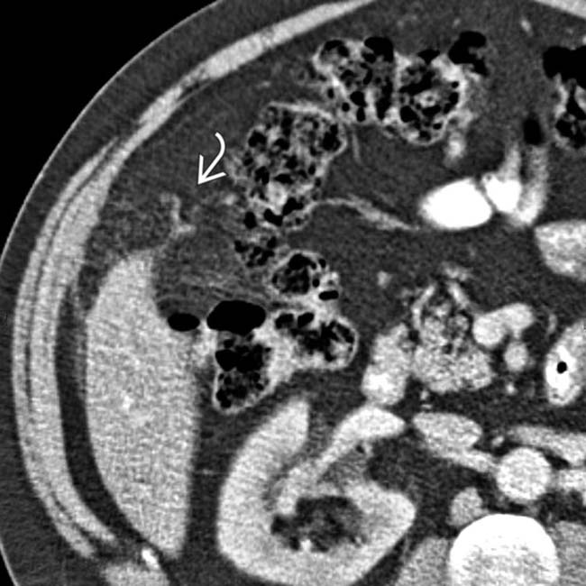
image
Axial CECT in the same patient shows slightly infiltrated fat within the lesion and within some of the surrounding omentum image.
image
Axial CECT shows the classic presentation and appearance of spontaneous omental infarction. Note the infiltrated ovoid fatty mass, which appears to be encapsulated image, overlying the ascending colon.
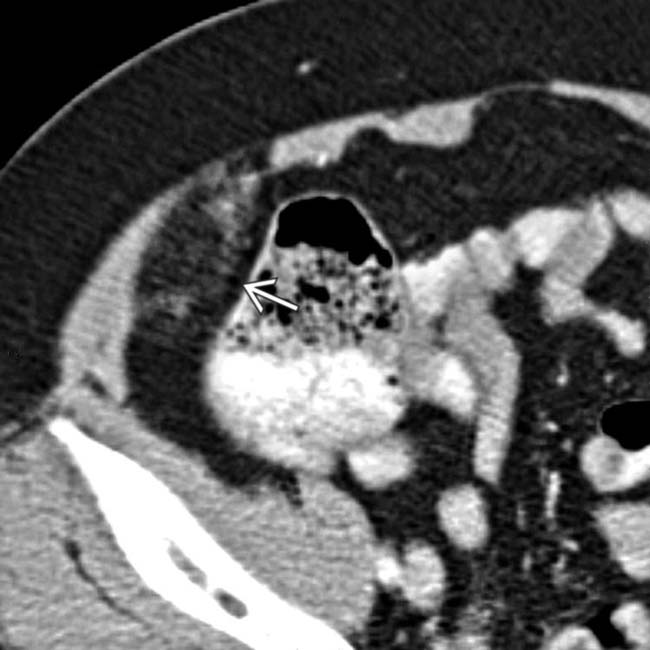
image
Axial CECT in the same patient demonstrates subtle mass effect on the cecum image.
image
Axial CECT shows postoperative fat necrosis and omental infarction following gastric surgery. Note 2 areas of edematous fat within the left omentum image.
image
Axial CECT in a higher plane of the same patient demonstrates soft tissue infiltration around areas of fat necrosis image.
image
Axial CECT shows infiltration and mass effect in the omentum adjacent to the sigmoid colon. Note the staple line image from prior sigmoid resection 2 months previously.
image
Axial CECT shows increased attenuation stranding in the omental fat image 
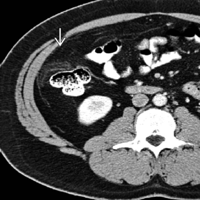
adjacent to the ascending colon. (Courtesy D. Green, MD.)
image
Axial NECT shows infiltration of omental fat image 
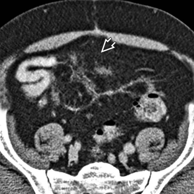
in a patient with an omental infarct. (Courtesy D. Avrin, MD.)
image
Axial CECT shows a heterogeneous mass of mixed fat density in the omentum image
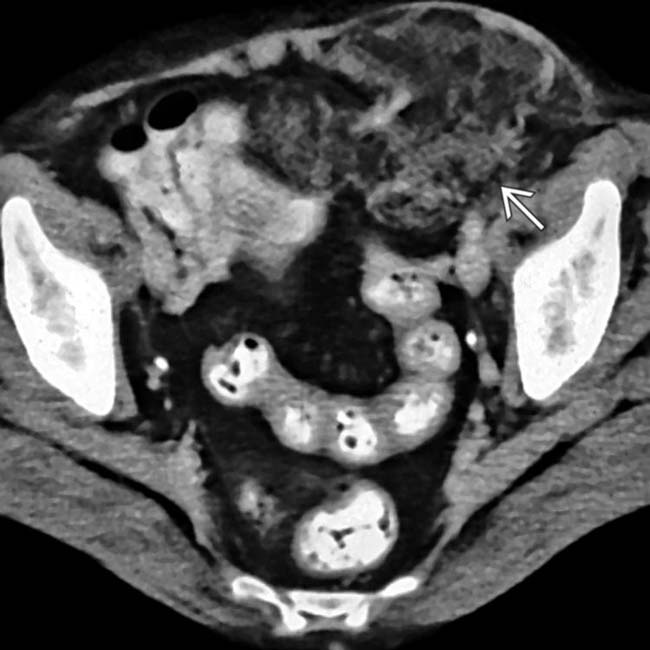
, displacing the anterior abdominal wall, representing a large omental infarct.
image
Axial CECT in a 5-year-old boy who presented with acute onset of abdominal pain with RUQ and epigastric tenderness demonstrates a mass of infiltrated fat image 
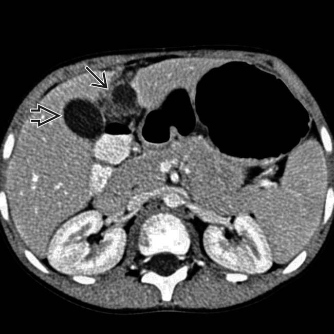
in the RUQ. The gallbladder image is normal.
image
Coronal CECT in the same patient is helpful in illustrating the relationship of the omental infarct image 
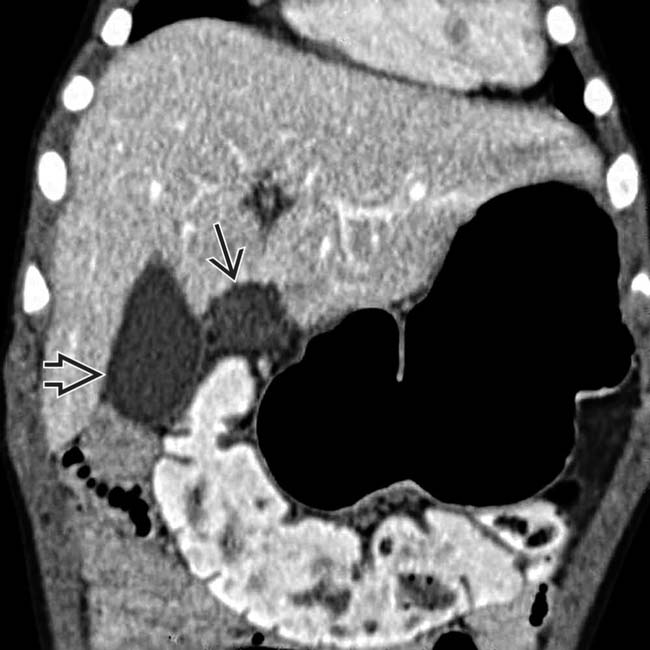
to the ascending colon, which is a characteristic feature. The gallbladder image is again clearly visualized as normal.
image
Axial CECT in a 54-year-old woman who presented with acute right abdominal pain illustrates a classic omental infarct image 
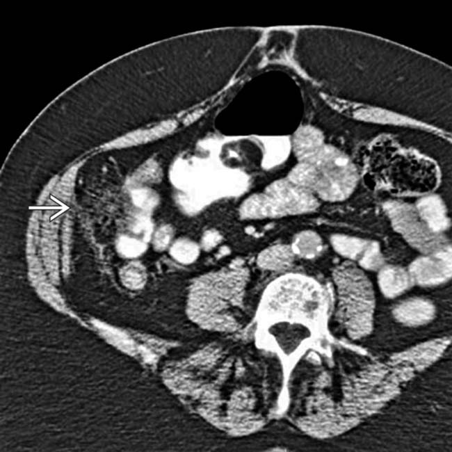
as a ball of “dirty fat” lying adjacent to the ascending colon. Appropriately, this patient was managed without surgery.
image
Coronal NECT in a 39-year-old woman presenting with acute right-sided abdominal pain shows the classic omental infarct image 
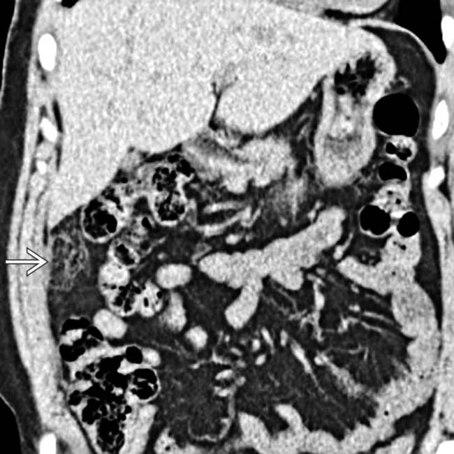
as a ball of “dirty fat” adjacent to the ascending colon.

SELECTED REFERENCES

1. Patlas, MN, et al. Cross-sectional imaging of nontraumatic peritoneal and mesenteric emergencies. Can Assoc Radiol J. 2013; 64(2):148–153.

Ryan, J, et al. Education and imaging: gastrointestinal: omental infarction. J Gastroenterol Hepatol. 2013; 28(2):378.

Kamaya, A, et al. Imaging manifestations of abdominal fat necrosis and its mimics. Radiographics. 2011; 31(7):2021–2034.

Kerr, SF, et al. Postoperative omental infarction following colonic resection. Clin Radiol. 2012; 67(2):134–139.

Itenberg, E, et al. Modern management of omental torsion and omental infarction: a surgeon’s perspective. J Surg Educ. 2010; 67(1):44–47.

Sasmal, PK, et al. Omental torsion and infarction: a diagnostic dilemma and its laparoscopic management. J Laparoendosc Adv Surg Tech A. 2010; 20(3):225–229.

Jeon, YS, et al. Is it from the mesentery or the omentum? MDCT features of various pathologic conditions in intraperitoneal fat planes. Surg Radiol Anat. 2009; 31(1):3–11.

Abdennasser el, K, et al. Omental torsion and infarction: CT appearance. Intern Med. 2008; 47(1):73–74.

Battaglia, L, et al. Simultaneous idiopathic segmental infarction of the great omentum and acute appendicitis: a rare association. World J Emerg Surg. 2008; 3:30.

Bestman, TJ, et al. An Unusual Complication after Roux-en-Y Gastric Bypass: Torsion and Infarction of the Divided Omentum. Obes Surg. 2008. [Epub ahead of print].

Cianci, R, et al. Idiopathic segmental infarction of the greater omentum diagnosed by unenhanced multidetector-row CT and treated successfully by laparoscopy. Emerg Radiol. 2008; 15(1):51–56.

Fu, CY, et al. Acute abdominal pain caused by spontaneous hemorrhagic infarction of a solitary plexiform neurofibroma of lesser omentum. Z Gastroenterol. 2008; 46(4):344–347.

Garg, AG, et al. Inflammatory fatty masses of the abdomen. Semin Ultrasound CT MR. 2008; 29(5):378–385.

Mihrshahi, S, et al. Primary idiopathic omental infarction in the adult. ANZ J Surg. 2008; 78(10):931–932.

Park, TU, et al. Omental infarction: Case series and review of the literature. J Emerg Med. 2008. [Epub ahead of print].

Ramos, CT, et al. Laparoscopic omentectomy for omental infarction after bicycle handlebar injury. J Laparoendosc Adv Surg Tech A. 2008; 18(2):327–329.

Rao, A, et al. Segmental omental infarction. Emerg Radiol. 2007; 14(3):195–197.

Coulier, B. Segmental omental infarction in childhood: a typical case diagnosed by CT allowing successful conservative treatment. Pediatr Radiol. 2006; 36(2):141–143.

Hirano, Y, et al. Left-sided omental torsion with inguinal hernia. World J Gastroenterol. 2006; 12(4):662–664.

Singh, AK, et al. Omental infarct: CT imaging features. Abdom Imaging. 2006.

Van Kerkhove, F, et al. Omental infarction in childhood. JBR-BTR. 2006; 89(4):198–200.

Bachar, GN, et al. Sonographic diagnosis of right segmental omental infarction. J Clin Ultrasound. 2005; 33(2):76–79.

Feo, CF, et al. Primary torsion of the greater omentum: a difficult diagnosis. Dig Dis Sci. 2005; 50(7):1283–1284.

Kerem, M, et al. Torsion of the greater omentum: preoperative computed tomographic diagnosis and therapeutic laparoscopy. JSLS. 2005; 9(4):494–496.

Loh, MH, et al. Omental infarction—a mimicker of acute appendicitis in children. J Pediatr Surg. 2005; 40(8):1224–1226.

Singh, AK, et al. Acute epiploic appendagitis and its mimics. Radiographics. 2005; 25(6):1521–1534.

van Breda Vriesman, AC, et al. Omental infarction: a self-limiting disease. AJR Am J Roentgenol. 2005; 185(1):280. [author reply 280-1].

Abadir, JS, et al. Accurate diagnosis of infarction of omentum and appendices epiploicae by computed tomography. Am Surg. 2004; 70(10):854–857.

Pereira, JM, et al. Disproportionate fat stranding: a helpful CT sign in patients with acute abdominal pain. Radiographics. 2004; 24(3):703–715.

Singh, AK, et al. Omental infarct: an unusual CT appearance after superior mesenteric artery occlusion. Emerg Radiol. 2004; 10(5):276–278.

Macari, M, et al. The acute right lower quadrant: CT evaluation. Radiol Clin North Am. 2003; 41(6):1117–1136.

Paroz, A, et al. Idiopathic segmental infarction of the greater omentum: a rare cause of acute abdomen. J Gastrointest Surg. 2003; 7(6):805–808.

Varjavandi, V, et al. Omental infarction: risk factors in children. J Pediatr Surg. 2003; 38(2):233–235.

Grattan-Smith, JD, et al. Omental infarction in pediatric patients: sonographic and CT findings. AJR Am J Roentgenol. 2002; 178(6):1537–1539.

McClure, MJ, et al. Radiological features of epiploic appendagitis and segmental omental infarction. Clin Radiol. 2001; 56(10):819–827.

Amin, Z, et al. A case of acute right-sided abdominal pain. Br J Radiol. 1999; 72(856):421–422.

Schlesinger, AE, et al. Sonographic appearance of omental infarction in children. Pediatr Radiol. 1999; 29(8):598–601.