Disorders of the contractile structures

Pain on resisted elevation
This, together with pain on active elevation, points to a lesion of one of the elevators of the shoulder – the levator scapulae or the trapezius muscle. Disorders of these structures are extremely rare.
If the pain is centrally located and is provoked by bilateral elevation of the arm, traction fracture of the spinous process of C7 and/or T1 is most likely. This presents as a stress fracture of one of the lower cervical spinous processes. The lesion should be suspected when bilateral limitation of about 90° on active elevation of the arm is found. Active elevation of both shoulders is also painful, as is resisted shoulder elevation. Passive elevation is painless. Movements of the neck are only slightly painful (see p. 237).
A stress fracture of the first rib is another possible cause. It gives rise to unilateral pain at the base of the neck not preceded by trauma. Active elevation of the arm cannot progress beyond the horizontal and is painful but passive elevation is normal. Pain is increased by active and resisted shoulder elevation and by active and passive side flexion of the neck to the opposite side. Resisted side flexion to the painful side is also painful (see p. 236).
Lesion of the levator scapulae muscle
Pain is felt unilaterally in the upper scapular area, mostly of the dominant shoulder.1 It is activity-related: neck movements as well as shoulder girdle or arm movements may be of influence. So are prolonged postures such as sitting with the head in slight flexion as happens during computer work.
On examination the pain is elicited during resisted elevation of the shoulder. The neck examination may be slightly positive. Palpation shows the lesion to lie at the insertion on the superior angle of the scapula.
The lesion usually lies at the tenoperiosteal insertion at the superior angle and the medial border of the scapula next to the superior angle and responds equally well to deep transverse friction or to infiltration with 20 mg of triamcinolone.
Lesion of the trapezius muscle
A muscular lesion in the trapezius is as uncommon as pain in the trapezius is common. Usually pain in that area is referred from the cervical spine: it is the most common localization of cervical dural pain (see p. 123). Because this phenomenon is often accompanied by extrasegmental tenderness, examination based on palpation will easily result in the wrong diagnosis of ‘trapezius syndrome’. Proper functional testing, however, usually turns out to be negative, showing that no focal lesion is present.
Muscular lesions do occur, however, but are very uncommon. They give rise to rather localized pain which can be provoked by testing against resistance: resisted elevation of the shoulder is painful, together with resisted neck extension and/or side flexion.
The lesion responds to local infiltration of a local anaesthetic or to deep friction.
Pain on resisted protraction
When the pain is felt anteriorly, this suggests that the lesion lies in the pectoralis minor or major muscle. Active protraction usually also elicits pain. When the pectoralis minor is at fault, resisted scapular depression is painful. If the lesion lies in the pectoralis major, resisted adduction and medial rotation of the arm is positive. Both lesions can be treated by procaine infiltrations or deep friction (see p. 260).
If the pain is located in the scapular area a lesion of the serratus anterior is probable.
Lesion of the serratus anterior muscle
On arm movement, the serratus anterior keeps the vertebral border of the scapula firmly attached to the thorax. In a lesion at its scapular insertion, unilateral scapular pain is felt on moving the arm. Because the pain is felt at the inner side of the scapula, the attention of the examiner is drawn to one or other structure in this area. Therefore it is discussed here, although it is likely to be encountered in the clinical examination of the shoulder.
Pain is felt on full active elevation of the arm, whereas passive elevation remains painless. Resisted abduction of the arm does not hurt. In this event, the patient is asked to bring the arm horizontal in front of the body. Resisted horizontal abduction is now performed in this position and elicits pain (Fig. 1).

Treatment is by deep transverse friction.
Technique: deep friction to the serratus anterior
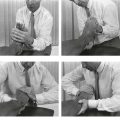
The patient lies prone, the ipsilateral arm in full medial rotation, the elbow flexed to 90°, the hand lying on the back. The therapist stands at the side and brings the arm further into medial rotation by using the forearm which lifts the medial edge of the scapula off the thorax. The thumb is now placed at the anterior aspect of the medial scapular margin. The other fingers are put on the posterior aspect of the scapula and provide counterpressure (Fig. 2). Massage is given along the scapular border by adduction–abduction movements of the arm, performed in a craniocaudal direction, for about 20 minutes, three times a week. Full cure is normally achieved in 3–5 weeks.

Pain on resisted approximation
If resisted approximation is painful but pain is not elicited on passive approximation, the lesion must lie in the rhomboid or in the trapezius. The response to infiltrations with procaine is good.
Weakness of scapular approximation
This is the result of neuritis of the spinal accessory nerve. Unilateral resisted approximation of the scapula is weak, together with a loss of about 5° of active elevation of the arm (see p. 524).
Pain on resisted depression
A positive result suggests that there is a problem in the subclavius, the pectoralis minor or the latissimus dorsi and is usually combined with pain on active and passive elevation, which demonstrates that one of the shoulder depressors is involved. A sprain of the subclavius is mostly likely. The patient complains of pain in the anterior claviculopectoral area. Alternatively, a sprain of the pectoralis minor may be responsible. Pain on resisted forward movement of the shoulder then establishes the diagnosis. When the latissimus dorsi is at fault, resisted adduction and medial rotation of the arm is also painful.
Lesion of the subclavius muscle
The patient complains of pain around the clavicle or in the upper pectoral area on certain activities or movements. Examination of the shoulder is negative which excludes the pectoralis major and the structures originating at the coracoid process (short head of biceps or coracobrachialis). Shoulder girdle examination shows the ‘contractile tissue pattern’: active and passive shoulder elevation is positive, as is resisted shoulder depression. On palpation a tender spot is found in the muscle belly of the subclavius.
Treatment of the subclavius is by deep transverse friction. The lesion usually does not respond to infiltration with procaine.
Technique: deep friction to the subclavius
The patient lies supine, the arm raised with the hand on the head. The therapist stands at the ipsilateral side and puts the middle finger, reinforced by the index, just below and parallel to the clavicle on the painful spot (Fig. 3). Friction is given by pronation–supination movements of the arm. This is performed for about 20 minutes on alternate days. The patient is usually cured in 10 sessions.

A summary of shoulder girdle disorders is outlined in Box 1.