Hernia most commonly contains loop of Ileum, although can rarely involve other pelvic viscera (i.e., bladder)

 May also be located between superior and middle fasciculi of obturator externus or between internal and external obturator muscles


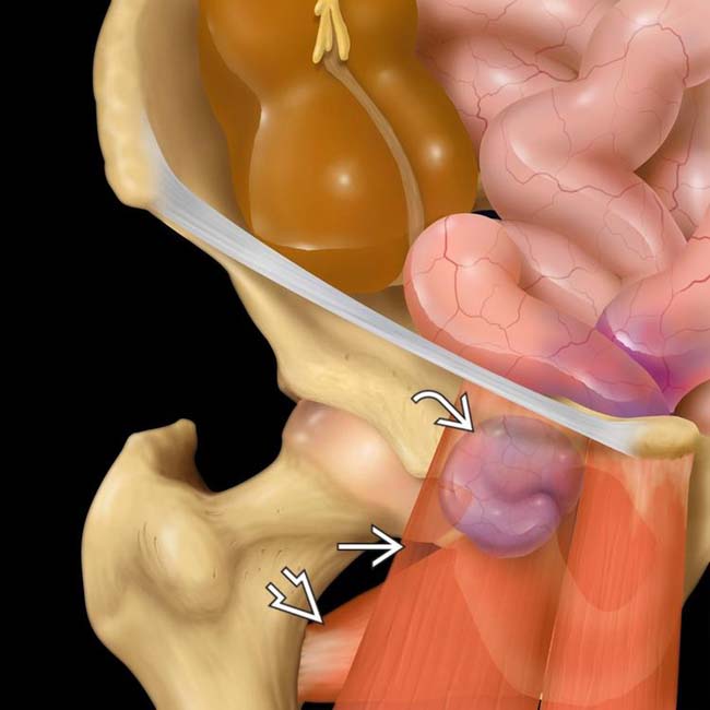
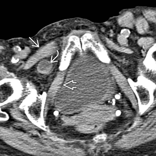
 lies deep to the pectineus muscle
  and superficial to the obturator externus muscle
  .
.
 . Obturator hernias most commonly contain herniated ileum, but other pelvic viscera can also herniate, as in this case.
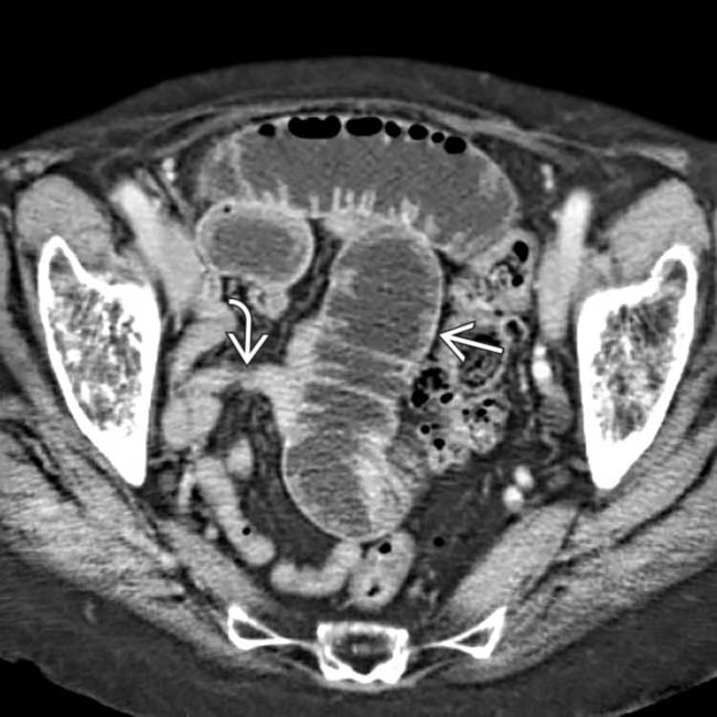
 and collapsed distal bowel
  .
.
 trapped between the obturator externus
  and the pectineus
  muscles. These are the classic imaging findings of an obturator hernia.


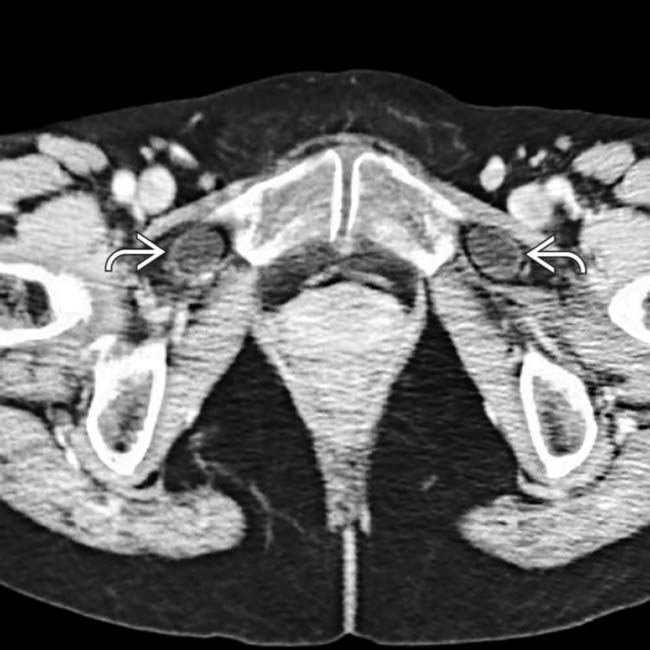
 lying between the pectineus
  and obturator muscles
  in a patient with a obturator hernia.
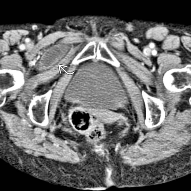
 between pectineus and obturator externus muscles.
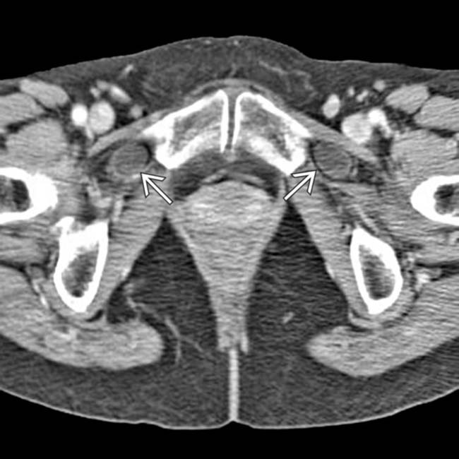
 . Bilateral obturator hernias are rare, accounting for only 6% of all cases.
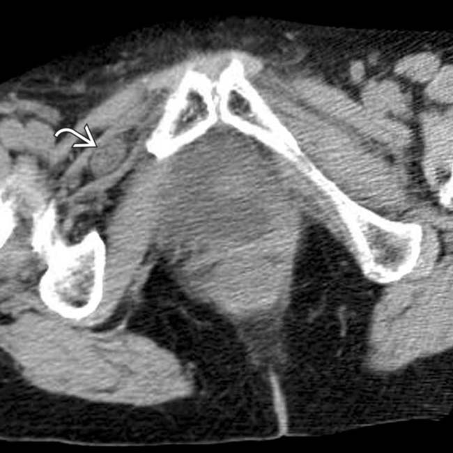
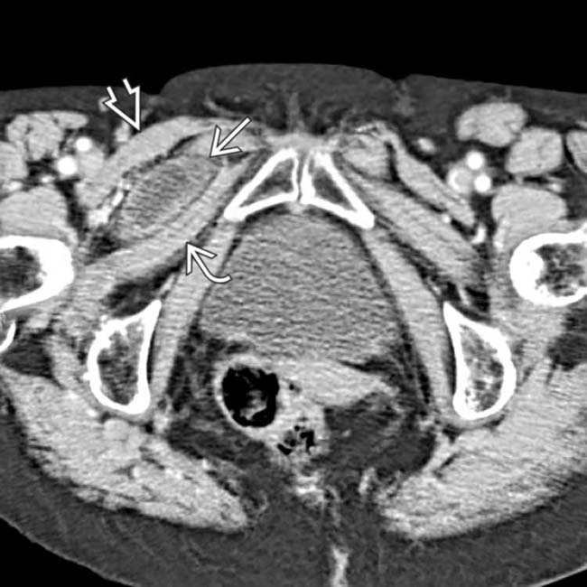
 entrapped within an obturator hernia.
 within the hernia. Only 1 wall of the bowel appears to be trapped.


