15 Obturator Block
Placement
Anatomy
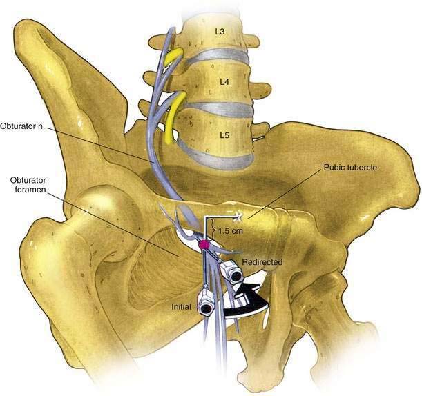
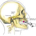
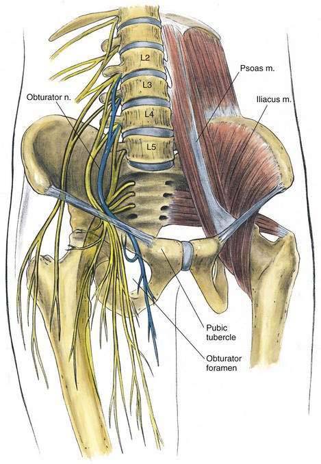
The obturator nerve emerges from the medial border of the psoas muscle at the pelvic brim and travels along the lateral aspect of the pelvis anterior to the obturator internus muscle and posterior to the iliac vessels and ureter. It enters the obturator canal cephalad and anterior to the obturator vessels, which are branches from the internal iliac vessels. In the obturator canal, the obturator nerve divides into anterior and posterior branches (Fig. 15-1). The anterior branch supplies the anterior adductor muscles and sends an articular branch to the hip joint and a cutaneous area on the medial aspect of the thigh. The posterior branch innervates the deep adductor muscles and sends an articular branch to the knee joint. In 10% of patients an accessory obturator nerve may be found.
Needle Puncture
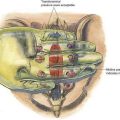
The pubic tubercle should be located and an “X” marked 1.5 cm caudad and 1.5 cm lateral to the tubercle (Fig. 15-2). The needle is inserted at this point, and at a depth of approximately 1.5 to 4 cm it contacts the horizontal ramus of the pubis. The needle is then withdrawn, redirected laterally in a horizontal plane, and inserted 2 to 3 cm deeper than the depth of the initial contact with bone. The needle tip now lies within the obturator canal (see Fig. 15-2) With the needle in this position, 10 to 15 mL of local anesthetic solution is injected while the needle is advanced and withdrawn slightly to ensure development of a “wall” of local anesthetic in the canal.