Normally Sterile Body Fluids, Bone and Bone Marrow, and Solid Tissues

Published on 08/02/2015 by admin

Filed under Basic Science

Last modified 22/04/2025

Print this page

rate 1 star rate 2 star rate 3 star rate 4 star rate 5 star
Your rating: none, Average: 0 (0 votes)

This article have been viewed 8937 times

Normally Sterile Body Fluids, Bone and Bone Marrow, and Solid Tissues

Objectives

1. Describe the fine main cavities of the human body; also name the membranes associated with these cavities and state the function of these membranes.

2. Define each of the following body cavity fluids, and explain the diagnostic culture methods for each: pleural fluid, pericardial fluid, peritoneal fluid, joint fluid, and dialysis fluid.

3. Define parietal and visceral pleura.

4. Define cellulitis; name the etiologic agents of this illness, and explain the associated risk factors for the development of disease.

5. Define pleural effusion; explain the difference between exudative pleural effusion and transudative pleural effusion.

6. Explain when a pleural effusion becomes an empyema and what medical condition contributes to the development of an empyema?

7. Define pericarditis and myocarditis; explain the physical conditions that may contribute to the accumulation of pericardial fluid.

8. Define peritonitis and differentiate between primary and secondary peritonitis.

9. Name the etiologic agents most commonly isolated from primary peritonitis cases in children, adults, sexually active females, and immunocompromised patients.

10. Define osteomyelitis; explain how this infection is transmitted, the diagnostic method, and the organisms most frequently responsible for this type of infection.

11. Explain the process for culturing organisms from the following specimens: bone, tissue, and bone marrow.

12. Correlate patient signs and symptoms with laboratory results to identify the etiologic agent associated with the body fluid, bone and bone marrow, and other solid tissue infection.

The human body is divided into five main body cavities: cranial, spinal, thoracic, abdominal, and pelvic. Each cavity is lined with membranes, and within the body wall and these membranes, or between the membranes and organs, are small spaces filled with minute amounts of fluid. The purpose of this fluid is to bathe the organs and membranes, reducing the friction between organs.

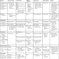
Bacteria, fungi, virus, or parasite can invade any body tissue or sterile body fluid site. Although from different areas of the body, all specimens discussed in this chapter are considered normally sterile. Therefore, even one colony of a potentially pathogenic microorganism may be significant. (Refer to Table 5-1 for a quick guide regarding collection, transport, and processing of specimens from sterile body sites.)

Specimens From Sterile Body Sites

Fluids

In response to infection, fluid may accumulate in any body cavity. Infected solid tissue often presents as cellulitis or with abscess formation. Areas of the body from which fluids are typically sent for microbiologic studies (in addition to blood and cerebrospinal fluid [see Chapters 68 and 71]) include those in Table 77-1.

TABLE 77-1

Microbiology Laboratory Body Fluid Collection Sites

Body Area Fluid Name(s)
Thorax Thoracentesis or pleural or empyema fluid
Abdominal cavity Paracentesis or ascitic or peritoneal fluid
Joint Synovial fluid
Pericardium Pericardial fluid

Pleural Fluid

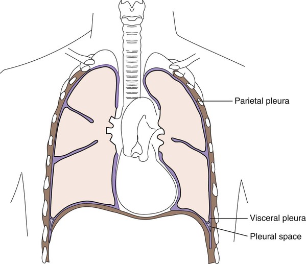
Lining the entire thoracic cavity (see Chapter 69) of the body is a serous membrane called the parietal pleura. Covering the outer surface of the lung is another membrane called the visceral pleura (Figure 77-1). Within the pleural space between the lung and chest wall is a small amount of fluid called pleural fluid that lubricates the surfaces of the pleura (the membranes surrounding the lungs and lining the chest cavity). Normally, equilibrium exists among the pleural membranes, but in certain disease states, such as cardiac, hepatic, or renal disease, excess amounts of this fluid can be produced and accumulates in the pleural space; this is known as a pleural effusion. Pleural effusions can either be exudative or transudative. Exudative pleural effusions are caused by inflammation, infection, and cancer, whereas transudative effusions are due to systemic changes, such as congestive heart failure.

Normal pleural fluid contains few or no cells and has a consistency similar to serum, but with a lower protein count. Pleural fluid containing numerous white blood cells is indicative of infections. Pleural fluid specimens are collected by thoracentesis, a procedure in which a needle is inserted through the chest wall into the pleural space and the excess fluid aspirated. This fluid is then submitted to the laboratory as thoracentesis fluid, pleural fluid, or empyema fluid. The fluid, or effusion, can then be analyzed for cell count, total protein, glucose, lactate dehydrogenase, amylase, cytology, and culture. The total protein and glucose results determine if the effusion is transudate or exudate. The patient’s serum or plasma glucose level is needed to compare with the results indicated in the body fluid. Several characteristics can be used to determine whether a fluid is a transudate or exudate (Table 77-2). When effusions are extremely purulent or full of pus, the effusion is referred to as an empyema. Empyema often arises as a complication of pneumonia, but other infections near the lung (e.g., subdiaphragmatic infection) may seed microorganisms into the pleural cavity. It has been estimated that 50% to 60% of patients develop empyema as a complication of pneumonia.

TABLE 77-2

Pleural Fluid Effusion Characteristics

  Transudate Exudate
Appearance Clear Cloudy
Specific Gravity <1.015 >1.015
Total Protein <3.0 mg/dL >3.0 mg/dL
LD Fluid: Serum Ratio <0.6 >0.6
Cholesterol <60 mg/dL >60 mg/dL
Cholesterol Fluid: Serum Ratio <0.3 >0.3
Bilirubin Fluid:Serum Ratio <0.6 >0.6
Total Protein Fluid: Serum Ratio <0.5 >0.6
White Blood Cells <1000/µL (all white blood cell types, all <50%) >1000/µL
Red Blood Cells <10,000/µL = because of traumatic tap >100,000/µL
Clotting Will not clot May clot

Modified from Strasinger SK, Di Lorenzo MS: Urinalysis and body fluids, ed 5, Philadelphia, 2008, F.A. Davis.

Peritoneal Fluid

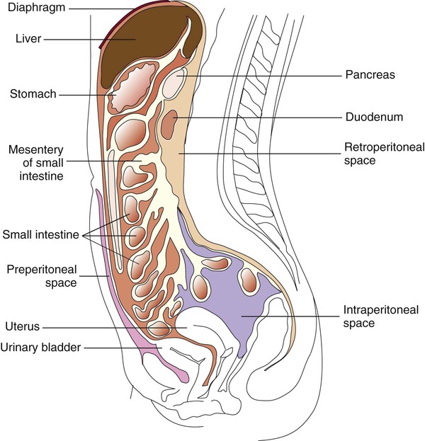
The peritoneum is a large, moist, continuous sheet of serous membrane lining the walls of the abdominal-pelvic cavity and the outer coat of the organs contained within the cavity (Figure 77-2). In the abdomen, these two membrane linings are separated by a space called the peritoneal cavity, which contains or abuts the liver, pancreas, spleen, stomach and intestinal tract, bladder, and fallopian tubes and ovaries. The kidneys occupy a retroperitoneal (behind the peritoneum) position. Within the healthy human peritoneal cavity is a small amount of fluid that maintains the surface moisture of the peritoneum. Normal peritoneal fluid may contain as many as 300 white blood cells per milliliter, but the protein content and specific gravity of the fluid are low. During an infectious or inflammatory process, increased amounts of fluid accumulate in the peritoneal cavity, a condition called ascites. Most cases of ascites are due to liver disease, and in severe cases, the abdomen is often distended. The fluid can be collected for testing by paracentesis (the insertion of a needle into the abdomen and removal of fluid). The peritoneal or ascites fluid can then be analyzed for amylase, protein, albumin, cell count, culture, and cytology. Often ascitic fluid contains an increased number of inflammatory cells and an elevated protein level.

Agents of infection gain access to the peritoneum through a perforation of the bowel, through infection within abdominal viscera, by way of the bloodstream, or by external inoculation (as in surgery or trauma). On occasion, as in pelvic inflammatory disease (PID), organisms travel through the natural channels of the fallopian tubes into the peritoneal cavity.

Primary Peritonitis.

Peritonitis results when the peritoneal membrane becomes inflamed and can be either primary or secondary. Primary peritonitis is rare and results when infection spreads from the blood and lymph nodes with no apparent evidence of infection. The organisms likely to be recovered from patient specimens with primary peritonitis vary with the patient’s age. The most common etiologic agents in children are Streptococcus pneumoniae and group A streptococci, Enterobacteriaceae, other gram-negative bacilli, and staphylococci. In adults, Escherichia coli is the most common bacterium, followed by S. pneumoniae and group A streptococci. Polymicrobic peritonitis is unusual in the absence of bowel perforation or rupture. Among sexually active young women, Neisseria gonorrhoeae and Chlamydia trachomatis are common etiologic agents of peritoneal infection, often in the form of a perihepatitis (inflammation of the surface of the liver, called Fitz-Hugh–Curtis syndrome). Tuberculous peritonitis occurs infrequently in the United States and is more likely to be found among individuals that have recently traveled in South America, Southeast Asia, or Africa. Fungal causes of peritonitis are not common, but Candida spp. may be recovered from immunosuppressed patients and patients receiving prolonged antibacterial therapy.

Secondary Peritonitis.

Secondary peritonitis is a complication of a perforated viscus (organ), surgery, traumatic injury, loss of bowel wall integrity following a destructive disease (e.g., ulcerative colitis, ruptured appendix, carcinoma), obstruction, or a preceding infection (liver abscess, salpingitis, septicemia). The nature, location, and etiology of the underlying process govern the agents recovered from peritoneal fluid. With PID as the background, gonococci, anaerobes, or chlamydiae are isolated. With peritonitis or intra-abdominal abscess, anaerobes generally are found in peritoneal fluid, usually together with Enterobacteriaceae and enterococci or other streptococci. In patients whose bowel flora has been altered by antimicrobial agents, more resistant gram-negative bacilli and Staphylococcus aureus may be encountered. Because anaerobes outnumber aerobes in the bowel by 1000-fold, it is not surprising that anaerobic organisms play a prominent role in intra-abdominal infection, perhaps acting synergistically with facultative bacteria. The organisms likely to be recovered include E. coli, the Bacteroides fragilis group, enterococci and other streptococci, Bilophila spp., other anaerobic gram-negative bacilli, anaerobic gram-positive cocci, and clostridia.

Peritoneal Dialysis Fluid

More than 900,000 patients with end-stage renal disease are maintained on continuous ambulatory peritoneal dialysis (CAPD). One in every 10 American adults, totaling more than 20 million, suffer from some type of chronic kidney disease. In this treatment, fluid is injected into the peritoneal cavity and subsequently removed, which allows exchange of salts and water and removal of various wastes in the absence of kidney function. Because the dialysate fluid is injected into the peritoneal cavity via a catheter, the break in the skin barrier places the dialysis patient at significant risk for infection. The average incidence of peritonitis in these patients is up to two episodes per year per patient. Peritonitis is diagnosed by the presence of two of the following: cloudy dialysate, abdominal pain, or a positive culture from dialysate. Although white blood cells are usually plentiful (a value of leukocytes >100/mL is usually indicative of infection), the number of organisms is usually too low for detection on Gram stain of the peritoneal fluid sediment unless a concentrating technique is used; fungi are more readily detected. Many recent studies show that improved sensitivity can be achieved by using automated blood culture systems in which 10 mL of fluid is inoculated into culture bottles.

Most infections originate from the patient’s own skin flora; Staphylococcus epidermidis and S. aureus are the most common etiologic agents, followed by streptococci, aerobic or facultative gram-negative bacilli, Candida spp., Corynebacterium spp., and others. The oxygen content of peritoneal dialysate is usually too high for the development of anaerobic infection. Among the gram-negative bacilli isolated, Pseudomonas spp., Acinetobacter spp., and the Enterobacteriaceae are frequently observed.

Pericardial Fluid

The heart and contiguous major blood vessels are surrounded by the pericardium, a protective tissue. The area between the epicardium, which is the membrane surrounding the heart muscle, and the pericardium is called the pericardial space and normally contains 15 to 20 mL of clear fluid. If an infectious agent is present within the fluid, the pericardium may become distended and tight, and eventually tamponade (interference with cardiac function and circulation) can ensue. Up to 500 mL of fluid can accumulate during infection, which may seriously complicate cardiac function.

Agents of pericarditis (inflammation of the pericardium) are usually viruses, especially Coxsackie virus. Parasites, bacteria, certain fungi, and noninfectious causes are also associated with this disease.

Myocarditis (inflammation of the heart muscle itself) may accompany or follow pericarditis. The pathogenesis of disease involves the host inflammatory response contributing to fluid buildup as well as cell and tissue damage. Common causes of myocarditis include viral infections with Coxsackie virus, echoviruses, or adenovirus. The most common etiologic agents of pericarditis and myocarditis are listed in Box 77-1. Other bacteria, fungi, and parasitic agents have been recovered from pericardial effusions.

Patients who develop pericarditis resulting from agents other than viruses are often immunocompromised or suffering from a chronic disease. An example is infective endocarditis, in which a myocardial abscess develops and then ruptures into the pericardial space.

Joint Fluid

Arthritis is an inflammation in a joint space. Infectious arthritis may involve any joint in the body. Infection of the joint usually occurs secondary to hematogenous spread of bacteria or, less often, fungi, as a direct extension of infection of the bone. It may also occur after injection of material, especially corticosteroids, into joints or after insertion of prosthetic material (e.g., total hip replacement). Although infectious arthritis usually occurs at a single site (monoarticular), a preexisting bacteremia or fungemia may seed more than one joint to establish polyarticular infection, particularly when multiple joints are diseased, such as in rheumatoid arthritis. In bacterial arthritis, the knees and hips are the most commonly affected joints in all age groups.

In addition to active infections associated with viable microorganisms within the joint, sterile, self-limited arthritis caused by antigen-antibody interactions may follow an episode of infection, such as meningococcal meningitis. When an etiologic agent cannot be isolated from an inflamed joint fluid specimen, either the absence of viable agents or inadequate transport or culturing procedures may be the cause. For example, even under the best circumstances, Borrelia burgdorferi is isolated from the joints of fewer than 20% of patients with Lyme disease. Nonspecific test results, such as increased white blood cell count, decreased glucose, or elevated protein, may indicate that an infectious agent is present but inconclusive.

Overall, Staphylococcus aureus is the most common etiologic agent of septic arthritis, accounting for approximately 70% of infections. In adults younger than 30 years of age, however, Neisseria gonorrhoeae is isolated most frequently. Haemophilus influenzae has been the most common agent of bacteremia in children younger than 2 years of age, and consequently it has been the most frequent cause of infectious arthritis in these patients, followed by S. aureus. The widespread use of H. influenzae type B vaccine should contribute to a change in this pattern. Streptococci, including groups A (Streptococcus pyogenes) and B (Streptococcus agalactiae), pneumococci, and viridans streptococci, are prominent among bacterial agents associated with infectious arthritis in patients of all ages. Among anaerobic bacteria, Bacteroides, including B. fragilis, may be recovered and Fusobacterium necrophorum, which usually involves more than one joint in the course of sepsis. Among people living in certain endemic areas of the United States and Europe, infectious arthritis is a prominent feature associated with Lyme disease. Chronic monoarticular arthritis is frequently due to mycobacteria, Nocardia asteroides, and fungi. Some of the more frequently encountered etiologic agents of infectious arthritis are listed in Box 77-2.

These agents act to stimulate a host inflammatory response, which is initially responsible for the pathology of the infection. Arthritis is also a symptom associated with infectious diseases caused by certain agents, such as Neisseria meningitidis, group A streptococci (rheumatic fever), and Streptobacillus moniliformis, in which the agent cannot be recovered from joint fluid. Presumably, antigen-antibody complexes formed during active infection accumulate in a joint, initiating an inflammatory response that is responsible for the ensuing damage.

Infections in prosthetic joints are usually associated with somewhat different etiologic agents than those in natural joints. After insertion of the prosthesis, organisms that gained access during the surgical procedure slowly multiply until they reach a critical mass and produce a host response. This may occur long after the initial surgery; approximately half of all prosthetic joint infections occur more than 1 year after surgery. Skin flora is the most common etiologic agent, with Staphylococcus epidermidis, other coagulase-negative staphylococci, Corynebacterium spp., and Propionibacterium spp. as the most common. However, Staphylococcus aureus is also a major pathogen in this infectious disease. Alternatively, organisms may reach joints during hematogenous spread from distant, infected sites.

Diagnosis of joint infections requires an aspiration of joint fluid for culture and microscopic examination. Inoculating the fluid directly into blood culture bottles may prevent the fluid from clotting. Some of the fluid may be Gram stained and inoculated onto blood as well as chocolate and anaerobic media. The use of AFB (acid fast bacteria) and fungal media must also be considered.

Bone

Bone Marrow Aspiration or Biopsy

Diagnosis of diseases, including brucellosis, histoplasmosis, blastomycosis, tuberculosis, and leishmaniasis, can sometimes be made by detection of the organisms in the bone marrow. Brucella spp. can be isolated on culture, as can fungi, but parasitic agents must be visualized in smears or sections made from bone marrow material. Many of the etiologic agents associated with disseminated infections in patients with human immunodeficiency virus (HIV) may be visualized or isolated from the bone marrow. Some of these organisms include cytomegalovirus, Cryptococcus neoformans, and Mycobacterium avium complex.

Bone Biopsy

A small piece of infected bone is occasionally sent to the microbiology laboratory to identify the etiologic agent of osteomyelitis (infection of bone). Patients develop osteomyelitis from hematogenous spread of an infectious agent, invasion of bone tissue from an adjacent site (e.g., joint infection, dental infection), breakdown of tissue caused by trauma or surgery, or lack of adequate circulation followed by colonization of a skin ulceration with microorganisms. Once established, infections in bone may progress toward chronicity, particularly if blood supply is insufficient in the affected area.

Staphylococcus aureus, seeded during bacteremia, is the most common etiologic agent of osteomyelitis among patients of all age groups. The toxins and enzymes produced by this bacterium, as well as its ability to adhere to smooth surfaces and produce a protective glycocalyx coating, seem to contribute to the organism’s pathogenicity. Osteomyelitis in younger patients is often associated with a single agent. Such infections are usually of hematogenous origin. Other organisms recovered from hematogenously acquired osteomyelitis include Salmonella spp., Haemophilus spp., Enterobacteriaceae, Pseudomonas spp., Fusobacterium necrophorum, and yeasts. S. aureus or P. aeruginosa is often recovered from cases in patients with drug addictions. Parasites or viruses are rarely, if ever, etiologic agents of osteomyelitis.

Bone biopsies from infections that have spread to a bone from a contiguous source or that are associated with poor circulation, especially in patients with diabetes, are likely to yield multiple isolates. Gram-negative bacilli are increasingly common among hospitalized patients; a break in the skin (surgery or intravenous line) may precede establishment of gram-negative osteomyelitis. Breaks in skin from other causes, such as a bite wound or trauma, also may be the initial event leading to underlying bone infection. For example, a human bite may lead to infection with Eikenella corrodens, whereas an animal bite may result in Pasteurella multocida osteomyelitis. Poor oral hygiene may lead to osteomyelitis of the jaw with Actinomyces spp., Capnocytophaga spp., and other oral flora, particularly anaerobes. Pigmented Prevotella and Porphyromonas, Fusobacterium, and Peptostreptococcus spp. are often involved. Pelvic infection in the female may result in a mixed aerobic and anaerobic osteomyelitis of the pubic bone.

Patients with neuropathy (pathologic changes in the peripheral nervous system) in the extremities, notably patients with diabetes, who may have poor circulation, may experience an unrecognized or notable trauma. They develop ulcers on the feet that do not heal, become infected, and may eventually progress to involve underlying bone. These infections are usually polymicrobial, involving anaerobic and aerobic bacteria. Prevotella or Porphyromonas, other gram-negative anaerobes, including the Bacteroides fragilis group, Peptostreptococcus spp., Staphylococcus aureus, and group A and other streptococci are frequently encountered.

Molecular testing, such as polymerase chain reaction, may be useful in determining the infectious organism associated with the patient’s condition when the laboratory is unable to recover the organism by traditional culture.

Solid Tissues

Pieces of tissue are removed from patients during surgical or needle biopsy procedures or may be collected at autopsy. Any agent of infection may cause disease in tissue, and laboratory practices should be adequate to recover bacteria, fungi, and viruses and detect the presence of parasites. Fastidious organisms (e.g., Brucella spp.) and agents of chronic disease (e.g., systemic fungi and mycobacteria) may require special media and long incubation periods for isolation. Some agents requiring special supportive or selective media are listed in Box 77-3.

Laboratory Diagnostic Procedures

Specimen Collection and Transport

Requirements for the collection and transport of specimens from sterile body sites vary because of the numerous types of specimens that can be collected and submitted to the laboratory for testing.

Fluids and Aspirates

Most specimens (pleural, peritoneal, pericardial, and synovial fluids) are collected by aspiration with a needle and syringe. Collecting pericardial fluid is not without risk to the patient because the sample is collected from the cavity immediately adjacent to the heart. Collection is performed by needle aspiration with electrocardiographic monitoring or as a surgical procedure. Laboratory personnel should be alerted in advance of the procedure, ensuring that the appropriate media, tissue culture media, and stain procedures are available immediately.

Body fluids from sterile sites should be transported to the laboratory in a sterile tube or airtight vial. From 1 to 5 mL of specimen is adequate for isolation of most bacteria, but the larger the specimen, the better, particularly for isolation of M. tuberculosis and fungi; at least 5 mL should be submitted for recovery of these organisms. Ten milliliters of fluid is recommended for the diagnosis of peritonitis. Anaerobic transport vials are available from several sources. These vials are prepared in an oxygen-free atmosphere and are sealed with a rubber septum or short stopper through which the fluid is injected. Transportation of fluid in a syringe capped with a sterile rubber stopper is not recommended. Most clinically significant anaerobic bacteria survive adequately in aerobic transport containers (e.g., sterile, screw-capped tubes) for short periods if the specimen is purulent and of adequate volume. However, collection in anaerobic transport media is recommended, and procedures vary in different laboratories. Specimens received in anaerobic transport vials should be inoculated to routine aerobic (an enriched broth, blood, chocolate, and sometimes MacConkey agar plates) and anaerobic media as quickly as possible. Specimens for recovery of fungi or mycobacteria may be transported in sterile, screw-capped tubes. At least 5 to 10 mL of fluid are required for adequate recovery of small numbers of organisms. If gonococci or chlamydia are suspected, additional aliquots should be sent to the laboratory for smears and appropriate cultures.

Percutaneous catheters are placed during many surgical procedures to prevent the accumulation of exudate and blood at the operative site. Often, the laboratory receives drainage fluids from these catheters for culture when signs and symptoms suggest infection. However, culture of such fluid is potentially misleading when the fluid becomes contaminated within the catheter or collection device, or when the fluid does not originate from a site of the infection. Everts and colleagues confirmed that direct aspiration of potentially infected fluid collections rather than catheter drainage fluid should be submitted for culture for the assessment of deep tissue infections in patients.

With respect to pericardial, pleural, synovial, and peritoneal fluids, the inoculation of blood culture broth bottles at the bedside or in the laboratory may be beneficial. An additional specimen should be submitted to the laboratory for a Gram stain. The specimen in the blood culture bottle is processed as a blood culture, facilitating the recovery of small numbers of organisms and diluting out the effects of antibiotics. Citrate or sodium polyanetholsulfonate (SPS) may be used as an anticoagulant. Specimens collected by percutaneous needle aspiration (paracentesis) or at the time of surgery should be inoculated into aerobic and anaerobic blood culture bottles immediately at the bedside.

Fluid from CAPD patients can be submitted to the laboratory in a sterile tube, urine cup, or the original bag. The bag is entered with a sterile needle and syringe to withdraw fluid for culture. Fluid should be directly inoculated into blood culture bottles (at least 20 mL [10 mL in each of two culture bottles]). Numerous studies indicate that in addition to blood culture bottles, an adult Isolator tube is a sensitive and specific method for culture.

Tissue

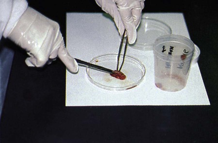
Tissue specimens are obtained following careful preparation of the skin. It is critical that biopsy specimens be collected aseptically and submitted to the microbiology laboratory in a sterile container. A wide-mouthed, screw-capped bottle or plastic container is recommended. Anaerobic organisms survive within infected tissue long enough to be recovered from culture. A small amount of sterile, nonbacteriostatic saline may be added to keep the specimen moist. Because homogenizing with a tissue grinder can destroy some organisms by the shearing forces generated during grinding, it is often best to use a sterile scissors and forceps to mince larger tissue specimens into small pieces suitable for culturing (Figure 77-3). Note that Legionella spp. may be inhibited by saline; a section of lung should be submitted without saline for Legionella isolation.

If anaerobic organisms are of concern, a small amount of tissue can be placed into a loosely capped, wide-mouthed plastic tube and sealed into an anaerobic pouch system, which also seals in moisture enough for survival of organisms in tissue until the specimen is plated. The surgeon should take responsibility for seeing that a second specimen is submitted to anatomic pathology for histologic studies. Formaldehyde-fixed tissue is not useful for recovery of viable microorganisms, although some organisms can be recovered after very short periods. Material from draining sinus tracts should include a portion of the tract’s wall obtained by deep curettage. Tissue from infective endocarditis should contain a portion of the valve and vegetation if the patient is undergoing valve replacement.

In some instances, contaminated material may be submitted for microbiologic examination. Specimens, such as tonsils or autopsy tissue, may be surface cauterized with a heated spatula or blanched by immersing in boiling water for 5 to 10 seconds to reduce surface contamination. The specimen may then be dissected with sterile instruments to permit culturing of the specimen’s center, which will not be affected by the heating. Alternatively, larger tissues may be cut in half with sterile scissors or a blade and the interior portion cultured for microbes.

Because surgical specimens are obtained at great risk and expense to the patient, and because supplementary specimens cannot be obtained easily, it is important that the laboratory save a portion of the original tissue (if enough material is available) in a small amount of sterile broth in the refrigerator and at –70° C (or, if necessary, at –20° C) for at least 4 weeks in case additional studies are indicated. If the entire tissue must be ground up for culture, a small amount of the suspension should be placed into a sterile tube and refrigerated.

Specimen Processing, Direct Examination, and Culture

Fluids and Aspirates

Techniques for laboratory processing of sterile body fluids are similar except for those previously discussed that are directly inoculated into blood culture bottles. Clear fluids may be concentrated by centrifugation or filtration, whereas purulent material can be inoculated directly to media. Any body fluid received in the laboratory that is already clotted must be homogenized to release trapped bacteria and minced or cut to release fungal cells. Either processing such specimens in a motorized tissue homogenizer or grinding them manually in a mortar and pestle or glass tissue grinder allows better recovery of bacteria. Hand grinding is often preferred, because motorized grinding can generate considerable heat and thereby kill microorganisms in the specimen. Grinding may lyse fungal elements; therefore, it is not recommended with specimens processed for fungi. Small amounts of whole material from a clot should be aseptically cut with a scalpel and placed directly onto media for isolation of fungi.

All fluids should be processed for direct microscopic examination. In general, if one organism is seen per oil immersion field, at least 105 organisms per milliliter of specimen are present. In such cases, often only a few organisms are present in normally sterile body fluids. Therefore, organisms must be concentrated in body fluids. For microscopic examination, cytocentrifugation (see Figure 71-4) should be used to prepare Gram-stained smears because organisms can be further concentrated up to 1000-fold. Body fluids should be concentrated by either filtration or high-speed centrifugation. Once the sample is concentrated, the supernatant is aseptically decanted or aspirated with a sterile pipette, leaving approximately 1 mL liquid in which to thoroughly mix the sediment. Vigorous vortexing or drawing the sediment up and down into a pipette several times is required to adequately suspend the sediment. This procedure should be done in a biologic safety cabinet. The suspension is used to inoculate media. Direct potassium hydroxide (KOH) or calcofluor white preparations for fungi and acid-fast stain for mycobacteria can also be performed. See Chapter 6 for detailed descriptions related to the preparation of smears for staining procedures.

Specimens for fungi should be examined by direct wet preparation or by preparing a separate smear for periodic acid-Schiff (PAS) staining in addition to Gram stain. Either 10% KOH or calcofluor white is recommended for visualization of fungal elements from a wet preparation. In addition to hyphal forms, material from the thoracic cavity may contain spherules of Coccidioides or budding yeast cells.

Lysis of leukocytes before concentration of CAPD effluents can significantly enhance recovery of organisms. Filtration of CAPD fluid through a 0.45-mm pore membrane filter allows a greater volume of fluid to be processed and usually yields better results. Because the numbers of infecting organisms may be low (fewer than 1 organism per 10 mL of fluid), a large quantity of fluid must be processed. Sediment obtained from at least 50 mL of fluid has been recommended. If the specimen is filtered, the filter should be cut aseptically into three pieces, one of which is placed on chocolate agar for incubation in 5% carbon dioxide, one on MacConkey agar, and the other on a blood agar plate for anaerobic incubation.

If fluids have been concentrated by centrifugation, the resulting sediment should be inoculated to an enrichment broth, blood, and chocolate agars. Because these specimens are from normally sterile sites, selective media are inadvisable because they may inhibit the growth of some organisms. Appropriate procedures for the isolation of anaerobes, mycobacteria, fungi, Chlamydia spp., and viruses should be used when such cultures are clinically indicated.

Bone

Clotted bone marrow aspirates or biopsies must be homogenized or ground to release trapped microorganisms. Specimens are inoculated to the same media as for other sterile body fluids. A special medium for enhancement of growth of Brucella spp. and incubation in 10% carbon dioxide may be needed. A portion of the specimen may be inoculated directly to fungal media. Sections are also made from biopsy material (bone) for fixation, staining, and examination (usually by anatomic pathologists) for the presence of mycobacterial, fungal, or parasitic agents. With respect to obtaining specimens from patients suspected of having osteomyelitis, cultures taken from open wound sites above infected bone or material taken from a draining sinus leading to an area of osteomyelitis may not reflect the actual etiologic agent of the underlying osteomyelitis. Cultures of samples of bone obtained during wound debridement surgery appear to be more useful for directing antibiotic therapy for better clinical outcome.

Diagnosis of prosthetic (artificial) joint infections is often difficult. Unfortunately, there is no universally accepted definition for the diagnosis of infection in the absence of microbiologic evidence because clinical symptoms such as pain do not differentiate infection from mechanical joint failure. There is no standardized approach to the laboratory diagnosis of these infections, and published data are conflicting. Further complicating the diagnosis is that the most common bacteria causing prosthesis infections are common skin contaminants such as coagulase-negative staphylococci. Some studies have reported that culture is relatively insensitive, possibly because of the organisms residing in biofilms, whereas polymerase chain reaction (PCR) assays were able to detect a majority of prosthetic joint infections. Atkins and colleagues recommended that five or six operative bone specimens be submitted for culture and that the cutoff for a definite diagnosis of infection be three or more of these specimens yielding the same organism. However, a recent study using PCR and culture using multiple media types and prolonged incubation found that appropriate culture was adequate to exclude bacterial infection in hip prostheses and PCR did not enhance diagnostic sensitivity for infection.

Normal bone is difficult to break up; however, most infected bone is soft and necrotic. Therefore, grinding the specimen in a mortar and pestle may break off some pieces. Small shavings from the most necrotic-looking areas of the bone specimen may sometimes be scraped off aseptically and inoculated to media. Pieces should be placed directly into media for recovery of fungi. Small bits of bone can be ground with sterile broth to form a suspension for bacteriologic and mycobacterial cultures. If anaerobes are to be recovered, all manipulations are best performed in an anaerobic chamber. If such an environment is unavailable, microbiologists should work quickly within a biosafety cabinet to inoculate prereduced anaerobic plates and broth with material from the bone.

Solid Tissue

Tissue should be manipulated in a laminar flow biologic safety cabinet. Processing tissue within an anaerobic chamber is even better. The microbiologist should cut through the infected area (which is often discolored) with a sterile scalpel blade. Half of the specimen can be used for fungal cultures and the other half for bacterial cultures. Both types of microbial agents should be considered in all tissue specimens. Some samples should also be sent to surgical pathology for histologic examination. Specimens should be cultured for viruses or acid-fast bacilli when requested. Material that is to be cultured for parasites should be finely minced or teased before inoculation into broth. Direct examination of stained tissue for parasites is often performed in the anatomic pathology lab. Imprint cultures of tissues may yield bacteriologic results identical to homogenates and may help differentiate microbial infection within the tissue’s center from surface colonization (growth only at the edge). Additional media can be inoculated for incubation at lower temperatures, which may facilitate recovery of certain systemic fungi and mycobacteria.

Tissue may also be inoculated to tissue culture cells for isolation of viruses. Brain, lung, spinal fluid, and blood are generally good specimens for viral isolation. Tissue may be examined by immunofluorescence for the presence of herpes simplex virus, varicella-zoster virus, cytomegalovirus, or rabies viral particles. Lung tissue should be examined by direct fluorescent antibody test for Legionella spp.

The tissues of all fetuses, premature infants, and babies who have died of an infectious process should be cultured for Listeria. Specimens of the brain, spinal fluid, blood, liver, and spleen are most likely to contain the organism. The isolation procedure may be reviewed in the reference by Seeliger and Cherry.