Nonneoplastic Pancreatic Cysts

Published on 19/07/2015 by admin

Filed under Radiology

Last modified 22/04/2025

Print this page

rate 1 star rate 2 star rate 3 star rate 4 star rate 5 star
Your rating: none, Average: 0 (0 votes)

This article have been viewed 1897 times

 Usually unilocular, well-defined cyst with sharp margin and thin imperceptible wall 

– Typically no internal complexity, septations, nodularity, or calcifications
– Usually no communication with pancreatic duct
– Usually single cyst but frequently multiple in patients with underlying syndrome
image Less commonly, imaging features can overlap with neoplastic pancreatic cysts, and lesions can demonstrate more complexity (multiloculation, calcifications, etc.)

– Lymphoepithelial cysts are more commonly complex (may have macroscopic fat) and can be intrapancreatic, abut pancreas, or appear exophytic

image May demonstrate signal loss on out-of-phase gradient-echo MR images (due to intracellular lipid)
image May be connected with pancreas by tiny, imperceptible stalk and appear exophytic or extrapancreatic

TOP DIFFERENTIAL DIAGNOSES

• Pancreatic pseudocyst
• Pancreatic intraductal papillary mucinous neoplasm (IPMN)
• Pancreatic serous cystadenoma
• Mucinous cystic neoplasm (MCN)
• Lymphangioma (mesenteric cyst)
• Cystic neuroendocrine tumor

PATHOLOGY

• Syndromes account for most nonneoplastic cysts diagnosed prospectively in clinical practice

image von Hippel-Lindau (VHL) disease, autosomal dominant polycystic kidney (ADPKD), and cystic fibrosis (CF)
• Isolated nonneoplastic cysts without a syndrome are far more rare in clinical practice

CLINICAL ISSUES

• ACR incidental findings committee suggests simple pancreatic cysts measuring ≤ 2 cm can be safely followed
• Simple pancreatic cysts in setting of a known syndrome (VHL, ADPKD, CF) are almost certainly benign
• Larger lesions or lesions with suspicious morphologic features often require EUS or cyst aspiration and consideration for surgical resection
image
(Left) Axial CECT in an asymptomatic patient demonstrates a simple-appearing, thin-walled cyst image 
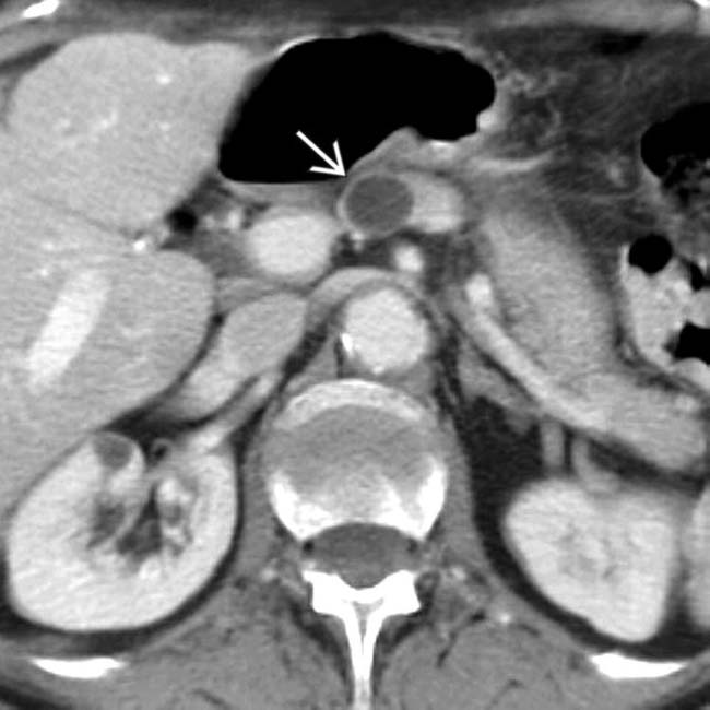
in the pancreatic neck.

image
(Right) Endoscopic ultrasound in the same patient shows a simple cyst image 
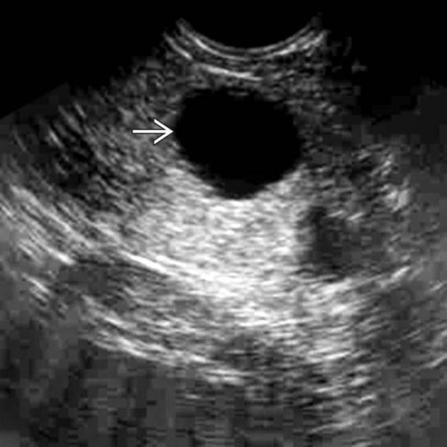
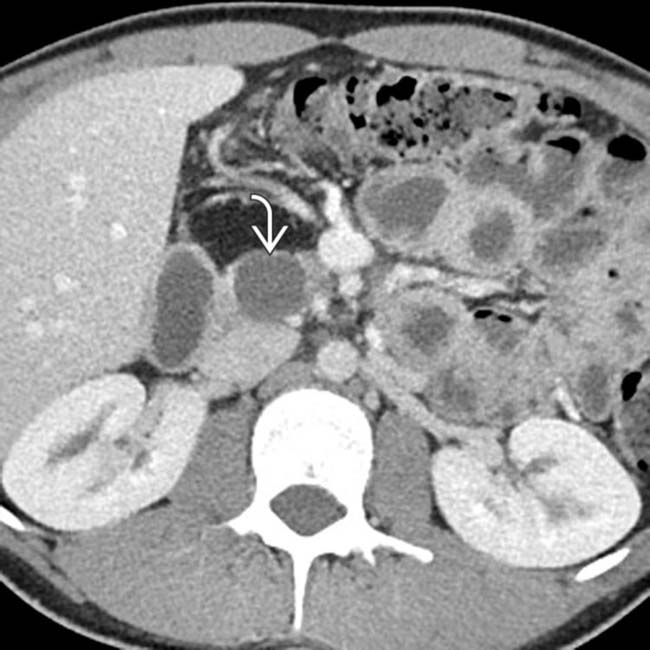
in the neck of the pancreas. There is no mural nodularity or other sign of complexity. Aspiration demonstrated clear serous fluid with no elevated tumor markers. It was elected to follow this cyst with sonography. It has remained stable for several years and is presumably a nonneoplastic simple cyst.
image
(Left) Axial CECT shows a small, simple-appearing cyst image 
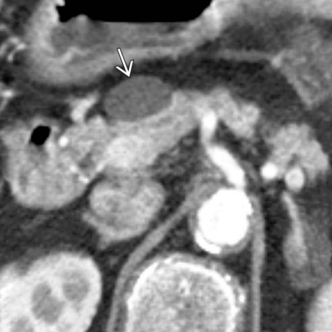
in the pancreatic head/neck. The wall of the cyst is imperceptible and there are no internal septations or other signs of complexity.

image
(Right) Endoscopic ultrasound in the same patient confirms the simple appearance of the cyst image
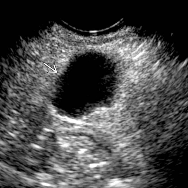
. Needle aspiration of the cyst demonstrated clear serous fluid with no malignant cells or elevated tumor markers, consistent with a nonneoplastic, simple cyst.

TERMINOLOGY

Synonyms

• Congenital, true, or epithelial pancreatic cyst

Definitions

• Group of nonneoplastic, noninflammatory, benign pancreatic cysts comprising several different histopathologic entities

IMAGING

General Features

• Best diagnostic clue

image Simple-appearing cyst with no septations or mural nodularity in a patient with no history of pancreatitis

– Consider strongly in patients with history of cystic fibrosis, autosomal dominant polycystic kidney disease (ADPKD), or von Hippel-Lindau (VHL)
• Size

image Usually quite small, although rarely can be much larger: Giant cysts as large as 15 cm in diameter reported
• Morphology

image Usually unilocular with round or oval shape, smooth thin wall, and absence of internal complexity
image Solitary or multiple (when associated with cystic syndromes)

CT Findings

• Imaging features can show some variability, since this category encompasses several histopathologically distinct types of nonneoplastic cysts

image Most nonneoplastic cysts are unilocular and well defined with a sharp margin and thin imperceptible wall
image Typically no internal complexity, septations, nodularity, or calcifications
image Usually no discernible communication with pancreatic duct
image Usually single isolated cyst, but often multiple in patients with underlying syndrome
• Less commonly, imaging features can overlap with neoplastic pancreatic cysts, and lesions can demonstrate more complexity (multiloculation, calcifications, etc.)

image Lymphoepithelial cysts have been described as more commonly demonstrating complexity (and even macroscopic fat) and may be either intrapancreatic, abut pancreas, or appear exophytic

– May be connected with pancreas by tiny, imperceptible stalk and appear exophytic or extrapancreatic
– Appear multilocular in 60% of cases

MR Findings

• Most nonneoplastic cysts are simple in appearance (hypointense on T1WI, hyperintense on T2WI, no enhancement or complexity)

image Lesions may demonstrate more complexity and be indistinguishable from a cystic neoplasm
image Lymphoepithelial cysts may demonstrate complexity and signal loss on out-of-phase gradient-echo images due to intracellular lipid
• Usually no communication with pancreatic duct on MRCP

image Rarely, some histopathologic subtypes of nonneoplastic cysts (i.e., retention cysts) may communicate with pancreatic duct

Ultrasonographic Findings

• Most often anechoic with no internal complexity or echoes

Radiographic Findings

• ERCP: Usually no communication between cyst and duct

Imaging Recommendations

• Best imaging tool

image CECT or MR followed by endoscopic US (EUS)

DIFFERENTIAL DIAGNOSIS

Pancreatic Pseudocyst

• More often complex in appearance with discrete wall
• Usually history or imaging stigmata of prior pancreatitis
• Much more common than nonneoplastic cysts, even in pediatric age group

Pancreatic Intraductal Papillary Mucinous Neoplasm (IPMN)

• Much more common than nonneoplastic cysts
• Side-branch IPMNs usually demonstrate communication with adjacent pancreatic duct
• If simple in appearance with no discernible communication to pancreatic duct, side-branch IPMN may not be distinguishable from nonneoplastic cyst

Pancreatic Serous Cystadenoma

• Classic appearance is honeycomb morphology with multiple (> 6) microcysts and central scar with calcification
• Unilocular or macrocystic variants of serous cystadenoma may be indistinguishable from true cysts except by histology
• Patients with VHL also have increased incidence of serous cystadenoma: Nonneoplastic cysts and serous cystadenoma may coexist

Mucinous Cystic Neoplasm (MCN)

• Complex multilocular cystic mass with a few macrocysts, usually in tail of pancreas

image Often will demonstrate thick wall, mural nodularity, or thick septations, particularly when malignant
• Unilocular, simple MCN may be indistinguishable from true cyst except by histology

Lymphangioma (Mesenteric Cyst)

• Multiseptated mesenteric or peripancreatic cystic mass abutting pancreas
• Often will have a “feathery” morphology on imaging with multiple locules
• Can be small and simple in appearance and might appear indistinguishable from primary nonneoplastic pancreatic cyst when located adjacent to pancreas

Cystic Neuroendocrine Tumor

• Usually demonstrates a surrounding rim of avid enhancement or hypervascular mural nodularity (most evident on arterial phase imaging)
• Patients with VHL also have increased incidence of neuroendocrine tumors: Nonneoplastic cysts and cystic neuroendocrine tumors may coexist

PATHOLOGY

General Features

• Syndromes account for most nonneoplastic cysts diagnosed prospectively in clinical practice 

image VHL
image ADPKD
image Beckwith-Wiedemann syndrome
image Cystic fibrosis
• Isolated nonneoplastic cysts without a syndrome are far more rare in clinical practice

Staging, Grading, & Classification

• While generally grouped together into a category of nonneoplastic pancreatic cysts, these cysts can be further subdivided into multiple distinct histopathologic subtypes

image Retention cysts: Cystic dilatation of pancreatic duct side branch due to intraluminal obstruction

– Can be congenital or due to obstruction of side branch by mucin, chronic pancreatitis, or calculi
– Communicate with pancreatic duct making differentiation from IPMN difficult
image “True” cysts: Lined by cuboidal epithelium and do not communicate with pancreatic duct

– Rare entity in adults, with only a few reports in literature, and more common in children
image Squamoid cysts: Lined by squamous epithelium
image Mucinous nonneoplastic cysts: Mucinous epithelium, no cellular atypia, and no communication with pancreatic duct

– Presence of mucin makes preoperative diagnosis with EUS cyst aspiration very difficult, and these lesions are almost always resected and diagnosed on postoperative histopathology
image Enterogenous cysts: Congenital cysts derived from foregut filled with serous and mucoid fluid
image Endometrial cysts: Extremely rare manifestation of endometriosis in pancreas
image Lymphoepithelial cysts:  Rare benign cysts that may be either intrapancreatic or extrapancreatic and are lined by squamous epithelium and layer of lymphoid tissue

– Filled with squamous material, lymphocytes, and keratinaceous debris but no atypia or neoplastic cells
– Usually occur in older males
image Acinar cell cystadenomas: Lined by benign acinar cells
image Pancreatic enteric duplication cysts: Rare and most common in children; can present with pancreatitis

Gross Pathologic & Surgical Features

• True epithelial lining (absent in pseudocysts)

CLINICAL ISSUES

Presentation

• Most common signs/symptoms

image Almost always an incidental finding in patients being imaged for other reasons
image Rarely symptomatic (pain, pancreatitis) due to size and mass effect
• Other signs/symptoms

image EUS-guided cyst aspiration yields fluid with low CEA, low amylase, and negative cytology

Demographics

• Epidemiology

image Nonneoplastic cysts are very rare, < 1% of all pancreatic cysts
• Lymphoepithelial cysts are most common in men between 5th and 6th decades
• Other types of nonneoplastic cysts vary in gender and age predispositions, but congenital nonneoplastic cysts can rarely be visualized in utero on prenatal US

Natural History & Prognosis

• While management of incidental pancreatic cysts is controversial and varies depending on individual specialty society, simple-appearing pancreatic cysts ≤ 2 cm in asymptomatic adults are rarely of any clinical significance

image ACR incidental findings committee in 2010 suggested simple pancreatic cysts measuring ≤ 2 cm could be safely followed
• Simple pancreatic cysts in setting of a known syndrome (VHL, cystic fibrosis, ADPKD) are almost certainly benign

image Careful attention must be paid in patients with VHL to differentiate nonneoplastic cysts from cystic neuroendocrine tumors

Treatment

• Most lesions can be safely followed with imaging surveillance
• Larger lesions or lesions with suspicious morphologic features often require EUS or cyst aspiration and consideration for surgical resection
• Nonneoplastic cysts are not uncommonly resected due to difficulty in differentiating them from cystic pancreatic neoplasms

DIAGNOSTIC CHECKLIST

Consider

• Nonneoplastic pancreatic cysts are much less common than both pancreatic pseudocysts and cystic pancreatic neoplasms
• Consider nonneoplastic cysts most strongly in patients with history of cystic fibrosis, ADPKD, Beckwith-Wiedemann, or VHL

Image Interpretation Pearls

• Based on imaging alone, nonneoplastic cysts cannot be reliably distinguished from simple-appearing neoplastic cysts (such as IPMNs)
• Endoscopic US with cyst aspiration can play a valuable role in risk stratifying pancreatic cysts with indeterminate imaging features
image
(Left) Axial CECT demonstrates innumerable tiny nonneoplastic cysts in the pancreas, as well as multiple left-sided renal cysts, a common constellation of findings in von Hippel-Lindau (VHL). VHL patients can often demonstrate diffuse cystic replacement of the pancreas.

image
(Right) Coronal MRCP with MIP reconstruction demonstrates extensive replacement of the entire pancreas with T2 bright cysts in a patient with VHL.
image
(Left) Axial CECT in a patient with VHL demonstrates an avidly enhancing left-sided renal cell carcinoma image
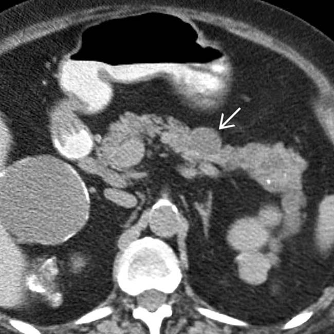
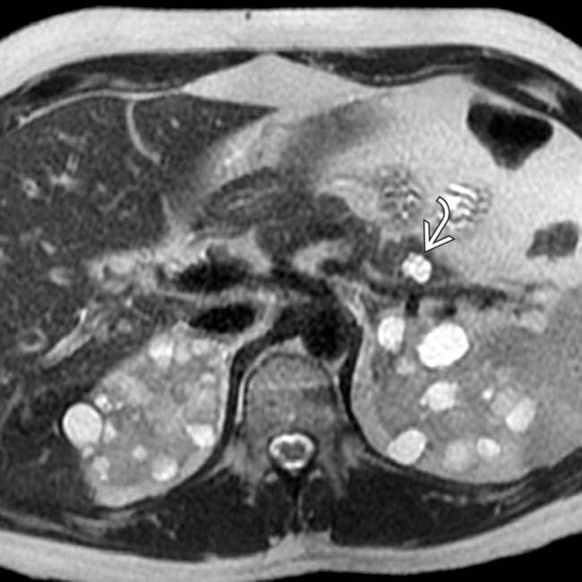
. The well-defined, simple-appearing cyst image in the pancreas almost certainly represents a nonneoplastic pancreatic cyst, very common in VHL patients.

image
(Right) Axial CECT in a patient with cystic fibrosis shows fatty replacement and pseudohypertrophy of the pancreas image
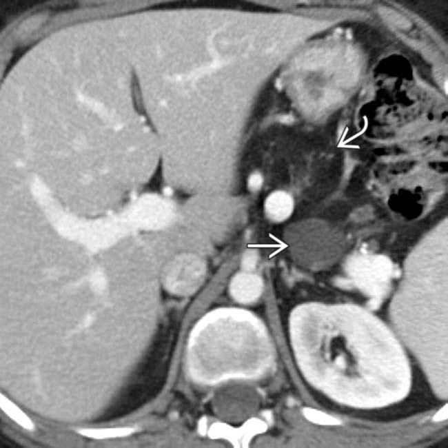
, characteristic of pancreatic involvement with this disease. Notice also the thin-walled retention cyst in the tail of the pancreas image.
image
(Left) Axial CECT in a patient with cystic fibrosis demonstrates a well-defined simple-appearing cyst image in the pancreatic head. Nonneoplastic cysts are a common finding in cystic fibrosis patients.

image
(Right) Axial CECT demonstrates a cyst in the pancreatic tail. Notice the internal complexity within the cyst, including possible mural nodularity image 
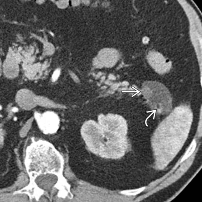
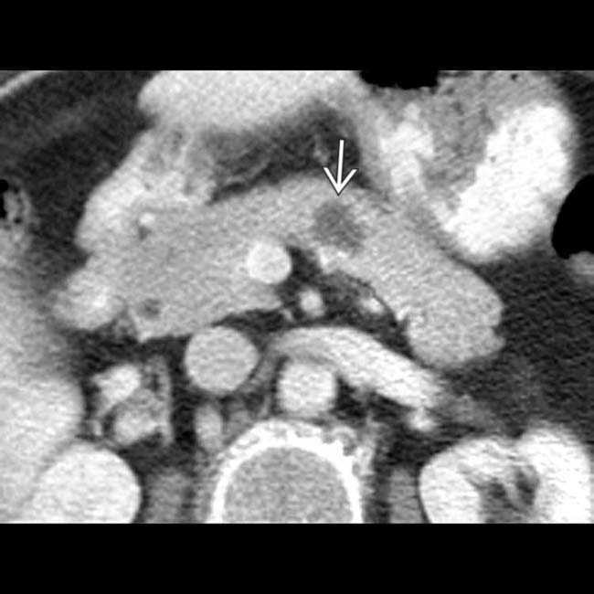
and a small focus of internal fat density image. This was found to be a lymphoepithelial cyst at resection, as these lesions can sometimes demonstrate internal fat.
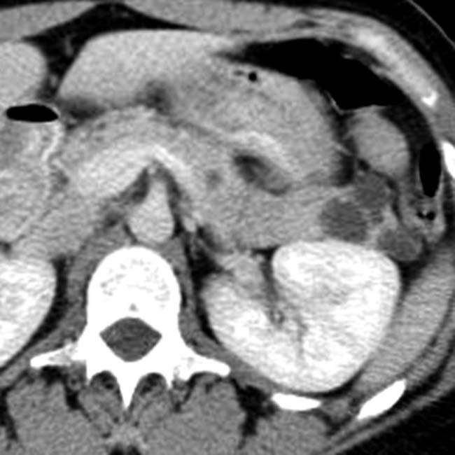
image
Axial CECT in a patient with von Hippel-Lindau syndrome shows several small cysts in the pancreatic tail.

image
Axial NECT in a patient with autosomal dominant polycystic kidney disease shows multiple renal cysts, some with calcified walls. Also present are several pancreatic cysts image.
image
Axial T2 MR demonstrates multiple simple T2-bright cysts in the left kidney, as well as a simple T2-bright nonneoplastic cyst image 
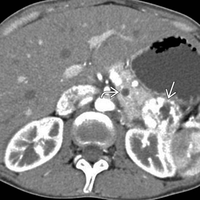
in the pancreas, compatible with the patient’s VHL.
image
Axial gadolinium-enhanced MR of the body and tail of the pancreas in a patient with von Hippel-Lindau syndrome shows multiple thin-walled pancreatic cysts image
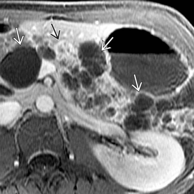
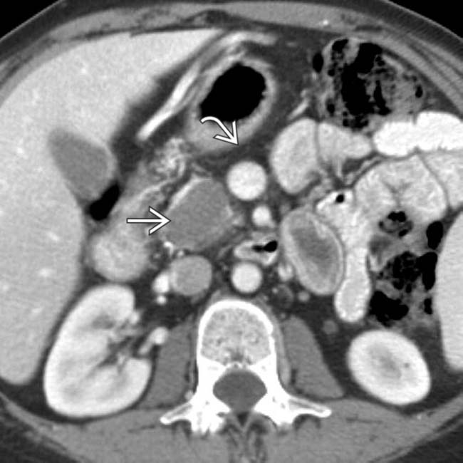
. In addition, note the small hypervascular neuroendocrine tumor image in the body of the pancreas.
image
Axial T2-weighted MR in the same patient shows high signal within the multiple thin-walled simple epithelial cysts image 
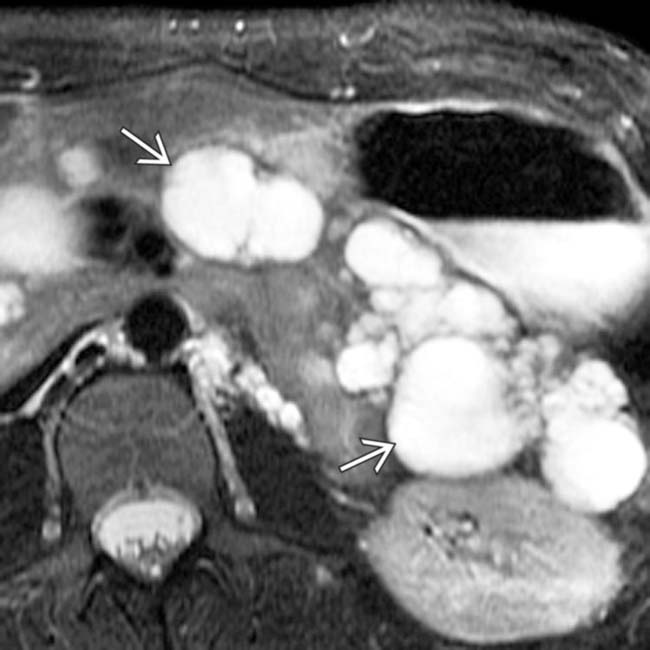
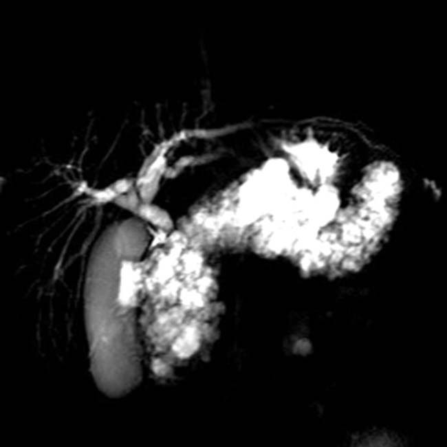
characteristic of pancreatic cystosis in this disease.
image
Axial CECT in a patient with von Hippel-Lindau syndrome shows multiple small pancreatic cysts image 
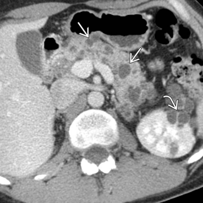
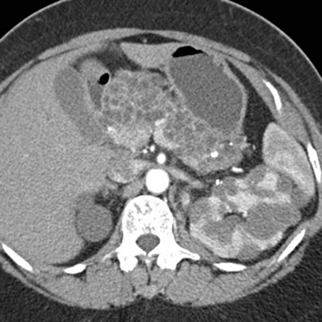
as well as multiple renal cysts image.
image
Axial CECT in the same patient at a more caudal level again shows multiple simple pancreatic cysts image 
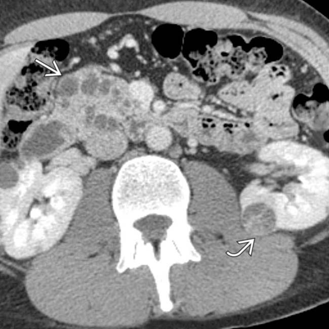
in the pancreatic head. In addition, there is a small cystic renal cell carcinoma in the left kidney image.
image
Axial CECT in a CF patient at the level of the head of the pancreas shows the fatty replacement and pseudohypertrophy of the pancreas image as well as a simple-appearing cyst image.
image
Axial CECT shows a water density cystic lesion image in the pancreatic body.
image
Endoscopic ultrasound in the same patient shows an anechoic cyst image 
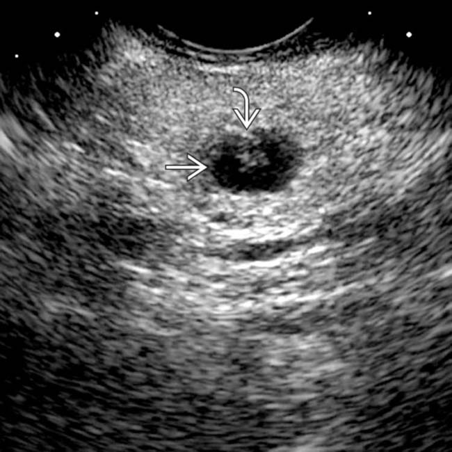
with a small mural nodule image. Needle aspiration yielded clear fluid with borderline elevated CEA and cellular atypia. This was interpreted as equivocal for malignancy, and a laparoscopic distal pancreatectomy was performed. The excised specimen showed an epithelial lining and was classified as a true cyst of the pancreas.

SELECTED REFERENCES

1. Al-Salem, AH, et al. Congenital pancreatic cyst: diagnosis and management. J Pediatr Gastroenterol Nutr. 2014; 59(4):e38–e40.

2. Assifi, MM, et al. Non-neoplastic epithelial cysts of the pancreas: a rare, benign entity. J Gastrointest Surg. 2014; 18(3):523–531.

Kim, WH, et al. Lymphoepithelial cyst of the pancreas: comparison of CT findings with other pancreatic cystic lesions. Abdom Imaging. 2013; 38(2):324–330.

Osiro, S, et al. Is preoperative diagnosis possible? A clinical and radiological review of lymphoepithelial cysts of the pancreas. JOP. 2013; 14(1):15–20.

de Castro, SM, et al. Evaluation of a selective management strategy of patients with primary cystic neoplasms of the pancreas. Int J Surg. 2011; 9(8):655–658.

Johnson, PT, et al. Common incidental findings on MDCT: survey of radiologist recommendations for patient management. J Am Coll Radiol. 2011; 8(11):762–767.

Megibow, AJ, et al. The incidental pancreatic cyst. Radiol Clin North Am. 2011; 49(2):349–359.

Molvar, C, et al. Nonneoplastic cystic lesions of pancreas: a practical clinical, histologic, and radiologic approach. Curr Probl Diagn Radiol. 2011; 40(4):141–148.

Wellner, UF, et al. Incidental cystic tumor in the pancreas: observe or operate? Gastroenterology. 2011; 140(5):e1–e2.

Berland, LL, et al. Managing incidental findings on abdominal CT: white paper of the ACR incidental findings committee. J Am Coll Radiol. 2010; 7(10):754–773.

Federle, et al. Diagnostic Imaging: Abdomen, 2nd edition. Salt Lake City: Amirsys, 2010.

Carboni, F, et al. Solitary true cyst of the pancreas in adults. A report of two cases. JOP. 2009; 10(4):429–431.

Ahlawat, SK. Lymphoepithelial cyst of pancreas. Role of endoscopic ultrasound guided fine needle aspiration. JOP. 2008; 9(2):230–234.

Goh, BK, et al. Non-neoplastic cystic and cystic-like lesions of the pancreas: may mimic pancreatic cystic neoplasms. ANZ J Surg. 2006; 76(5):325–331.