Nerve conduction studies and electromyography
Nerve conduction studies
Sensory studies
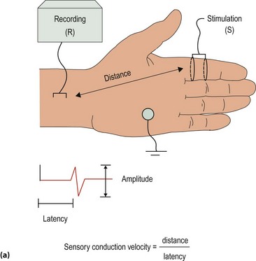
Sensory nerves are studied by stimulating the nerve at one point along it, for example the index finger, and recording at a distant site along the nerve, for example the median aspect of the wrist (Fig. 1a). By recording the time difference for the action potential to appear at two sites along the nerve and measuring the distance between them, the mean conduction velocity can be calculated across the segment of nerve between the two recording points. The amplitude of the action potential can be recorded from the oscilloscope screen. Sensory nerves can be studied orthodromically (distal to proximal) or antidromically (proximal to distal).
Motor studies
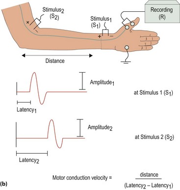
Motor studies involve stimulating the motor nerve at distal and proximal sites and recording the muscle action potentials (Fig. 1b). The amplitude of the muscle response (compound muscle action potential, CMAP) to supramaximal nerve stimulation can be measured. The time from the distal site of stimulation to the onset of the CMAP is the terminal or distal motor latency. This measure includes components of nerve conduction, neuromuscular transmission and muscle activation times. The difference in time to CMAP between the two sites of stimulation and the distance between is used to measure conduction velocity.
Interpretation of nerve conduction studies
There are two types of abnormality detected by nerve conduction studies:
 Focal: where a single nerve is affected, e.g. the median nerve at the wrist in carpal tunnel syndrome.
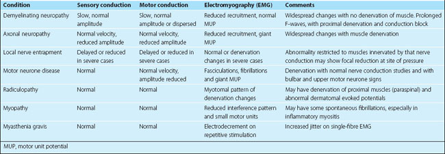
 Generalized neuropathy: this can be further characterized as axonal, if responses are small with normal velocity, or demyelinating, if there is prominent slowing of motor conduction (Fig. 2). The two changes can coexist. Typical patterns of abnormality are given in Table 1.
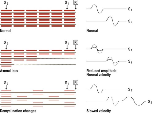

 unmyelinated fibres, ‘small fibre’ neuropathies and autonomic neuropathies, for which other tests may be valuable (p. 115)
| Technique | Method | Significance |
|---|---|---|
| F-wave | Supramaximal nerve stimulation causes antidromic anterior horn cell stimulation and a rebound wave down the motor neurone | Measures delay in proximal conduction, affected in demyelinating neuropathies and radiculopathies |
| H-wave | Electrophysiological equivalent of the tendon reflex; stimulation of motor neurone via sensory input | May be affected by proximal sensory disturbance or motor neurone dysfunction |
| Single-fibre EMG | A measure of the time difference of activation of two muscle fibres, innervated by branches of the same nerve, by using two very fine concentric electrodes | Great variability in the time difference (‘increased jitter’) is seen in disorders of neuromuscular transmission, e.g. myasthenia gravis |
| Thermal threshold | Measure patient’s ability to detect small temperature changes, produced by heating a small patch of skin, measured by a thermistor | Measure of small unmyelinated fibre function |
| Sympathetic skin response | Laser Doppler measurement of nail bed blood flow response to temperature change | Measures small unmyelinated sympathetic fibres |
Electromyography
There are three parts to the electromyographic examination of a muscle.
Spontaneous activity


Other situations
Neuromuscular junction disorder resulting from myasthenia gravis causes a failure of neuromuscular transmission with increasing use (p. 110). The electrophysiological equivalent of fatiguability is the decline in amplitude of the compound motor action potential on repetitive stimulation of the motor nerve. Single-fibre electromyography may also help in diagnosis (see Table 2).


