Nephrotic Syndrome

Published on 25/03/2015 by admin

Filed under Pediatrics

Last modified 22/04/2025

Print this page

rate 1 star rate 2 star rate 3 star rate 4 star rate 5 star
Your rating: none, Average: 0 (0 votes)

This article have been viewed 3102 times

Chapter 521 Nephrotic Syndrome

Nephrotic syndrome, a manifestation of glomerular disease, is characterized by nephrotic range proteinuria and the triad of clinical findings associated with large urinary losses of protein: hypoalbuminemia, edema, and hyperlipidemia. Nephrotic range proteinuria is defined as protein excretion of > 40 mg/m2/hr or a first morning protein : creatinine ratio of >2-3 : 1. The annual incidence is 2-3 cases per 100,000 children per year in most Western countries and higher in underdeveloped countries resulting predominantly from malaria. Though early referral to a pediatric nephrologist is recommended, once nephrotic syndrome has been diagnosed, management should be a collaborative effort between the nephrologist and primary care physician.

Etiology

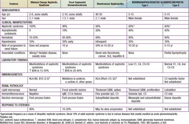
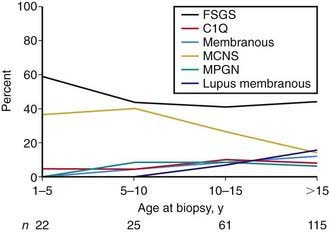
Most children with nephrotic syndrome have a form of primary or idiopathic nephrotic syndrome (Table 521-1). Glomerular lesions associated with idiopathic nephrotic syndrome include minimal change disease (the most common), focal segmental glomerulosclerosis, membranoproliferative glomerulonephritis, membranous nephropathy and diffuse mesangial proliferation (Table 521-2). These etiologies have different age distributions (Fig. 521-1).

Table 521-1 CAUSES OF CHILDHOOD NEPHROTIC SYNDROME

GENETIC DISORDERS

Nephrotic Syndrome (Typical)

Proteinuria with or Without Nephrotic Syndrome

Multisystem Syndromes with or Without Nephrotic Syndrome

Metabolic Disorders with or Without Nephrotic Syndrome

IDIOPATHIC NEPHROTIC SYNDROME

SECONDARY CAUSES

Infections

Drugs

Immunologic or Allergic Disorders

Associated with Malignant Disease

Glomerular Hyperfiltration

Note: Childhood nephrotic syndrome can also be consequence of inflammatory glomerular disorders, normally associated with features of nephritis, e.g., vasculitis, lupus nephritis, membranoproliferative glomerulonephritis, IgA nephropathy.

From Eddy AA, Symons JM: Nephrotic syndrome in childhood, Lancet 362:629–638, 2003.

Nephrotic syndrome may also be secondary to systemic diseases such as systemic lupus erythematosus, Henoch-Schönlein purpura, malignancy (lymphoma and leukemia), and infections (hepatitis, HIV, and malaria) (see Table 521-1).

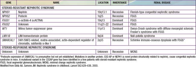
A number of hereditary proteinuria syndromes are caused by mutations in genes that encode critical protein components of the glomerular filtration apparatus (Table 521-3).

Pathophysiology

The underlying abnormality in nephrotic syndrome is an increased permeability of the glomerular capillary wall, which leads to massive proteinuria and hypoalbuminemia. On biopsy, the extensive effacement of podocyte foot processes (the hallmark of idiopathic nephrotic syndrome) suggests a pivotal role for the podocyte. Idiopathic nephrotic syndrome is associated with complex disturbances in the immune system, especially T cell–mediated immunity. In focal segmental glomerulosclerosis, a plasma factor, probably produced by a subset of activated lymphocytes, may be responsible for the increase in capillary wall permeability. Alternatively, mutations in podocyte proteins (podocin, α-actinin 4) and MYH9 (podocyte gene) are associated with focal segmental glomerulosclerosis (see Table 521-3). Steroid-resistant nephrotic syndrome can be associated with mutations in NPHS2 (podocin) and WT1 genes, as well as other components of the glomerular filtration apparatus, such as the slit pore, and include nephrin, NEPH1, and CD-2 associated protein.

Although the mechanism of edema formation in nephrotic syndrome is incompletely understood, it seems likely that in most instances, massive urinary protein loss leads to hypoalbuminemia, which causes a decrease in plasma oncotic pressure and transudation of fluid from the intravascular compartment to the interstitial space. The reduction in intravascular volume decreases renal perfusion pressure, activating the renin-angiotensin-aldosterone system, which stimulates tubular reabsorption of sodium. The reduced intravascular volume also stimulates the release of antidiuretic hormone, which enhances the reabsorption of water in the collecting duct.

This theory does not apply to all patients with nephrotic syndrome because some patients actually have increased intravascular volume with diminished plasma levels of renin and aldosterone. Therefore, other factors, including primary renal avidity for sodium and water, may be involved in the formation of edema in some patients with nephrotic syndrome.

In the nephrotic state, serum lipid levels (cholesterol, triglycerides) are elevated for 2 reasons. Hypoalbuminemia stimulates generalized hepatic protein synthesis, including synthesis of lipoproteins. This is also why a number of coagulation factors are increased, increasing the risk of thrombosis. In addition, lipid catabolism is diminished as a result of reduced plasma levels of lipoprotein lipase related to increased urinary losses of this enzyme.

Patients with nephrotic syndrome are at increased risk of infections (sepsis, peritonitis, pyelonephritis), especially with encapsulated organisms such as Streptococcus pneumoniae and Haemophilus influenza. Some reasons for this include loss of complement factor C3b, opsonins such as properdin factor B, and immunoglobulins in the urine. An additional risk factor is the use of immunosuppressive medications to treat nephrotic syndrome.

Nephrotic syndrome is a hypercoagulable state resulting from multiple factors: vascular stasis, an increase in hepatic production of fibrinogen and other clotting factors, decreased serum levels of anticoagulation factors, increased plasma platelet production (as an acute phase reactant), and increased platelet aggregation. The coagulopathy manifests with thromboembolic events.

521.1 Idiopathic Nephrotic Syndrome

Approximately 90% of children with nephrotic syndrome have idiopathic nephrotic syndrome. Idiopathic nephrotic syndrome is associated with primary glomerular disease without evidence of a specific systemic cause. Idiopathic nephrotic syndrome includes multiple histologic types: minimal change disease, mesangial proliferation, focal segmental glomerulosclerosis, membranous nephropathy, and membranoproliferative glomerulonephritis.

Pathology

In minimal change nephrotic syndrome (MCNS) (about 85% of total cases of nephrotic syndrome in children), the glomeruli appear normal or show a minimal increase in mesangial cells and matrix. Findings on immunofluorescence microscopy are typically negative, and electron microscopy simply reveals effacement of the epithelial cell foot processes. More than 95% of children with minimal change disease respond to corticosteroid therapy.

Mesangial proliferation is characterized by a diffuse increase in mesangial cells and matrix on light microscopy. Immunofluorescence microscopy might reveal trace to 1+ mesangial IgM and/or IgA staining. Electron microscopy reveals increased numbers of mesangial cells and matrix as well as effacement of the epithelial cell foot processes. Approximately 50% of patients with this histologic lesion respond to corticosteroid therapy.

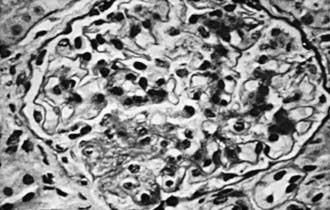
In focal segmental glomerulosclerosis (FSGS), glomeruli show lesions that are both focal (present only in a proportion of glomeruli) and segmental (localized to ≥1 intraglomerular tufts). The lesions consist of mesangial cell proliferation segmental scarring on light microscopy (Fig. 521-2 and see Table 521-2). Immunofluorescence microscopy is positive for IgM and C3 staining in the areas of segmental sclerosis. Electron microscopy demonstrates segmental scarring of the glomerular tuft with obliteration of the glomerular capillary lumen. Similar lesions may be seen secondary to HIV infection, vesicoureteral reflux, and intravenous use of heroin and other drugs of abuse. Only 20% of patients with FSGS respond to prednisone. The disease is often progressive, ultimately involving all glomeruli, and ultimately leads to end-stage renal disease in most patients.

Clinical Manifestations

The idiopathic nephrotic syndrome is more common in boys than in girls (2 : 1) and most commonly appears between the ages of 2 and 6 yr (see Fig. 521-1). However, it has been reported as early as 6 mo of age and throughout adulthood. MCNS is present in 85-90% of patients <6 yr of age. In contrast, only 20-30% of adolescents who present for the first time with nephrotic syndrome have MCNS. The more common cause of idiopathic nephrotic syndrome in this older age group is FSGS. The incidence of FSGS may be increasing; it may be more common in African-American, Hispanic, and Asian patients.

The initial episode of idiopathic nephrotic syndrome, as well as subsequent relapses, usually follows minor infections and, uncommonly, reactions to insect bites, bee stings, or poison ivy.

Children usually present with mild edema, which is initially noted around the eyes and in the lower extremities. Nephrotic syndrome can initially be misdiagnosed as an allergic disorder because of the periorbital swelling that decreases throughout the day. With time, the edema becomes generalized, with the development of ascites, pleural effusions, and genital edema. Anorexia, irritability, abdominal pain, and diarrhea are common. Important features of minimal change idiopathic nephrotic syndrome are the absence of hypertension and gross hematuria (previously termed nephritic features).

The differential diagnosis of the child with marked edema includes protein-losing enteropathy, hepatic failure, heart failure, acute or chronic glomerulonephritis, and protein malnutrition. A diagnosis other than MCNS should be considered in children <1 yr of age, a positive family history of nephrotic syndrome, presence of extrarenal findings (e.g., arthritis, rash, anemia), hypertension or pulmonary edema, acute or chronic renal insufficiency, and gross hematuria.

Treatment

Children with their first episode of nephrotic syndrome and mild to moderate edema may be managed as outpatients. Such outpatient management is not practiced in all major centers, because the time required for successful education of the family regarding all aspects of the condition can require a short period of hospitalization. The pathophysiology and treatment of nephrotic syndrome must be carefully reviewed with the family to enhance understanding of their child’s disease. The child’s parents must be able to recognize the signs and symptoms of the complications of the disease and its treatment and must be taught how to use and interpret the results of urinary dipstick testing for protein.

Children with onset of uncomplicated nephrotic syndrome between 1 and 8 yr of age are likely to have steroid-responsive MCNS, and steroid therapy may be initiated without a diagnostic renal biopsy. Children with features that make MCNS less likely (gross hematuria, hypertension, renal insufficiency, hypocomplementemia, or age <1 yr or >8 yr) should be considered for renal biopsy before treatment.

In children with presumed MCNS, prednisone should be administered (after confirming a negative PPD test and administering the polyvalent pneumococcal vaccine) at a dose of 60 mg/m2/day (maximum daily dose, 80 mg) in a single daily dose for 4-6 consecutive wk. An initial 6-wk course of daily steroid treatment leads to a significantly lower relapse rate than previously recommended shorter courses of daily therapy. About 80-90% of children respond to steroid therapy (clinical remission, diuresis, and urine trace or negative for protein for 3 consecutive days) within 3 wk. The vast majority of children who respond to prednisone therapy do so within the first 5 wk of treatment.

After the initial 6-wk course, the prednisone dose should be tapered to 40 mg/m2/day given every other day as a single daily dose for at least 4 wk. The alternate-day dose is then slowly tapered and discontinued over the next 1-2 mo. There is evidence that both an increased dose of steroids and a prolonged duration of therapy are important factors in reducing the risk of relapse. While planning the duration of steroid therapy, the side effects of prolonged corticosteroid administration must be kept in mind.

Children with severe symptomatic edema, including large pleural effusions, ascites, or severe genital edema, should be hospitalized. In addition to sodium restriction, fluid restriction may be necessary if the child is hyponatremic. A swollen scrotum may be elevated with pillows to enhance fluid removal by gravity. Diuresis may be augmented by the administration of loop diuretics (furosemide), orally or intravenously, although extreme caution should be exercised. Aggressive diuresis can lead to intravascular volume depletion and a significantly increased risk of intravascular thrombosis.

When a patient has significant generalized edema with evidence of intravascular volume depletion (e.g., hemoconcentration), IV administration of 25% albumin (0.5-1.0 g albumin/kg), as a slow infusion followed by furosemide (1-2 mg/kg/dose IV) is sometimes necessary. Such therapy should be used only in collaboration with a pediatric nephrologist and mandates close monitoring of volume status, blood pressure, serum electrolyte balance, and renal function. Symptomatic volume overload, with hypertension, heart failure, and pulmonary edema is a potential complication of parenteral albumin therapy, particularly when administered as rapid infusions.

Children who continue to have proteinuria (2+ or greater) after 8 wk of steroid therapy are considered steroid resistant, and a diagnostic renal biopsy should be performed.

Children with nephrotic syndrome should attend school and participate in physical activities as tolerated. At the initial presentation and during relapse, a low-sodium diet should be followed. This restriction may be lifted once the child enters remission. Although there are no data to support their safety or efficacy, oral diuretics are used by many clinicians to control edema in children with nephrotic syndrome. Because of the increased risks of thromboembolic complications, diuretic use should be reserved for patients with severe symptoms and must be closely monitored.

Many children with nephrotic syndrome experience at least 1 relapse (3-4+ proteinuria plus edema). Although relapse rates of 60-80% have been noted in the past, the relapse rate in children treated with longer initial steroid courses may be as low as 30-40%.

Relapses should be treated with 60 mg/m2/day (80 mg daily max) in a single AM dose until the child enters remission (urine trace or negative for protein for 3 consecutive days). The prednisone dose is then changed to alternate-day dosing as noted with initial therapy, and gradually tapered over 4-8 wk.

A subset of patients relapse while on alternate-day steroid therapy or within 28 days of completing a successful course of prednisone therapy. Such patients are termed steroid dependent. Patients who respond well to prednisone therapy but relapse ≥4 times in a 12-mo period are termed frequent relapsers. Children who fail to respond to prednisone therapy within 8 wk of therapy are termed steroid resistant. Steroid-resistant nephrotic syndrome is usually caused by FSGS (80%), MCNS, or mesangial proliferative glomerulonephritis.

Steroid-dependent patients, frequent relapsers, and steroid-resistant patients are candidates for alternative therapies, particularly if the child has severe corticosteroid toxicity (cushingoid appearance, hypertension, cataracts, and/or growth failure). Cyclophosphamide prolongs the duration of remission and reduces the number of relapses in children with frequently relapsing and steroid-dependent nephrotic syndrome. The potential side effects of the drug (neutropenia, disseminated varicella, hemorrhagic cystitis, alopecia, sterility, increased risk of future malignancy) should be carefully reviewed with the family before initiating treatment. Cyclophosphamide (2 mg/kg) is given as a single oral dose for a total duration of 8-12 wk. Alternate-day prednisone therapy is often continued during the course of cyclophosphamide administration. During cyclophosphamide therapy, the white blood cell count must be monitored weekly and the drug should be withheld if the count falls below 5,000/mm3. The cumulative threshold dose above which oligo- or azoospermia occurs in boys is >250 mg/kg.

Cyclosporine or tacrolimus are also effective in inducing and maintaining prolonged remissions in children with steroid-resistant nephrotic syndrome and are useful as steroid-sparing agents. Children must be monitored for side effects, including hypertension, nephrotoxicity, hirsutism, and gingival hyperplasia. Mycophenolate can maintain remission in children with steroid-dependent or frequently relapsing nephrotic syndrome. Levamisole, an anthelmintic agent with immunomodulating effects that has been shown to reduce the risk of relapse in comparison to prednisone, is not available in the USA.

Most children who respond to cyclosporine, tacrolimus, or mycophenolate therapy tend to relapse when the medication is discontinued. Angiotensin-converting enzyme (ACE) inhibitors and angiotensin II blockers may be helpful as adjunct therapy to reduce proteinuria in steroid-resistant patients.

Complications

Infection is a major complication of nephrotic syndrome. Children in relapse have increased susceptibility to bacterial infections because of urinary losses of immunoglobulins and properdin factor B, defective cell-mediated immunity, their immunosuppressive therapy, malnutrition, and edema or ascites acting as a potential culture medium. Spontaneous bacterial peritonitis is a common infection, although sepsis, pneumonia, cellulitis, and urinary tract infections may also be seen. Although Streptococcus pneumoniae is the most common organism causing peritonitis, gram-negative bacteria such as Escherichia coli may also be encountered. The patient’s caregivers should be counseled to seek medical attention if the child appears ill, has a fever, or complains of persistent abdominal pain. A high index of suspicion for bacterial peritonitis, prompt evaluation (including cultures of blood and peritoneal fluid), and early initiation of antibiotic therapy are critical.

Children with nephrotic syndrome should receive the 23-serotype pneumococcal vaccine (in addition to the 7-valent conjugate pneumococcal vaccine), given according to the routine childhood immunization schedule, ideally administered when the child is in remission and off daily prednisone therapy. Live virus vaccines should not be administered to children who are receiving daily or alternate-day high-dose steroids (≥2 mg/kg/day of prednisone or its equivalent, or ≥20 mg/day if the child weighs >10 kg). Vaccines can be administered after corticosteroid therapy has been discontinued for at least 1 mo. Nonimmune nephrotic children in relapse, if exposed to varicella, should receive varicella-zoster immunoglobulin (1 dose ≤96 hours after significant exposure). Influenza vaccine should be given on a yearly basis.

Children with nephrotic syndrome are also at increased risk of thromboembolic events. The incidence of this complication in children is 2-5%, which represents a much lower risk than that of adults with nephrotic syndrome. Both arterial and venous thromboses may be seen, including renal vein thrombosis, pulmonary embolus, sagittal sinus thrombosis, and thrombosis of indwelling arterial and venous catheters. The risk of thrombosis is related to increased prothrombotic factors (fibrinogen, thrombocytosis, hemoconcentration, relative immobilization) and decreased fibrinolytic factors (urinary losses of antithrombin III, proteins C and S). Prophylactic anticoagulation is not recommended in children unless a previous thromboembolic event has occurred. To minimize the risk of thromboembolic complications, aggressive use of diuretics and the use of indwelling catheters should be avoided if possible. Hyperlipidemia, particularly in patients with complicated nephrotic syndrome, may be a risk factor for cardiovascular disease; myocardial infarction is a rare complication in children. It has been suggested that 3-hydroxy-3-methylglutaryl coenzyme A (HMG-CoA) reductase-inhibiting drugs should be used to treat the hyperlipidemia seen in persistent nephrotic syndrome, but controlled data regarding risks or benefits are not available.

Monitoring of children with nephrotic syndrome is noted in Table 521-4.

Prognosis

Most children with steroid-responsive nephrotic syndrome have repeated relapses, which generally decrease in frequency as the child grows older. Although there is no proven way to predict an individual child’s course, children who respond rapidly to steroids and those who have no relapses during the first 6 mo after diagnosis are likely to follow an infrequently relapsing course. It is important to indicate to the family that the child with steroid-responsive nephrotic syndrome is unlikely to develop chronic kidney disease, that the disease is rarely hereditary, and that the child (in the absence of prolonged cyclophosphamide therapy) will remain fertile. To minimize the psychologic effects of the condition and its therapy, children with idiopathic nephritic syndrome should not be considered chronically ill and should participate in all age-appropriate childhood activities and maintain an unrestricted diet when in remission.

Children with steroid-resistant nephrotic syndrome, most often caused by FSGS, generally have a much poorer prognosis. These children develop progressive renal insufficiency, ultimately leading to end-stage renal disease requiring dialysis or kidney transplantation. Recurrent nephrotic syndrome develops in 30-50% of transplant recipients with FSGS. There have not been adequate randomized clinical trials in this subset of patients to guide therapy. A large NIH sponsored multicenter, randomized clinical trial is comparing cyclosporine with mycophenolate mofetil in the treatment of focal segmental glomerulosclerosis.

Bibliography

Abeyagunawardena AS, Trompeter RS. Increasing the dose of prednisolone during viral infections reduced the risk of relapse in nephrotic syndrome: a randomized controlled trial. Arch Dis Child. 2008;93:226-228.

Eddy AA, Symons JM. Nephrotic syndrome in childhood. Lancet. 2003;362:629-639.

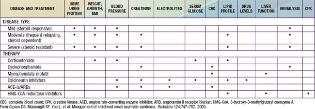
Gipson DS, Massengill SF, Yao L, et al. Management of childhood onset nephrotic syndrome. Pediatrics. 2009;124:747-757.

Hodson EM, Craig JC, Willis NS. Evidence based management of steroid sensitive nephrotic syndrome. Pediatr Nephrol. 2005;20:1523-1530.

Kapur G, Valentini RP, Imam AA, et al. Treatment of severe edema in children with nephrotic syndrome with diuretics alone—a prospective study. Clin J Am Soc Nephrol. 2009;4:907-913.

Kerlin BA, Blatt NB, Fuh B, et al. Epidemiology and risk factors for thromboembolic complications of childhood nephrotic syndrome: a Midwest Pediatric Nephrology Consortium (MWPNC) study. J Pediatr. 2009;155:105-110.

Kopp JB, Smith MW, Nelson GW, et al. MYH9 is a major-effect risk gene for focal segmental glomerulosclerosis. Nat Genet. 2008;40:1175-1184.

Leonard MB, Feldman HI, Shults J, et al. Long-term, high-dose glucocorticoids and bone mineral content in childhood glucocorticoid-sensitive nephrotic syndrome. N Engl J Med. 2004;351:868-875.

Loeffler K, Gowrishankar M, Yiu V. Tacrolimus therapy in pediatric patients with treatment-resistant nephrotic syndrome. Pediatr Nephrol. 2004;19:281-287.

Machuca E, Esquivel EL, Antignac C. Idiopathic nephrotic syndrome: genetic aspects. In: Avner ED, Harmon WE, Niaudet P, et al, editors. Pediatric nephrology. ed 6. Heidelberg, Germany: Springer-Verlag; 2009:643-666.

Mao J, Zhang Y, Du L, et al. NPHS1 and NPHS2 gene mutations in chinese children with sporadic nephrotic syndrome. Pediatr Res. 2007;61:117-122.

Niaudet P, Boyer O. Idiopathic nephrotic syndrome in children: clinical aspects. In: Avner ED, Harmon WE, Niaudet P, et al, editors. Pediatric nephrology. ed 6. Heidelberg, Germany: Springer-Verlag; 2009:667-702.

Focal Segmental Glomerulosclerosis Clinical Trial (FSGS-CT). sponsored by the National Institute of Diabetes and Digestive and Kidney Diseases (NIDDK). https://clinicaltrials.gov.

521.2 Secondary Nephrotic Syndrome

Nephrotic syndrome can occur as a secondary feature of many forms of glomerular disease. Membranous nephropathy, membranoproliferative glomerulonephritis, postinfectious glomerulonephritis, lupus nephritis, and Henoch-Schönlein purpura nephritis can all have a nephrotic component (see Tables 521-1 and 521-2). Secondary nephrotic syndrome should be suspected in patients >8 yr and those with hypertension, hematuria, renal dysfunction, extrarenal symptoms (rash, arthralgias, fever), or depressed serum complement levels.

In certain areas of the world, malaria and schistosomiasis are the leading causes of nephrotic syndrome. Other infectious agents associated with nephrotic syndrome include hepatitis B virus, hepatitis C virus, filaria, leprosy, and HIV.

Nephrotic syndrome has been associated with malignancy, particularly in the adult population. In patients with solid tumors, such as carcinomas of the lung and gastrointestinal tract, the renal pathology often resembles membranous glomerulopathy. Immune complexes composed of tumor antigens and tumor-specific antibodies presumably mediate the renal involvement. In patients with lymphomas, particularly Hodgkin lymphoma, the renal pathology most often resembles MCNS. The proposed mechanism of the nephrotic syndrome is that the lymphoma produces a lymphokine that increases permeability of the glomerular capillary wall. Nephrotic syndrome can develop before or after the malignancy is detected, resolve as the tumor regresses, and return if the tumor recurs.

Nephrotic syndrome has also developed during therapy with numerous drugs and chemicals. The histologic picture can resemble membranous glomerulopathy (penicillamine, captopril, gold, nonsteroidal anti-inflammatory drugs, mercury compounds), MCNS (probenecid, ethosuximide, methimazole, lithium), or proliferative glomerulonephritis (procainamide, chlorpropamide, phenytoin, trimethadione, paramethadione).

521.3 Congenital Nephrotic Syndrome

Nephrotic syndrome (massive proteinuria, hypoalbuminemia, edema, and hypercholesterolemia) has a poorer prognosis when it occurs in the 1st yr of life, when compared to nephrotic syndrome manifesting in childhood. Congenital nephrotic syndrome is defined as nephrotic syndrome manifesting at birth or within the first 3 mo of life. Congenital nephrotic syndrome may be classified as primary or as secondary to a number of etiologies such as in-utero infections (cytomegalovirus, toxoplasmosis, syphilis, hepatitis B and C, HIV), infantile systemic lupus erythematosus, or mercury exposure.

Primary congenital nephrotic syndrome is due to a variety of syndromes inherited as autosomal recessive disorders (see Table 521-3). A number of structural and functional abnormalities of the glomerular filtration barrier causing congenital nephrotic syndrome have been elucidated. The glomerular filtration barrier, which is both size and charge selective, is composed of 3 layers: the fenestrated endothelium, the glomerular basement membrane, and the podocyte foot processes. The foot processes are interconnected by bridging structures, the slit diaphragms, which act as a size selective filter, whereas the glomerular basement membrane restricts molecules based on their ionic charge.

In a large European cohort of children with congenital nephrotic syndrome, 85% demonstrated disease, causing mutations in 4 genes (NPHS1, NPHS2, WT1, and LAMB2), the first 3 of which encode components of the glomerular filtration barrier. The Finnish type of congenital nephritic syndrome is caused by mutations in the NPHS1 or NPHS2 genes, which encode nephrin and podocin, critical components of the slit diaphragm. Affected infants most commonly present at birth with edema due to massive proteinuria, and they are typically delivered with an enlarged placenta (>25% of the infant’s weight). Severe hypoalbuminemia, hyperlipidemia, and hypogammaglobulinemia result from loss of filtering selectivity at the glomerular filtration barrier. Prenatal diagnosis can be made by the presence of elevated maternal and amniotic α-fetoprotein levels.

Denys-Drash syndrome is caused by mutations in the WT1 gene, which results in abnormal podocyte function. Patients present with early-onset nephrotic syndrome, progressive renal insufficiency, ambiguous genitalia, and Wilms’ tumors.

Mutations in the LAMB2 gene, seen in Pierson syndrome, lead to abnormalities of β2 laminin, a critical component of glomerular and ocular basement membranes. In addition to congenital nephrotic syndrome, affected infants display bilateral microcoria (fixed narrowing of the pupil).

Regardless of the etiology of congenital nephrotic syndrome, diagnosis is made clinically in newborns or infants who demonstrate severe generalized edema, poor growth and nutrition with hypoalbuminemia, increased susceptibility to infections, hypothyroidism (due to urinary loss of thyroxin-binding globulin), and increased risk of thrombotic events. Most infants have progressive renal insufficiency.

Secondary congenital nephrotic syndrome can resolve with treatment of the underlying cause, such as syphilis (Table 521-5). The management of primary congenital nephrotic syndrome includes intensive supportive care with intravenous albumin and diuretics, regular administration of intravenous gamma-globulin, and aggressive nutritional support (often parenteral), while attempting to pharmacologically decrease urinary protein loss with angiotensin-converting enzyme inhibitors, angiotensin II receptor inhibitors, and prostaglandin synthesis inhibitors or even unilateral nephrectomy. If conservative management fails, and patients suffer from persistent anasarca or repeated severe infections, bilateral nephrectomies are performed and chronic dialysis is initiated. Renal transplantation is the definitive treatment of congenital nephrotic syndrome, though recurrence of the nephrotic syndrome has been reported to occur after transplantation.