[level-membership-for-pediatrics-category]
Chapter 382 Neoplasms of the Larynx, Trachea, and Bronchi
382.1 Vocal Nodules
Laryngopharyngeal reflux commonly exacerbates vocal abuse, adding to swelling of the vocal cords. When vocal abuse is the main factor, the voice is worse in the evenings; with laryngopharyngeal reflux, hoarseness is worse in the morning. An antireflux regimen is indicated (Chapter 315.1).
382.2 Recurrent Respiratory Papillomatosis
Papillomas are the most common respiratory tract neoplasms in children, occurring in 4.3/100,000. They are simply warts—benign tumors—caused by the human papillomavirus (HPV) (Chapter 258); the same pathology is found in condylomata acuminata (vaginal warts). HPV types 6 and 11 are most commonly associated with laryngeal disease. Fifty percent of recurrent respiratory papillomatosis (RRP) cases occur in children <5 yr, but the diagnosis may be made at any age; 67% of children with RRP are born to mothers who had condylomata during pregnancy or parturition. The risk for transmission is ∼1/500 vaginal births in mothers with active condylomata. Neonates have been reported to have RRP, suggesting intrauterine transmission of HPV.
Clinical Manifestations
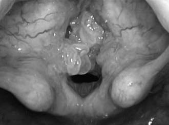
These benign squamous lesions can produce chronic hoarseness in the infant. Most occur in the larynx, specifically on the vocal cords, but in 31% these lesions occur in other areas of the respiratory tract: nose, pharynx (especially the uvula and posterior soft palate), trachea, bronchi, and lungs. As growth of the lesions on the vocal cords progresses, hoarseness increases and communication becomes difficult. Respiratory distress develops. Surgical excision is a quality of life issue that warrants removal to improve the voice. Intervention becomes a medical necessity when airway obstruction progresses (Fig. 382-1). Symptoms often occur first during sleep with symptoms typical of obstructive sleep apnea. Progressive respiratory distress during sleep, exertion, daily activities, and finally at rest indicates the need for surgical intervention.
382.6 Tracheal Neoplasms
Tracheal neoplasms manifest with stridor, wheezing, cough, or pneumonia and are rarely diagnosed until 75% of the lumen has been obstructed (Fig 382-2). Symptoms mimic asthma and are often misdiagnosed as such. Chest radiographs or airway films can identify the obstruction. Pulmonary function studies demonstrate an abnormal flow-volume loop. A mild response to bronchodilator therapy may be misleading. Rational treatment is based upon the histopathology.
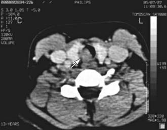
Figure 382-2 A CT scan of the trachea with a circumscribed intraluminal tracheal mass (arrow) in the tracheal wall.
(From Venizelos I, Papathomas T, Anagnostou E, et al: Pediatric inflammatory myofibroblastic tumor of the trachea: a case report and review of the literature, Pediatr Pulmonol 43:831–835, 2008.)
[/level-membership-for-pediatrics-category][not-level-membership-for-pediatrics-category]
Chapter 382 Neoplasms of the Larynx, Trachea, and Bronchi
382.1 Vocal Nodules
Laryngopharyngeal reflux commonly exacerbates vocal abuse, adding to swelling of the vocal cords. When vocal abuse is the main factor, the voice is worse in the evenings; with laryngopharyngeal reflux, hoarseness is worse in the morning. An antireflux regimen is indicated (Chapter 315.1).
382.2 Recurrent Respiratory Papillomatosis
Papillomas are the most common respiratory tract neoplasms in children, occurring in 4.3/100,000. They are simply warts—benign tumors—caused by the human papillomavirus (HPV) (Chapter 258); the same pathology is found in condylomata acuminata (vaginal warts). HPV types 6 and 11 are most commonly associated with laryngeal disease. Fifty percent of recurrent respiratory papillomatosis (RRP) cases occur in children <5 yr, but the diagnosis may be made at any age; 67% of children with RRP are born to mothers who had condylomata during pregnancy or parturition. The risk for transmission is ∼1/500 vaginal births in mothers with active condylomata. Neonates have been reported to have RRP, suggesting intrauterine transmission of HPV.
Clinical Manifestations
These benign squamous lesions can produce chronic hoarseness in the infant. Most occur in the larynx, specifically on the vocal cords, but in 31% these lesions occur in other areas of the respiratory tract: nose, pharynx (especially the uvula and posterior soft palate), trachea, bronchi, and lungs. As growth of the lesions on the vocal cords progresses, hoarseness increases and communication becomes difficult. Respiratory distress develops. Surgical excision is a quality of life issue that warrants removal to improve the voice. Intervention becomes a medical necessity when airway obstruction progresses (Fig. 382-1). Symptoms often occur first during sleep with symptoms typical of obstructive sleep apnea. Progressive respiratory distress during sleep, exertion, daily activities, and finally at rest indicates the need for surgical intervention.
[/not-level-membership-for-pediatrics-category]
