Neoplasms of Bone

Published on 25/03/2015 by admin

Filed under Pediatrics

Last modified 22/04/2025

Print this page

rate 1 star rate 2 star rate 3 star rate 4 star rate 5 star
Your rating: none, Average: 0 (0 votes)

This article have been viewed 2062 times

Chapter 495 Neoplasms of Bone

495.1 Malignant Tumors of Bone

Carola A.S. Arndt

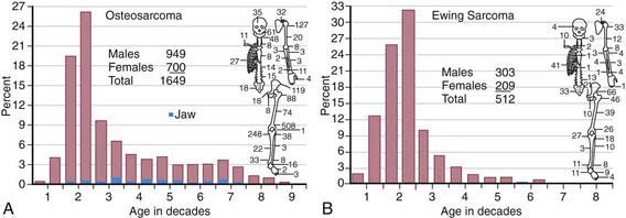
The annual incidence of malignant bone tumors in the USA is approximately 7 cases/million white children <14 yr of age, with a slightly lower incidence in African-American children. Osteosarcoma is the most common primary malignant bone tumor in children and adolescents, followed by Ewing sarcoma (Table 495-1; Fig. 495-1). In children <10 yr of age, Ewing sarcoma is more common than osteosarcoma. Both tumor types are most likely to occur in the second decade of life.

Table 495-1 COMPARISON OF FEATURES OF OSTEOSARCOMA AND THE EWING FAMILY OF TUMORS

FEATURE OSTEOSARCOMA EWING FAMILY OF TUMORS
Age Second decade Second decade
Race All races Primarily whites
Sex (M : F) 1.5 : 1 1.5 : 1
Cell Spindle cell–producing osteoid Undifferentiated small round cell, probably of neural origin
Predisposition Retinoblastoma, Li-Fraumeni syndrome, Paget disease, radiotherapy None known
Site Metaphyses of long bones Diaphyses of long bones, flat bones
Presentation Local pain and swelling; often, history of injury Local pain and swelling; fever
Radiographic findings Sclerotic destruction (less commonly lytic); sunburst pattern Primarily lytic, multilaminar periosteal reaction (“onion-skinning”)
Differential diagnosis Ewing sarcoma, osteomyelitis Osteomyelitis, eosinophilic granuloma, lymphoma, neuroblastoma, rhabdomyosarcoma
Metastasis Lungs, bones Lungs, bones
Treatment Chemotherapy Chemotherapy
Ablative surgery of primary tumor Radiotherapy and/or surgery of primary tumor
Outcome Without metastases, 70% cured; with metastases at diagnosis, ≤20% survival Without metastases, 60% cured; with metastases at diagnosis, 20-30% survival
image

Figure 495-1 A, Age and skeletal distribution of 1,649 cases of osteosarcoma in the Mayo Clinic files. B, Age and skeletal distribution of 512 cases of Ewing sarcoma in the Mayo Clinic files.

(From Unni KK, editor: Dahlin’s bone tumors: general aspects and data on 11,087 cases, ed 5, Philadelphia, 1996, Lippincott-Raven. Reprinted by permission of the Mayo Foundation.)

Osteosarcoma

Pathogenesis

Although the cause of osteosarcoma is unknown, certain genetic or acquired conditions predispose patients to development of osteosarcoma. Patients with hereditary retinoblastoma have a significantly increased risk for development of osteosarcoma. The sites of osteosarcoma in these patients were initially thought to be located only in previously irradiated areas, but later studies have shown them to arise in sites far from the radiation field. Predisposition to development of osteosarcoma in these patients may be related to loss of heterozygosity of the RB gene. Osteosarcoma also occurs in the Li-Fraumeni syndrome, which is a familial cancer syndrome associated with germline mutations of the p53 gene. Kindreds with Li-Fraumeni syndrome have a spectrum of malignancies in 1st-degree relatives, including carcinoma of the breast, soft tissue sarcomas, brain tumors, leukemia, adrenal cortical carcinoma, and other malignancies. Rothmund-Thomson syndrome is a rare syndrome associated with short stature, skin telangiectasia, small hands and feet, hypoplasticity or absence of the thumbs, and a high risk of osteosarcoma. Osteosarcoma also can be induced by irradiation for Ewing sarcoma, craniospinal irradiation for brain tumors, or high-dose irradiation for other malignancies. Other benign conditions that can be associated with malignant transformation to osteosarcoma include Paget disease, enchondromatosis, multiple hereditary exostoses, and fibrous dysplasia.

The pathologic diagnosis of osteosarcoma is made by demonstration of a highly malignant, pleomorphic, spindle cell neoplasm associated with the formation of malignant osteoid and bone. There are four pathologic subtypes of conventional high-grade osteosarcoma: osteoblastic, fibroblastic, chondroblastic, and telangiectatic. No significant differences in outcome are associated with the various subtypes, although the chondroblastic component of that subtype may not respond as well to chemotherapy. The role in prognosis of various genes such as drug resistance-related genes, tumor suppressor genes, and genes related to apoptosis is being evaluated.

Telangiectatic osteosarcoma may be confused with aneurysmal bone cyst because of its lytic appearance on radiography. High-grade osteosarcoma typically arises in the diaphyseal region of long bones and invades the medullary cavity. It also may be associated with a soft tissue mass. Two variants of osteosarcoma, parosteal and periosteal osteosarcoma, should be distinguished from conventional osteosarcoma because of their characteristic clinical features. Parosteal osteosarcoma is a low-grade, well-differentiated tumor that does not invade the medullary cavity and most commonly is found in the posterior aspect of the distal femur. Surgical resection alone often is curative in this lesion, which has a low propensity for metastatic spread. Periosteal osteosarcoma is a rare variant that arises on the surface of the bone but has a higher rate of metastatic spread than the parosteal type and an intermediate prognosis.

Diagnosis

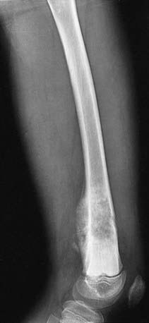
Bone tumor should be suspected in a patient who presents with deep bone pain often causing nighttime awakening in whom there is a palpable mass, and radiographs demonstrate a lesion. The lesion may be mixed lytic and blastic in appearance, but new bone formation is usually visible. The classic radiographic appearance of osteosarcoma is the sunburst pattern (Fig. 495-2). When osteosarcoma is suspected, the patient should be referred to a center with experience in managing bone tumors. The biopsy and the surgery should be performed by the same surgeon so that the incisional biopsy site can be placed in a manner that will not compromise the ultimate limb salvage procedure. Tissue usually is obtained for molecular and biologic studies at the time of the initial biopsy. Before biopsy, MRI of the primary lesion and the entire bone should be performed to evaluate the tumor for its proximity to nerves and blood vessels, soft tissue and joint extension, and skip lesions. The metastatic work-up, which should be performed before biopsy, includes CT of the chest and radionuclide bone scanning to evaluate for lung and bone metastases, respectively. The differential diagnosis of a lytic bone lesion includes histiocytosis, Ewing sarcoma, lymphoma, and bone cyst.

Treatment

With chemotherapy and surgery, the 5-yr disease-free survival rate of patients with nonmetastatic extremity osteosarcoma is 65-75%. Complete surgical resection of the tumor is important for cure. The current approach is to treat patients with preoperative chemotherapy in an attempt to facilitate limb salvage operations and to treat micrometastatic disease immediately. Up to 80% of patients are able to undergo limb salvage operations after initial chemotherapy. Some institutions use intra-arterial chemotherapy to infuse chemotherapy directly into an artery feeding the tumor, although this approach has not been shown to be better than conventional intravenous chemotherapy. It is important to resume chemotherapy as soon as possible after surgery. Lung metastases present at diagnosis should be resected by thoracotomies at some time during the course of treatment. Active agents currently in use in multidrug chemotherapy regimens for conventional osteosarcoma include doxorubicin, cisplatin, methotrexate, and ifosfamide.

One of the most important prognostic factors in osteosarcoma is the histologic response to chemotherapy. An international cooperative group is performing a randomized trial of the postoperative addition of high-dose ifosfamide with etoposide to standard three-drug therapy with cisplatin, doxorubicin, and methotrexate to improve the outcome of patients with a poor histologic response. Patients with good histologic response will be randomized to the addition of PEGylated interferon-α2b. For patients with metastatic disease, a new approach currently being investigated is the addition of zoledronic acid, a bisphosphonate, to intensive chemotherapy. After limb salvage surgery, intensive rehabilitation and physical therapy are necessary to ensure maximal functional outcome.

For patients who require amputation, early prosthetic fitting and gait training are essential to enable them to resume normal activities as soon as possible. Before definitive surgery, patients with tumors on weight-bearing bones should be instructed to use crutches to avoid stressing the weakened bones and causing pathologic fracture. The role of chemotherapy in parosteal and periosteal osteosarcomas is not well defined.

Ewing Sarcoma

Diagnosis

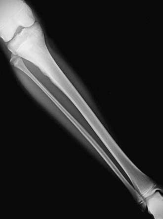
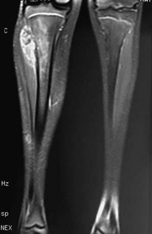
The diagnosis of Ewing sarcoma should be suspected in a patient who presents with pain and swelling, with or without systemic symptoms, and with a radiographic appearance of a primarily lytic bone lesion with periosteal reaction, the characteristic onion-skinning (Fig. 495-3). A large associated soft tissue mass often is visualized on MRI or CT (Fig. 495-4). The differential diagnosis includes osteosarcoma, osteomyelitis, Langerhans cell histiocytosis, primary lymphoma of bone, metastatic neuroblastoma, or rhabdomyosarcoma in the case of a pure soft tissue lesion. Patients should be referred to a center with experience in managing bone tumors for evaluation and biopsy. Thorough evaluation for metastatic disease includes CT of the chest, radionuclide bone scan, and bone marrow aspiration and biopsy specimens from at least two sites. MRI of the tumor and the entire length of involved bone should be performed to determine the exact extension of the soft tissue and bony mass and the proximity of tumor to neurovascular structures. To avoid compromising an ultimate potential for limb salvage by a poorly planned biopsy incision, the same surgeon should perform the biopsy and the surgical procedure. CT-guided biopsy of the lesion often provides diagnostic tissue. It is important to obtain adequate tissue for special stains, cytogenetics, and molecular studies.

Treatment

Tumors of the Ewing sarcoma family are best managed with a comprehensive multidisciplinary approach in which the surgeon, chemotherapist, and radiation oncologist plan therapy. Multiagent chemotherapy is important because it can shrink the tumor rapidly and is usually given before local control is attempted. The addition of ifosfamide and etoposide to the conventional agents vincristine, doxorubicin, and cyclophosphamide improves the outcome for patients with nonmetastatic Ewing sarcoma. Chemotherapy usually causes dramatic shrinkage of the soft tissue mass and rapid, significant pain relief. One completed randomized study for patients with nonmetastatic Ewing sarcoma showed a statistically significantly better outcome when patients were treated on a 14-day schedule than on a 21-day schedule. Current studies are evaluating the addition of topoisomerase inhibitors to standard chemotherapy, and plans are under way to investigate the role of insulin-like growth factor receptor inhibitors for certain groups of patients. An international cooperative group trial is evaluating whether myeloablative chemotherapy and stem cell rescue is superior to chemotherapy with lung irradiation for patients with pulmonary metastases. Myeloablative chemotherapy for patients with extremely high risk disease (bone and marrow metastases) also is being studied, as are approaches using angiogenesis inhibitors on a standard chemotherapy backbone. Ewing sarcoma is considered a radiosensitive tumor, and local control may be achieved with irradiation or surgery. Radiation therapy is associated with a risk of radiation-induced second malignancies, especially osteosarcoma, as well as failure of bone growth in skeletally immature patients. Many centers prefer surgical resection, if possible, to achieve local control. It is important to provide the patient with crutches if the tumor is in a weight-bearing bone, to avoid a pathologic fracture before definitive local control. Chemotherapy should be resumed as soon as possible after surgery.

495.2 Benign Tumors and Tumor-like Processes of Bone

Carola A.S. Arndt

Benign bone lesions in children are common in comparison with the relatively rare malignant neoplasms of bone and present diagnostic challenges. Some, although histologically benign, can be life-threatening. No single element in the history or diagnostic test is sufficient to rule out malignancies or suggest nonneoplastic conditions. A broad range of diagnostic possibilities must be considered when confronted with an unknown bone lesion. Benign lesions may be painless or painful, especially if a pathologic fracture is impending. Night pain that awakens a child suggests malignancy; relief of such pain with aspirin is common with benign lesions such as osteoid osteomas. Rapidly enlarging lesions usually are associated with malignancy, but several benign lesions, such as aneurysmal bone cysts, can enlarge faster than most malignancies. Several conditions, such as osteomyelitis, can simulate the appearance of benign bone tumors.

Many benign bone tumors are diagnosed incidentally or after pathologic fracture. Management of these fractures is similar to that of nonpathologic fractures in the same location. It is unusual for benign bone tumors to interfere with fracture healing. Likewise, the fractures rarely result in changes or healing of these tumors, which usually are treated after the fracture has healed.

Radiographs of any suspected bone lesion should always be obtained in two planes. Additional studies may be necessary to help arrive at the correct diagnosis and to guide treatment. Although these lesions are benign, many do require intervention.

Osteochondroma (exostosis) is one of the most common benign bone tumors in children. Because many are completely asymptomatic and unrecognized, the true incidence of this lesion is unknown. Most osteochondromas develop in childhood, arising from the metaphysis of a long bone, particularly the distal femur, proximal humerus, and proximal tibia. The lesion enlarges with the child until skeletal maturity. Most are discovered at 5-15 yr of age, when the child or parent notices a bony, nonpainful mass. Some are discovered because they are irritated by pressure during athletic or other activities. Osteochondromas appear radiographically as stalks or broad-based projections from the surface of the bone, usually in a direction away from the adjacent joint. Invariably, the lesion is radiographically smaller than suggested by palpation because the cartilage cap covering the lesion is not seen. This cartilage cap may be up to 1 cm thick. Both the cortex of the bone and the marrow space of the involved bone are continuous with the lesion. Malignant degeneration of a chondrosarcoma is rare in children but occurs in as many as 1% of adults. Routine removal is not performed unless the lesion is large enough to cause symptoms or if the lesion grows rapidly.

Multiple hereditary exostoses is a related but rare condition characterized by the presence of multiple osteochondromas. Severely involved children can have short stature, limb-length inequality, premature partial physeal arrests, and deformity of both the upper and lower extremities. These children must be monitored carefully during growth.

Enchondroma is a benign lesion of hyaline cartilage that occurs centrally in the bone. Most of these lesions are asymptomatic and occur in the hands. Most are discovered incidentally, although pathologic fractures often lead to the diagnosis. Radiographically, the lesions occupy the medullary canal, are radiolucent, and are sharply marginated. Punctate or stippled calcification may be present within the lesion, but this is much more common in adults than in children. Almost all enchondromas are solitary. Most can simply be observed, with curettage and bone grafting reserved for lesions that are symptomatic or large enough to weaken the bone structurally. Multifocal involvement is referred to as Ollier disease and can result in bone dysplasia, short stature, limb-length inequality, and joint deformity. Surgery may be necessary to correct or prevent such deformities. When multiple enchondromas are associated with angiomas of the soft tissue, the condition is referred to as Maffucci syndrome. A high rate of malignant transformation has been reported in both of these multifocal conditions.

Chondroblastoma is a rare lesion usually found in the epiphysis of long bones. Most patients present in the second decade with complaints of mild to moderate pain in the adjacent joint. Common sites include the hip, shoulder, and knee. Muscle atrophy and local tenderness may be the only clinical findings. The lesion appears radiographically as a sharply marginated radiolucency within the epiphysis or apophysis, occasionally with metaphyseal extension across the physis. Proximity to the joint can cause deformity of the subchondral bone, an effusion, or erosion into the joint. Recognition is important because most lesions can be cured with curettage and bone grafting before joint destruction occurs.

Chondromyxoid fibroma is an uncommon benign bone tumor in children. This metaphyseal lesion usually causes pain and local tenderness. The lesion occasionally is asymptomatic. Chondromyxoid fibroma appears radiographically as eccentric, lobular, metaphyseal radiolucency with sharp, sclerotic, and scalloped margins. The lower extremity is involved most often. Treatment usually consists of curettage and bone grafting or en bloc resection.

Osteoid osteoma is a small benign bone tumor. Most of these tumors are diagnosed between 5-20 yr of age. The clinical pattern is characteristic, consisting of unremitting and gradually increasing pain that often is worst at night and is relieved by aspirin. Boys are affected more often than girls. Any bone can be involved, but the most common sites are the proximal femur and tibia. Vertebral lesions can cause scoliosis or symptoms that mimic a neurologic disorder. Examination can reveal a limp, atrophy, and weakness when the lower extremity is involved. Palpation and range of motion do not alter the discomfort. Radiographs are distinctive, showing a round or oval metaphyseal or diaphyseal lucency (0.5-1.0 cm diameter) surrounded by sclerotic bone. The central lucency, or nidus, shows intense uptake on bone scan. About 25% of osteoid osteomas are not visualized on plain radiographs but can be identified with CT. Because of the small size of the lesion and its location adjacent to thick cortical bone, MRI is poor at detecting osteoid osteomas. Treatment is directed at removing the lesion. This can involve en bloc excision, curettage, or percutaneous CT-guided ablation of the nidus. Patients with mild pain may be treated with salicylates. Some lesions resolve spontaneously after skeletal maturity.

Osteoblastoma is a locally destructive, progressively growing lesion of bone with a predilection for the vertebrae, although almost any bone may be involved. Most patients note the insidious onset of dull aching pain, which may be present for months before they seek medical attention. Spinal lesions can cause neurologic symptoms or deficits. The radiographic appearance is variable and less distinctive than that of other benign bone tumors. About 25% show features suggesting a malignant neoplasm, making biopsy necessary in many cases. Expansile spinal lesions often involve the posterior elements. Treatment involves curettage and bone grafting or en bloc excision, taking care to preserve nerve roots when treating spinal lesions. Surgical stabilization of the spine may be necessary.

Fibromas (nonossifying fibroma, fibrous cortical defect, metaphyseal fibrous defect) are fibrous lesions of bone that occur in 40% of children >2 yr of age. They most likely represent a defect in ossification rather than a neoplasm and usually are asymptomatic. Most are discovered incidentally when radiographs are taken for other reasons, usually to rule out a fracture after trauma. Occasional pathologic fractures can occur through rare large lesions. Physical examination usually is unrevealing. Radiographs show a sharply marginated eccentric lucency in the metaphyseal cortex. Lesions may be multilocular and expansile, with extension from the cortex into the medullary bone. The long axis of the lesion runs parallel to that of the bone. Approximately 50% are bilateral or multiple. Because of the characteristic radiographic appearance, most lesions do not require biopsy or treatment. Spontaneous regression can be expected after skeletal maturity. Curettage and bone grafting may be recommended for lesions occupying >50% of the bone diameter because of the risk of a pathologic fracture.

Unicameral bone cysts can occur at any age in childhood but are rare in children <3 yr of age and after skeletal maturity. The cause of these fluid-filled lesions is unknown. Some resolve spontaneously after skeletal maturity is reached. Most are asymptomatic until diagnosis, which usually follows a pathologic fracture. Such fractures can occur with relatively minor trauma, such as with throwing or catching a ball. Unicameral bone cysts appear radiographically as solitary, centrally located lesions within the medullary portion of the bone. These cysts are most common in the proximal humerus or femur. They often extend to (but not through) the physis and are sharply marginated. The cortex expands, but that does not exceed the width of the adjacent physis. Treatment involves allowing the pathologic fracture to heal, followed by aspiration and injection with methylprednisolone or bone marrow. Repeat injections, curettage, and bone grafting occasionally are necessary to treat recurrent lesions.

Aneurysmal bone cyst is a reactive lesion of bone seen in persons <20 yr of age. The lesion is characterized by cavernous spaces filled with blood and solid aggregates of tissue. Although the femur, tibia, and spine are most commonly involved, this progressively growing, expansile lesion develops in any bone. Pain and swelling are common. Spinal involvement can lead to cord or nerve root compression and associated neurologic symptoms, including paralysis. Radiographs show eccentric lytic destruction and expansion of the metaphysis surrounded by a thin sclerotic rim of bone. Posterior elements of the spine are involved more commonly than the vertebral body. Unlike most other benign bone tumors, which usually are confined to a single bone, aneurysmal bone cysts can involve adjacent vertebrae. Rapid growth is characteristic and can lead to confusion with malignant neoplasms. Treatment consists of curettage and bone grafting or excision. Spinal lesions can require stabilization after excision. As with other benign tumors, attempts are made to preserve nerve roots and other vital structures. Recurrence after surgical treatment occurs in 20-30% of patients, is more common in younger than older children, and usually occurs in the first 1-2 yr after treatment.

Fibrous dysplasia is a developmental abnormality characterized by fibrous replacement of cancellous bone. Lesions may be solitary or multifocal (polyostotic), relatively stable, or progressively more severe. Most children are asymptomatic, although those with skull involvement might have swelling or exophthalmos. Pain and limp are characteristic of proximal femoral involvement. Limb-length discrepancy, bowing of the tibia or femur, and pathologic fractures may be presenting complaints. The triad of polyostotic disease, precocious puberty, and cutaneous pigmentation is known as Albright syndrome. Radiographic features of fibrous dysplasia include a lytic or ground-glass expansile lesion of the metaphysis or diaphysis. The lesion is sharply marginated and often is surrounded by a thick rim of sclerotic bone. Bowing, especially of the proximal femur, may be present. Treatment usually involves observation. Surgery is indicated for patients with progressive deformity, pain, or impending pathologic fractures. Bone grafting is not as successful in the treatment of fibrous dysplasia as with other benign tumors because the lesion often recurs within the grafted bone. Reconstructive surgical techniques often are necessary to provide stability.

Osteofibrous dysplasia affects children 1-10 yr of age. This lesion usually involves the tibia. It is clinically, radiographically, and histologically distinct from fibrous dysplasia. Most children present with anterior swelling or enlargement of the leg. Progression is unlikely after 10 yr of age. Radiographs show solitary or multiple lucent cortical diaphyseal lesions surrounded by sclerosis. Anterior bowing of the tibia often is present. The radiographic appearance closely resembles that of adamantinoma, a malignant neoplasm, making biopsy more common than with other benign bone tumors. Treatment involves observation. Some lesions heal spontaneously. Excision and bone grafting should be delayed until the child is >10 yr of age because of a high recurrence rate before this age. Pathologic fractures heal with immobilization.

Eosinophilic granuloma is a monostotic or polyostotic disease with no extraskeletal involvement. This latter finding distinguishes eosinophilic granuloma from the other forms of Langerhans’ cell histiocytosis (Hand-Schüller-Christian or Letterer-Siwe variants), which can have a less favorable prognosis (Chapter 501). Eosinophilic granuloma usually occurs during the first 3 decades of life and is most common in boys 5-10 yr of age. The skull is most commonly affected, but any bone may be involved. Patients usually present with local pain and swelling. Marked tenderness and warmth often are present in the area of the involved bone. Spinal lesions can cause pain, stiffness, and occasional neurologic symptoms. The radiographic appearance of the skeletal lesions is similar in all forms of Langerhans’ cell histiocytosis but is variable enough to mimic many other benign and malignant lesions of bone. The radiolucent lesions have well-defined or irregular margins with expansion of the involved bone and periosteal new bone formation. Spine involvement can cause uniform compression or flattening of the vertebral body. A skeletal survey is warranted because polyostotic involvement and the typical skull lesions strongly suggest the diagnosis of eosinophilic granuloma. Biopsy often is necessary to confirm the diagnosis because of the broad radiographic differential diagnosis. Treatment includes curettage and bone grafting, low-dose radiation therapy, or corticosteroid injection. Observation for symptomatic lesions is reasonable because most osseous lesions heal spontaneously and do not recur. Children with bone lesions should be evaluated for visceral involvement because treatment of Hand-Schüller-Christian disease and Letterer-Siwe disease is more complex and often systemic.

Bibliography

Arndt CAS, Crist WM. Common musculoskeletal tumors of childhood and adolescence. N Engl J Med. 1999;341:342-352.

Balamuth N, Woner R. Ewing’s sarcoma. Lancet Oncol. 2010;11(2):184-192.

Campanacci M, Capanna R, Picci P. Unicameral and aneurysmal bone cysts. Clin Orthop Relat Res. 1986;204:25-36.

Campanacci M, Laus M. Osteofibrous dysplasia of the tibia and fibula. J Bone Joint Surg Am. 1981;63:367-375.

Dahlin DC, Ivins JC. Benign chondroblastoma: a study of 125 cases. Cancer. 1972;30:401-413.

De Alava E, Gerald WL. Molecular biology of the Ewing’s sarcoma/primitive neuroectodermal tumor family. J Clin Oncol. 2000;18:204-213.

Freiberg AA, Loder RT, Heidelberger KT. Aneurysmal bone cysts in young children. J Pediatr Orthop. 1994;14:86-91.

Ginsberg JP, Woo SY, Johnson ME, et al. Ewing’s sarcoma family of tumors. In: Pizzo PA, Poplack DG, editors. Principles and practice of pediatric oncology. ed 4. Philadelphia: Lippincott Williams & Wilkins; 2009:973-1016.

Gorlick R, Anderson P, Andrulis I, et al. Biology of childhood osteogenic sarcoma and potential targets for therapeutic development: meeting summary. Clin Cancer Res. 2003;9:5442-5453.

Grier H, Krailo M, Tarbell N, et al. Addition of ifosfamide and etoposide to standard chemotherapy for Ewing’s sarcoma and primitive neuroectodermal tumor of bone. N Engl J Med. 2003;348:694-701.

Grier RJ. Surgical options for children with osteosarcoma. Lancet Oncol. 2005;6:85-92.

Kneisl JS, Simon MA. Medical management compared with operative treatment for osteoid osteoma. J Bone Joint Surg Am. 1992;74:179-185.

Marina N, Gebhardt M, Teot L, et al. Biology and therapeutic advances for pediatric osteosarcoma. Oncologist. 2004;9:422-441.

Mavrogenis AF, Papagelopoulos PJ, Soucacos PN. Skeletal osteochondromas revisited. Orthopedics. 2008;31:ii.

Meyers P, Schwartz C, Krailo M, et al. Osteosarcoma: the addition of muramyl tripeptide to chemotherapy improves overall survival—a report from the Children’s Oncology Group. J Clin Oncol. 2008;26:633-638.

Rodriguez-Galindo C, Sount SL, Pappo AS. Treatment of Ewing sarcoma family of tumors: current status and outlook for the future. Med Pediatr Oncol. 2003;40:276-287.

Schmale GA, Conrad EUIII, Raskind WH. The natural history of hereditary multiple exostoses. J Bone Joint Surg Am. 1994;76:986-992.