Neonatology

Published on 21/03/2015 by admin

Filed under Pediatrics

Last modified 22/04/2025

Print this page

rate 1 star rate 2 star rate 3 star rate 4 star rate 5 star
Your rating: none, Average: 0 (0 votes)

This article have been viewed 1455 times

Chapter 11 Neonatology

Short Cases

The neonatal examination

A thorough perinatal examination is the most commonly performed systematic assessment in paediatric practice. All medical undergraduates, graduates and paediatric postgraduates should be able to perform a thorough ‘baby check’. If there is ever a shortage of children with impressive signs around examination time, there is always a ready supply of neonates in level 2 nurseries, ideally suited to having a comprehensive baby check performed. The examination is standard irrespective of the gestation of the infant, although the interpretation of neurobehavioural findings requires a thorough knowledge of the variations between different gestational ages. The gestational age can be assessed by the Ballard method; this is indicated where the gestational age is in doubt, but is fairly complicated and not covered in this section. The object of this section is to provide a framework for the perinatal examination that is easy to remember. Only a limited sample of possible findings is given here, as a comprehensive discussion could double this book’s extent. Entire books can be, and have been, devoted to the examination of the newborn.

In hospital practice, ideally three checks are performed. An initial screening check is performed in the delivery room, assessing the baby’s adaptation to the extrauterine environment. This includes assessing the Apgar scores at 1 and 5 minutes, noting any life-threatening conditions, obvious dysmorphic features (e.g. Down syndrome), gross congenital anomalies (e.g. neural tube defects), any birth injuries and counting the umbilical cord vessels, making sure there are two arteries and one vein.

The second check should be the later comprehensive examination, within the first 24 hours of life. This is the check that must be done, irrespective of the introduction, in a neonate. The order of the examination is variable, depending on what the baby is doing at the time. Generally babies are quieter at the beginning of the examination, so always auscultate the heart first, before palpating anywhere else and disturbing the baby. The check may be approached in a somewhat opportunistic fashion: if the baby’s eyes are open when first approached, check the eyes and red light reflexes (with the ophthalmoscope set at +10); if the baby yawns or cries with a wide open mouth, take your torch and check the pharynx. Do not palpate or move the baby until the cardiorespiratory auscultation is complete: crying does not interfere very much with the remainder of the check.

After listening to the heart, listen to the chest and then palpate the abdomen and the femoral arterial pulsations in particular. Then re-inspect, checking the baby from head to toe, but leave the less pleasant aspects until later (such as checking the mouth and palate with a spatula, checking the red reflex when the baby will not open his or her eyes, checking the hips). A detailed approach is given below.

A third check is required. This may be done in hospital on day 5, but is usually performed by the local doctor at 5–10 days of age. This examination should repeat the comprehensive examination, but with particular emphasis on the following:

At this examination the urine and stool outputs are noted, and growth parameters are repeated as well, checking for adequate weight gain and feeding.

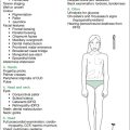
Inspect: growth, colour, respirations, posture, movements, cry

The initial thorough examination commences with inspection. Note the baby’s weight, length, head circumference and nutritional status (muscle bulk, fat). Note the colour (peripheral cyanosis is common) and the respiratory rate (normal being 40–60 breaths per minute; periodic breathing is normal) and depth (retraction indicates some respiratory distress). Causes of tachypnoea include primary respiratory disorders (e.g. hyaline membrane disease, transient tachypnoea of the newborn, meconium aspiration, congenital pneumonia, pneumothorax), space-occupying lesions (e.g. diaphragmatic hernia, congenital cystic lung lesions) or non-respiratory systems: cardiac (e.g. shunts, left-heart obstructive lesions, myocardial disease, arrhythmias), metabolic (e.g. hypoglycaemia), infective (e.g. septicaemia) or haematological causes (e.g. anaemia, polycythaemia). Stridor may be noted with upper airway obstruction. It may be purely inspiratory if the obstruction is extrathoracic, and may involve expiration if intrathoracic. The causes are usually divided into site, intraluminal (e.g. meconium, mucus, blood or milk), intramural (e.g. subglottic oedema from overenthusiastic tracheal suctioning) or extramural (e.g. vascular ring). For more details, see the short case on stridor in Chapter 15 (The respiratory system).

Posture, quality of movement and nature of cry are noted. The gestation of the infant must be considered with respect to posture. Normal full-term neonates usually lie with their hips abducted and slightly flexed, their knees flexed and their upper limbs adducted, with flexion at the elbow and clenching of the fists (not tight), with fingers covering the thumb. Term babies with significant hypotonia may lie with their hips and thighs in the ‘frog’s legs position’ (see the floppy infant case in Chapter 13, Neurology). Babies with serious intracranial pathology may lie with extended necks (opisthotonic posture), obligate flexion of thumbs or scissored lower limbs. Very premature infants (less than 30 weeks) normally have minimal flexor tone, like a rag doll; by 34 weeks, flexor tone appears in the lower limbs, and by 36 weeks in the upper limbs as well. A formal gestational assessment should be undertaken if the gestation is in doubt and the infant seems hypotonic, so as not to overinterpret normal findings of prematurity.

Normal movements may include alternating movements in all limbs, myoclonic jerks when asleep (sleep myoclonus) and jaw tremors when crying. Paucity of movement can occur in neuromuscular diseases (see the floppy infant case in Chapter 13). Involuntary movements can include tremulousness or ‘jitters’ (which can be stopped by gentle pressure) and seizures (may be subtle, tonic or more often focal, irrespective of the underlying pathology, and cannot be stopped by pressure, clearly differentiating them from jitters). Jitters can occur with hypoglycaemia, hypocalcaemia, sepsis and neonatal abstinence syndrome (NAS). Seizures can be due to neonatal encephalopathy (formally termed hypoxic ischaemic encephalopathy), intracranial haemorrhage, meningitis, metabolic causes (hypoglycaemia/hyponatraemia/hypocalcaemia/hypomagnesaemia; hypernatraemia/hyperammonaemia; kernicterus, various inborn errors, pyridoxine deficiency), developmental brain disorders and NAS, or can be idiopathic.

Asymmetric movement can occur secondary to cerebral malformations or intrauterine cerebrovascular accidents, or with birth injuries such as intracranial haemorrhage, or brachial plexus lesions such as Erb (C5,6) and Klumpke (C8,T1) palsies. Remember the myotomes of the upper limbs by the children’s nursery rhyme ‘five, six, pick up sticks [flex biceps], seven, eight, lay them straight [extension predominantly due to C7]’.

The cry is noted. A high-pitched cry goes with cerebral irritation, as in neonatal encephalopathy or meningitis. A hoarse cry can occur with upper airway obstruction and a cat cry occurs with the uncommon cri du chat syndrome (5p-syndrome).

Remember that you have to be opportunistic while a baby is settled. Do not examine the eyes or pharynx before other aspects of the examination that require a quiet baby. A suggested head-to-toe examination procedure is given below. The baby will determine in which order this occurs, however.

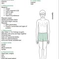
Systematic examination: head to toe

Head, neck and upper limbs

Initial inspection should detect any dysmorphic features. The head is examined for any traumatic injuries (abrasions, depressed fractures, haematomata or lacerations) and swellings (central [caput succedaneum] or to one [or both] side[s], stopping at suture lines [cephalhaematomata]). Fontanelles and sutures are palpated to assess size. They may be widened with increased intracranial pressure, as with hydrocephalus, or narrowed (or undetectable) and immobile with craniostynostosis.

The anterior fontanelle is highly variable in its shape and size. The head circumference (HC) must be measured, ideally with a paper (non-stretching) tape measure, around the head at the widest point: at term, the normal range is 32–37 cm. A useful rule of thumb is HC (cm) = height/2 + 10.

Next, the eyes are checked (if the baby allows this at the time), including red light reflexes. Make sure that the eyes are examined. Even a baby who refuses to open the eyes at all (even on parental report) should have the corneas visualised (forcibly) to exclude glaucoma (which causes them to appear enlarged and very hazy). The nose is inspected. The metal condensation test can show bilateral nasal patency. A shiny pink lesion in the nostril can be a frontal lobe encephalocoele (not mucus, so do not try to suck it out—the author has seen an encephalocoele being assaulted with a suction catheter for several minutes in just such a case). The lips, gums, tongue, palate and oropharynx must be thoroughly assessed, including using a torch and spatula. A submucous cleft can easily be missed if direct inspection is not performed. The size of the tongue is also noted: macroglossia can be a clue to hypothyroidism or to Beckwith syndrome.

The size and structure of the chin are noted. Micrognathia (mandibular hypoplasia) can occur separately or as part of the Pierre Robin sequence. At this point, the suck can be checked by placing the little finger in the baby’s mouth. The rooting reflex does not need to be tested. The ears are then checked for symmetry and positioning: deviations in each can occur with several syndromes.

The neck is then inspected for redundant skin posterolaterally, or ‘web neck’ (Noonan and Turner syndromes), and checked for any masses (e.g. lymphatic malformation or ‘cystic hygroma’) or fistulae (e.g. branchial), asymmetry (transient as in persistent fetal posturing; permanent as in the maldeveloped cervical vertebrae of Klippel–Feil syndrome) and range of movement (torticollis from sternocleidomastoid fibrosis/tumour).

Check the hands, forearms and arms from a dysmorphic viewpoint (see the case on the dysmorphic child in Chapter 9, Genetics and dysmorphology).

Cardiorespiratory system

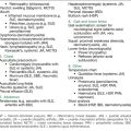
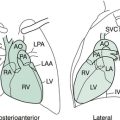
The conditions that must not be missed include coarctation (and other critical left-sided obstructive lesions such as hypoplastic left-heart syndrome, critical aortic stenosis and aortic interruption) and pulmonary hypertension, as evidenced by an active right ventricle (the presence of which, beyond day 3, may be associated with a significant systemic-to-pulmonary shunt, or an obstructed left-heart lesion, as above). A warm stethoscope helps to keep the infant quiet during auscultation. Localise the apex by auscultation. Note the heart rate (normal is 120–160 beats per minute). A murmur on day 1 can be due to a closing ductus arteriosus. This should disappear over the following days. Murmurs due to valvular lesions and vessel abnormalities (e.g. coarctation) will be present on day 1 also. Septal defects are unlikely to be heard on day 1, as there is minimal pressure differential between the left and right sides. As pressure changes occur (right ventricular pressure dropping, and left ventricular pressure increasing) the left side becomes dominant, and left-to-right shunts due to ventricular and atrial septal defects become audible after a few days. The second heart sound may be loud, and single or closely split, with pulmonary hypertension. After thorough auscultation, including all sites of possible radiation of any murmurs, listen to the chest. Finally, palpation of the femoral arteries and comparison with the brachial pulses is crucial to allow detection of the obstructed left-heart problems noted above.

Abdomen and genitalia

After inspection, gentle palpation for any masses or organomegaly is performed. The normal liver edge may be felt 1–2 cm below the costal margin in the midclavicular line, with the baby supine. The liver span at birth is around 2–2.5 cm. The kidneys may be easily palpable, especially the left one. Remove the nappy to check the groin, the genitalia and the anus. Any swelling in the groin is abnormal (e.g. indirect inguinal hernia).

In male babies, the scrotum is prominent, and should contain both testes (which should be of similar size). Hydrocoeles are common but often transient. Note the position of the urethral orifice (to detect epi- or hypospadias).

In female babies, the labia majora are prominent, and mucosal tags are frequent. There may be creamy vaginal discharge, replaced by day 3 with bleeding (pseudomenses). The labia should be spread to exclude imperforate hymen or other abnormalities (e.g. cysts of vaginal wall).

Ambiguous genitalia may represent virilised females (e.g. congenital adrenal hyperplasia [CAH], aromatase deficiency), inadequately virilised males (e.g. testicular defects, disorders of androgen production or action) or chromosomal defects. Take particular note of the phallus size, the position of orifices, labioscrotal fusion, pigmentation (CAH) and whether any gonads are palpable. For more detail, see the short case on ambiguous genitalia in Chapter 7 (Endocrinology). The baby’s anus must be evaluated for patency and position.

Hip examination

The hip examination is most important—to detect developmental dysplasia of the hip (DDH, previously called congenital dislocation of the hip [CDH]). DDH can be difficult to detect on just one examination, even for very experienced clinicians, as the femoral head could be enlocated on that occasion. Thus, examination of the hip should be re-performed after the initial assessment. Asymmetric skin creases in the buttocks may occur with DDH, but are not a reliable sign of this. Similarly, unilateral DDH can be accompanied by a weaker femoral pulse to palpation on the affected side, but this is uncommon.

There are two standard manoeuvres:

To remember which test is which, Ortolani’s Opens Out the lower limbs, Barlow’s Bumps Back the femoral head. You need remember only one of these, the other being the opposite. Often, these manoeuvres will detect a ‘click’ or ‘snap’, which is ligamentous or muscular in origin and usually of no significance if the hip feels otherwise stable. If the clinical examination is equivocal, or normal but there is a history of breech presentation, or a first-degree relative with DDH or significant oligohydramnios, hip ultrasound is indicated at 6 weeks.

Nervous system and spine

After having inspected for posture, symmetry and quality of movement, check the palmar grasp, pull the baby to sitting position, and note tone, degree of head control and head lag. Next, the baby can be positioned supine along the forearm, with the hand, palm upwards, supporting the neck and head and lifting the head gently. This support is transiently withdrawn by suddenly dorsiflexing the hand, eliciting the Moro (startle) reflex in a gentle fashion. The Moro reflex may demonstrate asymmetry of limb movement (e.g. with brachial plexus injury) or of facial expression (e.g. facial nerve palsy), or may be absent with significant intracranial pathology. If there are any concerns in any of the above areas, tap out the deep tendon reflexes.

The baby is then held in ventral suspension (normal being a convexity), and the spine is inspected carefully for abnormal curvature (e.g. scoliosis from vertebral anomalies), midline birthmarks, masses, pits or tufts of hair (e.g. diastematomyelia). Sacral or sacrococcygeal pits and sinuses are common and of no clinical importance. The anus can be inspected at this time if previously omitted. The Galant (truncal incurvation) reflex can then be checked by stroking down one side of the spine from the neck to the coccyx; the spine should curve towards that side. The Galant reflex gives some indication of segmental integrity from T2 to S1.

Next, the stepping and walking reflexes can be tested. A supported baby will make walking movements when the feet are placed on a firm surface. The baby may then have the dorsum of each foot placed against the underside of a ledge, and will normally step up on to the ledge.

This completes the neurological assessment. Testing of deep tendon reflexes is not required.

Skin

Many benign, transient rashes may affect neonates and often worry new parents. The most common rash noted in the first days of life is erythema toxicum neonatorum, appearing on any part of the body except the palms and soles as erythematous macules, usually with a central white or yellow papule. These ‘pustules’ are sterile, containing eosinophils. Milia are pinhead-sized white papules on the nose, forehead and cheeks, containing keratogenous material. Miliaria is due to obstructed eccrine sweat glands, usually seen on the forehead, scalp and skin folds.

Several pigmented lesions are common, especially the Mongolian spot, a blue–grey area over the lumbosacral region seen in most Asian and black babies, which can be mistaken for a bruise initially. Multiple café-au-lait spots occur in neurofibromatosis and several other syndromal diagnoses. Hypopigmented macules occur in tuberous sclerosus.

Vascular birthmarks are common, and there are two distinct types:

To reiterate, unpleasant aspects of the baby check may be best left until last, but must not be forgotten. These include the inspection of the palate (using spatula and torch), checking the hips (some babies seem to hate this), the Moro reflex and checking the eyes if this was not possible earlier.

The 6-week check

The procedure for the examination of the infant at the 6-week check is essentially the same as for the perinatal examination. By 6 weeks, most term infants should be able to smile responsively (spontaneous smiling occurs later, from 6 weeks to 5 months), and some primitive reflexes have been fully incorporated (stepping and walking reflexes). The baby is now able to lift the head momentarily when prone (usually by 3 weeks) and can hold the head in line with the trunk when held in ventral suspension.

The key areas to re-examine include growth parameters, particularly weight gain (normally 150–200 g per week), head circumference (e.g. to exclude hydrocephalus), cardiovascular system (murmurs and femoral pulses) for defects that may not have been apparent at discharge from hospital (e.g. septal defects, coarctation), hip examination (you can never examine the hips too often, as DDH can still present late), and eye examination to exclude cataracts or causes of leucocoria (white eye) that may have been overlooked or more difficult to detect in the earlier examination, and to check the ability to fix and follow to the midline (normally occurs by 5 weeks). If there is any jaundice, biliary atresia must be borne in mind, and direct and indirect bilirubin levels checked. Other, new problems may have arisen (e.g. nappy rashes, haemangiomata, umbilical granulomata). As with the neonatal examination, leaving the unpleasant parts until the end can be a good idea. The 6-week check is also a good time to discuss the importance of upcoming immunisation.