Neisseria meningitidis (Meningococcus)

Published on 27/03/2015 by admin

Filed under Pediatrics

Last modified 22/04/2025

Print this page

rate 1 star rate 2 star rate 3 star rate 4 star rate 5 star
Your rating: none, Average: 0 (0 votes)

This article have been viewed 1614 times

Chapter 184 Neisseria meningitidis (Meningococcus)

Neisseria meningitidis (also referred to as meningococcus) lives as a commensal in the nasopharynx of humans and is typically carried by 10% or more of the population at any one time. Relatively rarely the organism enters the bloodstream and may cause devastating disease. Why invasive meningococcal disease develops in a small proportion of exposed individuals is still largely not understood. Paradoxically, N. meningitidis also is unique for its ability to cause epidemic bacterial meningitis and sepsis. Although the last major meningococcal epidemic in the USA was in the 1940s, the organism remains an important cause of serious endemic disease in the country and of epidemic disease throughout the world. Despite advances in critical care medicine, previously healthy children and adolescents continue to succumb to fulminant meningococcal disease.

Epidemiology

Meningococci are transmitted by aerosol droplets or through contact with respiratory secretions, such as through kissing or sharing a drinking glass. The organism is not thought to survive for long periods in the environment, and transmission is decreased during periods of high ambient ultraviolet B radiation. Viral respiratory infections (influenza), exposure to tobacco smoke, marijuana use, bar patronage, binge drinking, attendance at nightclubs, and freshmen college students living in dormitories are all associated with increased rates of meningococcal carriage or disease. Respiratory viruses and/or exposure to smoke may alter the mucosal surface and enhance bacterial binding and/or decrease clearance of the organism from the nasopharynx.

Meningococcal disease is a global problem. Disease incidence rates are highly cyclic. After a decade of relatively high incidence in the 1990s, rates in the USA have steadily decreased. Over the last 10 years, the annual incidence averaged ≈1- 2/100,000 population, resulting in ≈2000 to 3500 culture-confirmed cases per year. The actual number of cases likely was higher, because in countries such as the United Kingdom, where polymerase chain reaction (PCR) methods are used routinely for diagnosis of suspected cases, only 50% of PCR-confirmed cases are culture-confirmed. In the USA, most cases of meningococcal disease are sporadic. Small outbreaks in elementary or secondary schools or colleges account for <2% of all cases.

The highest age-incidence of meningococcal disease occurs in infants <1 yr old (average annual rates of 5-9/100,000 population). The high rate in this age group is not entirely understood. It may be attributable to immature alternative and lectin complement pathways and to lack of acquired serum antibodies. In the absence of immunization, incidence rates decline by age 2-4 yr (1-2/100,000), with a further decline after age 4 yr (0.5/100,000). A secondary peak in incidence occurs among adolescents (1-3/100,000), which may be related to increased exposure from social activities.

In the USA, the majority of cases of disease in the first year of life is caused by capsular group B strains. After age 1 yr, disease is roughly equally distributed among group B, C, and Y strains. In most other industrialized countries group B strains predominate at all ages, in part because of introduction of routine group C meningococcal conjugate vaccination in infants and/or toddlers. For reasons not understood, disease in children caused by group Y strains was uncommon in the USA before the 1990s and remains relatively uncommon outside the country.

Since World War II, disease from group A strains has been largely confined to developing countries. The highest incidence of group A disease is in sub-Saharan Africa, with annual endemic rates of 10-25/ 100,000. Every 7 to 10 yr this region experiences large group A pandemics with annual rates as high as 1000/100,000. The onset of cases in the sub-Saharan region typically begins during the dry season and subsides with the rainy season, and may reemerge the following dry season. Endemic and epidemic meningococcal disease in this region has also been caused by group W-135 and X strains. These strains are infrequent causes of disease in other areas of the world, although W-135 isolates have been associated with outbreaks among pilgrims returning from the Hajj.

Pathogenesis

After exposure to meningococci, attachment of an organism to nasopharyngeal mucosal cells is mediated by specific bacterial adhesins. Multiple adhesins have been identified, but among the most important are pili and two opacity-associated proteins, Opa and Opc. CD46 and other, unidentified host cell receptors mediate pilus attachment. Opa and Opc interact with heparin sulfate proteoglycans and extracellular matrix proteins such as fibronectin and vitronectin. There are also specific receptor interactions, the most important being carcino-embryonic antigen cell adhesion molecule (CEACAM) proteins. Contact between the bacteria and host cells initiates internalization of the bacteria within membrane-bound vesicles. These molecular events lead to replication of the organism and establishment of an asymptomatic carrier state.

Although carriage can persist for weeks to months, onset of invasive meningococcal disease usually occurs within a few days to a week after acquisition of the organism. Development of disease depends on the virulence of the organism, innate susceptibility of the host, and presence or absence of serum antibodies capable of activating complement-mediated bacteriolysis and/or opsonophagocytosis. The strains responsible for invasive disease are always encapsulated and are usually derived from a limited number of so-called hypervirulent genetic lineages. Although these strains can be found in asymptomatic carriers, the majority of carrier strains either are nonencapsulated or are encapsulated organisms derived from diverse genetic lineages, many of which rarely cause disease.

The most important virulence determinant is the presence of a capsular polysaccharide, which enhances resistance of the organism to killing by normal human serum and helps resist opsonophagocytic killing. Additionally, endotoxin (lipopolysaccharide) has an essential role in stimulating cytokines and activating coagulation and bleeding, which are the clinical hallmarks of severe meningococcal sepsis. The ability of the organism to scavenge iron from human transferrin and lactoferrin and to bind human factor H (fH), a downregulating molecule in the complement cascade, are additional important mechanisms that allow meningococci to evade innate host defenses and to survive and grow in human serum or blood.

The severity of meningococcal disease is related to the circulating level of endotoxin in the bloodstream. During bacterial growth, outer membrane blebs, which are rich in endotoxin, are released. Meningococcal endotoxin is composed of lipopolysaccharide—also referred to as lipo-oligosaccharide (LOS) because of the presence of repeating short saccharides instead of long-chain saccharides characteristic of endotoxins of many other gram-negative bacteria. The lipid A portion of meningococcal LOS is responsible for the toxicity of the molecule, which is sensed by host cells through Toll-like receptors (TLRs), most notably TLR4 in association with an accessory protein, MD-2. Stimulation of TLR4 activates genes via pathways related to nuclear factor-κB (NF-κB), which leads to production of multiple proinflammatory cytokines including tumor necrosis factor-α (TNF-α), interleukin-1β (IL-1β), IL-6, and IL-8. Subsequently both the extrinsic (by way of induction of tissue factor expression on endothelial cells and monocytes) and intrinsic pathways of coagulation are activated. Progression of capillary leak and disseminated intravascular coagulopathy (DIC) can lead to multiple organ system failure, septic shock, and death. Following initiation of antibiotic therapy, circulating LOS and TNF-α levels can increase transiently as a result of rapid bacterial lysis, which then decreases with clearance of viable microbes. Activation of the complement and clotting cascades can continue well beyond this point, especially in fulminant cases.

Diffuse vasculitis and DIC are common with meningococcemia. Leukocyte-rich fibrin clots are seen in small vessels, including arterioles and capillaries. The resulting focal hemorrhage and necrosis that initially manifest as purpura in the skin may occur in any organ. The heart, central nervous system, skin, mucous and serous membranes, and adrenal glands are affected in most fatal cases, and microbes are often present in these lesions. Myocarditis is present in >50% of patients who die of meningococcal disease. Diffuse adrenal hemorrhage without vasculitis, the Waterhouse-Friderichsen syndrome, is common during fulminant meningococcemia. Meningitis is characterized by acute inflammatory cells in the leptomeninges and perivascular spaces. Focal cerebritis is uncommon.

About 10% of cases of meningococcal meningitis are caused by naturally occurring LOS mutants with penta-acylated instead of hexa-acylated lipid A. The penta-acylated mutant is poorly recognized by human TLR4 and, as a result, has attenuated endotoxin activity. Patients with meningitis caused by penta-acylated mutant strains are reported to have milder clinical syndromes, including decreased coagulopathy, than patients infected by strains that have the more common form of LOS with hexa-acylated lipid A.

Immunity

Naturally acquired serum antibodies to meningococci are elicited by asymptomatic carriage of pathogenic and nonpathogenic strains as well as by carriage of antigenically related species such as Neisseria lactamica. Bactericidal antibodies are produced against capsular polysaccharide and outer membrane proteins. Immunoglobulin M (IgM), IgG, and IgA responses are induced within a few weeks after nasopharyngeal colonization. Ongoing natural exposures may help maintain immunity.

The role of complement-mediated serum bactericidal antibodies is protective in military recruits exposed to epidemic group C meningococcal disease. Recruits with serum bactericidal titers of 1 : 4 or greater were protected from disease. The importance of serum bactericidal antibody also is underscored by a greatly increased risk of acquiring meningococcal disease in persons with inherited late complement component deficiencies (C5-C9), who lack bactericidal activity because of an inability to form a complement membrane attack complex. However, vaccine-induced antibodies in patients with late complement component deficiencies have opsonic activity, and in one study, meningococcal polysaccharide vaccination decreased the incidence of meningococcal disease among C5-C9 deficient individuals. These observations support an independent contribution of opsonophagocytic activity to protection against meningococcal disease and provide the rationale for the recommendation to immunize complement-deficient patients with meningococcal vaccines.

Host Factors

Persons with inherited deficiencies of properdin, factor D, or terminal complement components have up to a 1000-fold higher risk for development of meningococcal disease than complement-sufficient persons. The risk of meningococcal disease is also increased in patients with acquired complement deficiencies associated with diseases such as nephrotic syndrome, systemic lupus erythematosus, and hepatic failure.

Among persons with complement deficiencies, meningococcal disease is more prevalent during late childhood and adolescence, when carriage rates are higher than in children <10 yr; meningococcal infections may be recurrent. Although meningococcal disease can occasionally be overwhelming in patients with late complement component deficiency, cases are more typically described as being less severe than in complement-sufficient persons, perhaps reflecting the fact that these cases are often caused by unusual capsular groups such as W-135 and X. Although protective against early infection, extensive complement activation and bacteriolysis may contribute to the pathogenesis of severe disease once bacterial invasion has occurred.

A large number of host genetic factors appear to affect the risk and/or severity of meningococcal disease. The molecules implicated involved polymorphisms at epithelial surfaces, the complement cascade, pattern recognition receptors, clotting factors, or inflammatory mediators. To date, the strongest associations implicate genetic variation in complement regulators, particularly genes encoding mannose-binding protein (MBL), which is part of the lectin complement pathway, or in factor H, which is a down-regulator in the complement cascade. Factor H binds specifically to the surface of N. meningitidis, which enhances resistance to complement-killing of the bacteria and is critical for evasion of host defenses. Most other studies to identify susceptibility genes enrolled relatively small numbers of patients, and the results have not yet been confirmed or validated. Children with the IgG receptor allotype, FcγRIIa R/R131 (i.e., homozygous for arginine at position 131) are reported to have increased severity of meningococcal disease. One reason may be that neutrophils with this Fc receptor allotype exhibit less effective opsonophagocytosis than those with allotypes containing histidine at this position. Plasminogen activator converts plasminogen into its active form, plasmin, which elicits fibrinolysis. Functional polymorphisms in the promoter region of the gene for plasminogen-activator-inhibitor-1, which result in higher inhibitor levels and decreased fibrinolysis, have been associated with increased severity of meningococcal disease. The presence of factor V Leiden, which is known to increase the risk of thrombosis, also may exacerbate meningococcal purpura fulminans.

Clinical Manifestations

The spectrum of meningococcal disease varies widely, and recognized patterns include bacteremia without sepsis, meningococcemia without meningitis, meningitis with or without meningococcemia, and chronic infection. At least 80% of cases have overt clinical signs. Occult meningococcal bacteremia often manifests as fever with or without associated symptoms that suggest minor viral infections. Resolution of bacteremia may occur without antibiotics, but sustained bacteremia leads to meningitis in ≈60% of cases and to distant infection of other tissues. N. meningitidis is isolated from blood in about 65% of patients with meningococcal infections, from cerebrospinal fluid (CSF) in about 50% of patients, and from joint fluid in 1% of patients.

Acute meningococcemia may initially mimic illnesses caused by viruses or other bacteria, causing pharyngitis, fever, myalgias, weakness, vomiting, diarrhea, and/or headache. A fine maculopapular rash is evident in about 7% of cases, with onset typically early in the course of infection. Limb pain, myalgias, or refusal to walk occurs often and is the primary complaint in 7% of otherwise clinically unsuspected cases. Cold hands or feet and abnormal skin color are also early signs. In fulminant meningococcemia, the disease progresses rapidly over several hours from fever without other signs to septic shock characterized by prominent petechiae and purpura (purpura fulminans), hypotension, DIC, acidosis, adrenal hemorrhage, renal failure, myocardial failure, and coma (Fig. 184-1). Meningitis may or may not be present.

image

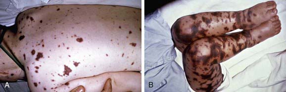
Figure 184-1 A, Purpuric rash in a 3 yr old with meningococcemia. B, Purpura fulminans in an 11 mo old with meningococcemia.

(From Thompson ED, Herzog KD: Fever and rash. In Zaoutis L, Chiang V, editors: Comprehensive pediatric hospital medicine, Philadelphia, 2007, Mosby, p 332, Figs. 62-6 and 62-7.)

Meningococcal meningitis is indistinguishable from meningitis due to other bacteria. Headache, photophobia, lethargy, vomiting, nuchal rigidity, and other signs of meningeal irritation are typically present. Seizures and focal neurologic signs occur less frequently than in patients with meningitis due to Streptococcus pneumoniae or Haemophilus influenzae type b. A meningoencephalitis-like picture can occur that may be associated with rapidly progressive cerebral edema, which may be more common with capsular group A infection.

Among 402 patients <21 yr old reported in three case series of all types of invasive meningococcal disease during the 1980s to early 2000s, about 80% presented with fever, 40% had hypotension or decreased peripheral perfusion, and 50% had petechiae and/or purpura. Purpura fulminans developed in 16%. Other presenting symptoms and signs included emesis (34%), lethargy (30%), irritability (21%), diarrhea (6%), rhinorrhea (10%), seizure (6%), and septic arthritis (8%). Radiographic evidence of pneumonia was present initially in 8% of patients in one series. Pleural effusion or empyema occurred in 15% of cases with meningococcal pneumonia and mechanical ventilation was required in 26% and vasopressor support in 35%. Nonsuppurative (presumed immune complex) arthritis developed in 4-6%. Uncommon manifestations of meningococcal disease include endocarditis, purulent pericarditis, pneumonia, endophthalmitis, mesenteric lymphadenitis, osteomyelitis, sinusitis, otitis media, and periorbital cellulitis. Primary purulent conjunctivitis can lead to invasive disease. N. meningitidis infections of the genitourinary tract are rare, but urethritis, cervicitis, vulvovaginitis, orchitis, and proctitis may occur.

Chronic meningococcemia, which occurs rarely, is characterized by fever, nontoxic appearance, arthralgias, headache, and a maculopapular to pustular rash, often with a hemorrhagic component. Symptoms are intermittent, with a mean duration of illness of 6-8 wk. Blood culture results are usually positive but cultures may initially be sterile. Chronic meningococcemia may spontaneously resolve, but meningitis can develop in untreated cases.

Diagnosis

Definitive diagnosis of meningococcal disease is established by isolation of N. meningitidis from a normally sterile body fluid such as blood, CSF, or synovial fluid. Meningococci are sometimes identified in Gram stain preparation and/or culture of petechial or purpuric skin lesions and occasionally are seen on Gram stain of the buffy coat layer of a spun blood sample. Culture results often are negative if the patient has been treated with antibiotics prior to collection of the culture specimen. Isolation of the organism from the nasopharynx is not diagnostic for invasive disease.

In patients with meningococcal meningitis, the cellular and chemical characteristics of the CSF are those of acute bacterial meningitis, showing gram-negative diplococci on Gram stain in 75% of cases. CSF culture results may be positive in patients with meningococcemia in the absence of CSF pleocytosis or clinical evidence of meningitis; conversely, positive CSF specimens that are positive for Gram stain are sometimes culture negative. Over-decolorized pneumococci in Gram stain preparations can be mistaken for meningococci, and, therefore, empirical therapy should not be narrowed to N. meningitidis infection on the basis of Gram stain findings alone.

Detection of capsular polysaccharide antigens by rapid latex agglutination tests in CSF can support the diagnosis in cases clinically consistent with meningococcal disease and is found in 53-90% of cases. Because false-positive results are reported and results do not significantly affect clinical practice, latex agglutination tests are not routinely recommended. These tests are most useful when their results are positive in the setting of partially treated infections in which Gram stain and culture results are negative. On the other hand, latex agglutination tests of serum or urine are not clinically useful. Capsular antigen tests are not reliable for group B strains because of cross reactions with other bacterial species (Escherichia coli K1 antigen). PCR-based assays for detection of meningococci in blood and CSF have been developed, and multiplex PCR assays that detect several bacterial species associated with meningitis, including the meningococcus, are under development.

Other laboratory findings include leukocytopenia or leukocytosis, often with increased percentages of neutrophils and band forms, thrombocytopenia, proteinuria, and hematuria. Elevations of erythrocyte sedimentation rate (ESR) and C-reactive protein, hypoalbuminemia, hypocalcemia, and metabolic acidosis, often with increased lactate levels, are common. Patients with DIC have decreased serum concentrations of prothrombin and fibrinogen and prolonged coagulation times.

Treatment

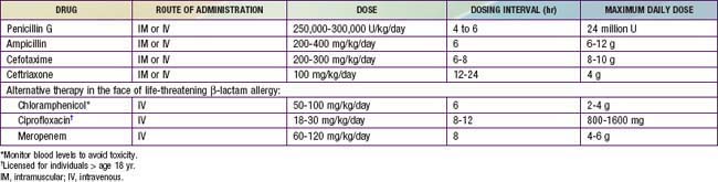
Empirical therapy should be initiated immediately for possible invasive meningococcal infections. β-Lactam antibiotics are the drugs of choice. Because of concerns about penicillin- or cephalosporin-resistant S. pneumoniae, intravenous (IV) vancomycin (60 mg/kg/day, divided in four doses, each dose given every 6 hr) should be added empirically as a second drug as part of initial empiric regimens for bacterial meningitis of unknown cause (Chapter 595.1). More specific therapy for meningococcal disease may be initiated when culture and antibiotic susceptibility results become available (Table 184-1). Although ciprofloxacin may be an alternative to cephalosporins for treatment of meningococcal infection, ciprofloxacin-resistant meningococci have been identified. Therapy in children is generally continued for 5-7 days.

Early treatment of meningococcal infections may prevent serious sequelae, but timely early diagnosis is often difficult in the absence of petechial or purpuric skin findings. High fever and leukocytosis with increased neutrophil and band counts are common in older children and adolescents with otherwise unsuspected meningococcal infection. Empiric outpatient treatment with careful follow-up of selected patients during meningococcal outbreaks and of nontoxic children with petechial rashes can be considered after blood culture specimens are obtained. Most of the latter do not have meningococcal infection.

Isolates of N. meningitidis with decreased susceptibility to penicillin (minimal inhibitory concentration of penicillin of 0.1-1.0 mg/mL) have been reported from Europe, Africa, Canada, and the USA. Decreased susceptibility is caused, at least in part, by altered penicillin-binding protein 2 and does not appear to adversely affect the response to therapy. In 2006 such strains represented ≈4% of isolates in the USA. Routine susceptibility testing of meningococcal isolates is not performed by many U.S. clinical microbiology laboratories.

Optimal supportive care is essential. Many adjunctive therapies have been attempted, but to date, none has shown clear benefit in children. Dexamethasone therapy for 2 to 4 days, with the first dose given before or during the initiation of antibiotic therapy, has decreased mortality in adults with S. pneumoniae meningitis; the benefit in patients with meningococcal meningitis has not been firmly established. Anticoagulant or fibrinolytic agents and vasodilators have been used with variable success in anecdotal reports. Activated protein C therapy is not recommended for infants with severe sepsis and purpura fulminans because of the increased risk of intracranial hemorrhage associated with its use.

Most children with meningococcal disease who do not require intubation or vasopressor support show ready response to antibiotics plus supportive care and demonstrate clinical improvement within 24-72 hr. Those requiring mechanical ventilation and other critical care interventions often have prolonged and complicated courses that may require hospitalization for weeks. Children with severe disease who show poor response to aggressive fluid and inotropic therapies may have adrenal insufficiency and may benefit from hydrocortisone supplementation. Extracorporeal membrane oxygenation, plasmapheresis, and hyperbaric oxygen have been described anecdotally as having limited success.

Complications

Acute complications of severe meningococcal disease are related to the vasculitis, DIC, and hypotension. Focal skin infarctions usually heal but can become secondarily infected, resulting in significant scarring and requiring skin grafting. The dry gangrene of extremities often seen with purpura fulminans may necessitate amputations. Adrenal hemorrhage, endophthalmitis, arthritis, endocarditis, pericarditis, myocarditis, pneumonia, lung abscess, peritonitis, and renal infarcts can occur during acute infection. Avascular necrosis of epiphyses and epiphyseal-metaphyseal defects can result from the generalized DIC and may lead to growth disturbances and late skeletal deformities.

Deafness is the most frequent neurologic sequela of meningitis, occurring in 5-10% of children. Cerebral arterial or venous thrombosis with resultant cerebral infarction can occur in severe cases. Meningococcal meningitis is rarely complicated by subdural effusion or empyema or by brain abscess. Other rare neurologic sequelae include ataxia, seizures, blindness, cranial nerve palsies, hemiparesis or quadriparesis, and obstructive hydrocephalus. The last often manifests 3-4 wk after onset of illness.

Nonsuppurative complications of meningococcal disease appear to be immune complex mediated and become apparent 4-9 days after the onset of illness. Arthritis and cutaneous vasculitis (erythema nodosum) are most common. The arthritis usually is monoarticular or oligoarticular, involves large joints, and is associated with sterile effusions that respond to nonsteroidal anti-inflammatory agents. Long-term sequelae are uncommon. Because most patients with meningococcal meningitis become afebrile by the 7th hospital day, persistence or recrudescence of fever after 5 days of antibiotics warrants evaluation for immune complex–mediated complications.

Reactivation of latent herpes simplex virus infections (primarily herpes labialis) is common during meningococcal infection.

Prevention

Close contacts of patients with meningococcal disease are at increased risk for infection. Antibiotic prophylaxis is indicated for household, daycare, and nursery school contacts and for anyone who has had contact with the patient’s oral secretions during the 7 days before onset of illness. Prophylaxis of contacts should be offered as soon as possible (Table 184-2), ideally within 24 hr of diagnosis of the patient. Because prophylaxis is not 100% effective, close contacts should be carefully monitored and brought to medical attention if they experience fever. Prophylaxis is not routinely recommended for medical personnel except those with intimate exposure, such as through mouth-to-mouth resuscitation, intubation, or suctioning before antibiotic therapy was begun.

Table 184-2 ANTIBIOTIC PROPHYLAXIS TO PREVENT NEISSERIA MENINGITIDIS INFECTION

DRUG DOSE DURATION
Rifampin:   2 days (4 doses)
Infants <1 mo 5 mg/kg PO every 12 hr  
Children >1 mo 10mg/kg PO every 12hr  
Adults 600 mg PO every 12 hr  
Ceftriaxone:    
Children <15 yr 125 mg IM 1 dose
Children >15 yr 250 mg IM 1 dose
Ciprofloxacin, persons >18 yr 500 mg PO 1 dose

IM, intramuscular; PO, by mouth.

Neither penicillin nor ampicillin treatment eradicates nasopharyngeal carriage; patients with meningococcal infection treated with penicillin or ampicillin should receive prophylaxis before hospital discharge. Droplet precautions should be observed for hospitalized patients for 24 hr after initiation of effective therapy. All confirmed or probable cases of meningococcal infection must be reported to the local public health department.

Vaccination

As of October 2010, three quadrivalent meningococcal vaccines containing capsular groups A, C, W-135, and Y are licensed in the USA. MPSV4 (Menomune, Sanfi Pasteur) contains only purified polysaccharides. The other two are conjugates of either purified polysaccharides coupled with diphtheria toxoid (MCV4-DT, Menactra, Sanofi Pasteur), or oligosaccharides coupled with a mutant, nontoxic diphtheria toxin, CRM197 (MenACWY-CRM, Menveo, Novartis Vaccines). All three vaccines are safe and effective. The conjugate vaccines more frequently cause transient fever and local redness, pain, or swelling at the injection site than MPSV4, which is attributed to the presence of the carrier proteins in the conjugate vaccines.

Following a single dose in otherwise healthy adolescents, all three vaccines elicit serum bactericidal antibody titers that peak at about 4-6 wk. These titers reflect the ability of the sera to kill the bacteria in the presence of complement. By 3 yr, half of vaccinated adolescents have serum titers <1 : 4 when measured with human complement. Titers of 1 : 4 or greater with human complement are considered protective.

Both meningococcal conjugate vaccines are more immunogenic in children than MPSV4 and, at all ages, the conjugate vaccines elicit serum bactericidal antibodies of superior quality (avidity) and persistence. The conjugate vaccines also prime for immunologic memory, which results in booster responses to a second injection. Vaccination also has the potential to decrease meningococcal carriage. In contrast, MPSV4 vaccination induces immunologic hyporesponsiveness to a subsequent injection of MPSV4 or conjugate vaccine. There is no evidence that MPSV4 vaccination decreases carriage. For all of these reasons, use of either conjugate vaccine is preferred over MPSV4.

In the USA, routine meningococcal vaccination is recommended for all children beginning at age 11 yr. In this age group, about 75% of meningococcal disease is caused by strains with capsular groups C, Y, or W-135 and therefore is potentially vaccine preventable. On the basis of reviews of age-related immunogenicity, disease burden, and cost effectiveness, the Advisory Committee on Immunization Practices (ACIP) and the American Academy of Pediatrics (AAP) do not recommend routine meningococcal vaccination for children < 11 yr. Beginning at age 2 yr, vaccination should be given to children with underlying conditions associated with increased risk of meningococcal disease (Table 184-3). As of October 2010, MPSV4 and MCV4-DT are the only vaccines approved by the FDA for use in this age group. MenACWY-CRM, however, is reported to be safe and immunogenic in children 2-10 yr, and regulatory approval by the FDA for this age group is under consideration.

The majority of otherwise healthy adolescents who were immunized at age 11-12 yr will have undetectable serum bactericidal titers by age 16 yr. Those engaging in social activities common to this age group, such as intimate kissing, smoking, or nightclub attendance, are at increased risk of exposure to N. meningitidis. At the October 2010 ACIP meeting, the Committee voted in favor of a booster dose of meningococcal conjugate vaccine for all adolescents aged 16 yr who received a first dose at age 11-12 yr (see later).

In the United Kingdom, the combination of a mass catch-up group C conjugate immunization campaign in 1999 and introduction of routine infant group C conjugate immunization resulted in a 95% decrease in group C disease. Similar results have been reported in other European countries and in Canada. Decreased nasopharyngeal carriage of group C strains among both vaccinees and nonvaccinees (herd immunity) also has been documented in the United Kingdom. In the USA, MenACWY-CRM and a new combination conjugate vaccine, meningococcal C,Y and Haemophilus influenzae type b, are likely to receive regulatory approval for use in infants and toddlers, the age groups with the highest incidence of meningococcal disease. In the meantime, the U.S. strategy targets adolescents. Immunization at this age may decrease carriage and have an indirect effect on lowering the incidence of disease in younger age groups that are not currently targeted for routine meningococcal immunization.

The group B polysaccharide capsule cross-reacts with glycosylated protein antigens present in brain, heart, and kidney and therefore is not a safe vaccine antigen. Several counties (Cuba, Norway, and New Zealand) successfully controlled group B epidemics by immunizing with tailor-made outer membrane vesicle (OMV) vaccines prepared from the respective epidemic strains. The principal limitation of OMV vaccines is that the bactericidal antibody responses are directed against PorA, which is antigenically variable. OMV vaccines are not available in the USA, where group B organisms demonstrate considerable PorA diversity. Promising alternative approaches for prevention of group B disease include two recombinant protein vaccines. Both vaccines are based on a novel antigen called factor H-binding protein (fHbp), given either alone (two antigenic variants) or in combination with two other recombinant proteins and an OMV vaccine. These vaccines are currently undergoing evaluation of safety and immunogenicity in infants, toddlers, and adolescents.

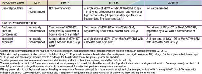
Recommendations for meningococcal vaccination can be found in Table 184-3. MCV4-DT or MenACWY-CRM as a single dose is routinely recommended for all adolescents at 11-12 yr at the preadolescent visit, and adolescents at age 15 yr or high school entry if not previously vaccinated. MPSV4 remains an acceptable alternative for this age group when conjugate vaccines are unavailable. MCV4-DT or MenACWY-CRM and the Tdap (tetanus and diphtheria toxoids and acellular pertussis booster) vaccine should be administered at separate injection sites to adolescents during the same visit if both vaccines are indicated. If this is not feasible, the meningococcal conjugate vaccines and Tdap can be administered in either sequence with a minimum interval of 1 mo between vaccines. MenACWY-CRM also can be administered at separate injection sites with Tdap and HPV vaccines. Either conjugate vaccine is also recommended for all incoming college freshmen living in dormitories who have not been previously immunized with a meningococcal vaccine. Many colleges and universities, and some states, have mandated meningococcal immunization of all matriculating freshmen. Because of waning immunity, otherwise healthy adolescents who received a first dose at age 11-12 yr should receive a booster dose of a meningococcal conjugate vaccine at 16 yr of age. For those given a first dose at age 13-15 yr, and who have not yet reached their 21st birthday, the booster dose should be given 5 yr after the first dose.

Two doses of MCV4-DT separated by 8 wk are recommended for children beginning at age 2 yr who are at increased risk of meningococcal disease because of immune or anatomic defects. These include children with anatomic or functional asplenia, complement component deficiencies, and those infected with HIV. Persons with immune or anatomic defects who have previously been vaccinated with a single dose should receive a booster dose at the earliest opportunity and then continue to receive boosters. For those immunized at 2-6 yr of age, a booster dose can be given after 3 yr. For those initially vaccinated at 7 yr of age or older, the booster dose can be given after 5 yr.

A single dose of MCV4-DT is recommended beginning at age 2 yr for otherwise healthy children traveling to areas of the world with high endemic or epidemic meningococcal disease rates such as sub-Saharan Africa. For children who continue to be at increased risk of exposure, a booster dose can be given after 3-5 yr (see above).

Cases of Guillain-Barré syndrome (GBS) with onset temporally associated with the administration of MCV4-DT had been reported to the Vaccine Adverse Events Reporting System. The data do not permit exclusion of a small increase in risk for GBS over what would be expected in the absence of a meningococcal conjugate vaccination program. A possible risk of GBS should be discussed during the informed consent process. Except in children at high risk for development of meningococcal disease, MCV4 vaccination should be avoided when there is a past medical history of GBS.

Bibliography

Allport T, Read L, Nadel S, et al. Critical illness and amputation in meningococcal septicemia: is life worth saving? Pediatrics. 2008;122:629-632.

Centers for Disease Control and Prevention. Licensure of a meningococcal conjugate vaccine (Menveo) and guidance for use—Advisory Committee on Immunization Practices (ACIP), 2010. MMWR Morbid Mortal Wkly Rep. 2010;59:273.

Centers for Disease Control and Prevention. Updated recommendation from the Advisory Committee on Immunization Practices (ACIP) for revaccination of persons at prolonged increased risk for meningococcal disease. MMWR Morbid Mortal Wkly Rep. 2009;58:1042-1043.

Centers for Disease Control and Prevention. Report from the Advisory Committee on Immunization Practices (ACIP): decision not to recommend routine vaccination of all children aged 2–10 years with quadrivalent meningococcal conjugate vaccine (MCV4). MMWR Morbid Mortal Wkly Rep. 2008;57:462-465.

Centers for Disease Control and Prevention. Revised recommendations of the Advisory Committee on Immunization Practices to vaccinate all persons aged 11–18 years with meningococcal conjugate vaccine. MMWR Morbid Mortal Wkly Rep. 2007;56:794-795.

Centers for Disease Control and Prevention. Updated recommendation from the Advisory Committee on Immunization Practices (ACIP) for revaccination of persons at prolonged increased risk for meningococcal disease. MMWR Morb Mortal Wkly Rep. 2009;58:1042-1043.

Centers for Disease Control and Prevention. Licensure of a meningococcal conjugate vaccine (Menveo) and guidance for use—Advisory Committee on Immunization Practices (ACIP), 2010. MMWR Morb Mortal Wkly Rep. 2009;59:273.

Cohn AC, MacNeil JR, Harrison LH, et al. Changes in Neisseria meningitidis disease epidemiology in the United States, 1998–2007: implications for prevention of meningococcal disease. Clin Infect Dis. 2010;50:184-193.

Davila S, Wright VJ, Chuen Khor C, et al. Genome-wide association study identifies variants in the CFH region associated with host susceptibility to meningococcal disease. Nat Genet. 2010;42:772-776.

Gardner P. Clinical practice: prevention of meningococcal disease. N Engl J Med. 2006;355:1466-1473.

Granoff DM. Relative importance of complement-mediated serum bactericidal and opsonic activity for protection against meningococcal disease. Vaccine. 2009;27(Suppl 2):B117-B125.

Granoff DM, Harrison L, Borrow R. Meningococcal vaccines. In: Plotkin SA, Offit P, Orenstein WA, editors. Vaccines. ed 5. Philadelphia: Saunders Elsevier; 2008:399-434.

Harrison LH, Kreiner CJ, Shutt KA, et al. Risk factors for meningococcal disease in student in grades 9–12. Pediatr Infect Dis J. 2008;27:193-199.

Hart CA, Thomson AP. Meningococcal disease and its management in children. BMJ. 2006;333:685-690.

Heckenberg SGB, de Gans J, Brouwer MC, et al. Clinical features, outcome, and meningococcal genotype in 258 adults with meningococcal meningitis. Medicine. 2008;87:185-192.

Maiden MC, Ibarz-Pavon AB, Urwin R, et al. Impact of meningococcal serogroup C conjugate vaccines on carriage and herd immunity. J Infect Dis. 2008;197:737-743.

Nascimento-Carvalho CM, Moreno-Carvalho OA. Changing the diagnostic framework of meningococcal disease. Lancet. 2006;367:371-372.

Snape MD, Perrett KP, Ford KJ, et al. Immunogenicity of a tetravalent meningococcal glycoconjugate vaccine in infants. JAMA. 2008;299:173-184.

Thompson MJ, Ninis N, Perera R, et al. Clinical recognition of meningococcal disease in children and adolescents. Lancet. 2006;367:397-403.

Vu DM, Welsch JA, Zuno-Mitchell P, et al. Antibody persistence 3 years after immunization of adolescents with quadrivalent meningococcal conjugate vaccine. J Infect Dis. 2006;193:821-828.

Wu HM, Harcourt BH, Hatcher CP, et al. Emergence of ciprofloxacin-resistant Neisseria meningitidis in North America. N Engl J Med. 2009;360:886-892.

Wright V, Hibber M, Levin M. Genetic polymorphisms in host response to meningococcal infection: the role of susceptibility and severity genes. Vaccine. 2009;27(Suppl 2):B90-B102.